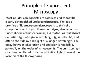
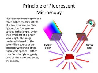
Sir George G. Stokes
The phenomenon of
fluorescence was known by
the middle of the nineteenth
century. British scientist Sir
George G. Stokes first made
the observation that the
mineral fluorspar exhibits
fluorescence when
illuminated with ultraviolet
light, and he coined the
word "fluorescence"
Differences between Conventional and
Fluorescent Microscope
Differences between Conventional and
Fluorescent Microscope
The Conventional
microscope uses
visible light (400-700
nanometers) to
illuminate and
produce a magnified
image of a sample.
A fluorescence microscope,
uses a much higher
intensity light source
which excites a
fluorescent species in a
sample of interest. This
fluorescent species in turn
emits a lower energy light
of a longer wavelength
that produces the
magnified image instead
of the original light
source.
What is Fluorescence?
Fluorescence is light
produced by a substance
when it is stimulated by
another light.
Fluorescence is called
"cold light" because it
does not come from a hot
source like an
incandescent light bulb.
What is Fluorescence
Microscopy?
Fluorescence microscopy is a unique way of using a
microscope to discover facts about specimens that often are not
shown by standard bright field microscopy. In bright field
microscopy, specimens are illuminated from outside, below or
above, and dark objects are seen against a light background. In
fluorescence microscopy, specimens are self-illuminated by
internal light, so bright objects are seen in vivid color against a
dark background. Bright objects against dark backgrounds are
more easily seen. This characteristic of fluorescence microscopy
makes it very sensitive and specific.
Principle of Fluorescent
Microscopy
Most cellular components are colorless and cannot be
clearly distinguished under a microscope. The basic
premise of fluorescence microscopy is to stain the
components with dyes. Fluorescent dyes, also known as
fluorophores of fluorochromes, are molecules that absorb
excitation light at a given wavelength (generally UV), and
after a short delay emit light at a longer wavelength. The
delay between absorption and emission is negligible,
generally on the order of nanoseconds. The emission light
can then be filtered from the excitation light to reveal the
location of the fluorophores.
JABLONSKI DIAGRAM
Principle of Fluorescent
Microscopy
Fluorescence microscopy uses a
much higher intensity light to
illuminate the sample. This
light excites fluorescence
species in the sample, which
then emit light of a longer
wavelength. The image
produced is based on the
second light source or the
emission wavelength of the
fluorescent species -- rather
than from the light originally
used to illuminate, and excite,
the sample.
Works on Faster Transmission
of Light
Fluorescence, describes
light emission that
continues only during the
absorption of the
excitation light. The time
interval between absorption of
excitation light and emission of
re-radiated light in
fluorescence is of
extraordinarily short duration,
usually less than a millionth of
a second.
Works on Principles of Light
Pathways
Specifically, a dichroic
mirror is used to separate
the excitation and
emission light paths.
Within the objective, the
excitation/emission share
the same optics. In a
fluorescence microscope,
the dichroic mirror
separates the light paths.
Advantages of Fluorescent
Microscopy
• Fluorescence microscopy is the most popular method for
studying the dynamic behavior exhibited in live cell imaging.
This stems from its ability to isolate individual proteins with a
high degree of specificity amidst non-fluorescing material.
• The sensitivity is high enough to detect as few as 50
molecules per cubic micrometer.
• Different molecules can now be stained with different
colors,
allowing multiple types of molecule to be tracked
simultaneously. These factors combine to give fluorescence
microscopy a clear advantage over other optical imaging
techniques, for both in vitro and in vivo imaging.
Fluorescence Microscope
Fluorescence
microscopy by
epi-illumination is
the most commonly
used method today
because it is simple to
do, needs relatively
simple equipment,
and is efficient.
Epifluorescence Microscopy
Epifluorescence microscopy is a
method of fluorescence
microscopy that is widely used in
life sciences The excitatory light is
passed from above (or, for
inverted microscopes, from
below), through the objective lens
and then onto the specimen
instead of passing it first through
the specimen. The fluorescence in
the specimen gives rise to emitted
light which is focused to the
detector by the same objective that
is used for the excitation
The Specimens to be Stained
Most specimens for
fluorescence microscopy
must be stained.
Fluorescent stains are
called "fluorochromes."
Acridine orange,
auramine O, and
fluorescent antibody (FA)
are the fluorochromes
used most.

Fluorescence microscopy

  • 1.
    Sir George G.Stokes The phenomenon of fluorescence was known by the middle of the nineteenth century. British scientist Sir George G. Stokes first made the observation that the mineral fluorspar exhibits fluorescence when illuminated with ultraviolet light, and he coined the word "fluorescence"
  • 2.
    Differences between Conventionaland Fluorescent Microscope
  • 3.
    Differences between Conventionaland Fluorescent Microscope The Conventional microscope uses visible light (400-700 nanometers) to illuminate and produce a magnified image of a sample. A fluorescence microscope, uses a much higher intensity light source which excites a fluorescent species in a sample of interest. This fluorescent species in turn emits a lower energy light of a longer wavelength that produces the magnified image instead of the original light source.
  • 4.
    What is Fluorescence? Fluorescenceis light produced by a substance when it is stimulated by another light. Fluorescence is called "cold light" because it does not come from a hot source like an incandescent light bulb.
  • 5.
    What is Fluorescence Microscopy? Fluorescencemicroscopy is a unique way of using a microscope to discover facts about specimens that often are not shown by standard bright field microscopy. In bright field microscopy, specimens are illuminated from outside, below or above, and dark objects are seen against a light background. In fluorescence microscopy, specimens are self-illuminated by internal light, so bright objects are seen in vivid color against a dark background. Bright objects against dark backgrounds are more easily seen. This characteristic of fluorescence microscopy makes it very sensitive and specific.
  • 6.
    Principle of Fluorescent Microscopy Mostcellular components are colorless and cannot be clearly distinguished under a microscope. The basic premise of fluorescence microscopy is to stain the components with dyes. Fluorescent dyes, also known as fluorophores of fluorochromes, are molecules that absorb excitation light at a given wavelength (generally UV), and after a short delay emit light at a longer wavelength. The delay between absorption and emission is negligible, generally on the order of nanoseconds. The emission light can then be filtered from the excitation light to reveal the location of the fluorophores.
  • 7.
  • 8.
    Principle of Fluorescent Microscopy Fluorescencemicroscopy uses a much higher intensity light to illuminate the sample. This light excites fluorescence species in the sample, which then emit light of a longer wavelength. The image produced is based on the second light source or the emission wavelength of the fluorescent species -- rather than from the light originally used to illuminate, and excite, the sample.
  • 9.
    Works on FasterTransmission of Light Fluorescence, describes light emission that continues only during the absorption of the excitation light. The time interval between absorption of excitation light and emission of re-radiated light in fluorescence is of extraordinarily short duration, usually less than a millionth of a second.
  • 10.
    Works on Principlesof Light Pathways Specifically, a dichroic mirror is used to separate the excitation and emission light paths. Within the objective, the excitation/emission share the same optics. In a fluorescence microscope, the dichroic mirror separates the light paths.
  • 11.
    Advantages of Fluorescent Microscopy •Fluorescence microscopy is the most popular method for studying the dynamic behavior exhibited in live cell imaging. This stems from its ability to isolate individual proteins with a high degree of specificity amidst non-fluorescing material. • The sensitivity is high enough to detect as few as 50 molecules per cubic micrometer. • Different molecules can now be stained with different colors, allowing multiple types of molecule to be tracked simultaneously. These factors combine to give fluorescence microscopy a clear advantage over other optical imaging techniques, for both in vitro and in vivo imaging.
  • 12.
    Fluorescence Microscope Fluorescence microscopy by epi-illuminationis the most commonly used method today because it is simple to do, needs relatively simple equipment, and is efficient.
  • 13.
    Epifluorescence Microscopy Epifluorescence microscopyis a method of fluorescence microscopy that is widely used in life sciences The excitatory light is passed from above (or, for inverted microscopes, from below), through the objective lens and then onto the specimen instead of passing it first through the specimen. The fluorescence in the specimen gives rise to emitted light which is focused to the detector by the same objective that is used for the excitation
  • 14.
    The Specimens tobe Stained Most specimens for fluorescence microscopy must be stained. Fluorescent stains are called "fluorochromes." Acridine orange, auramine O, and fluorescent antibody (FA) are the fluorochromes used most.