SlideShare a Scribd company logo
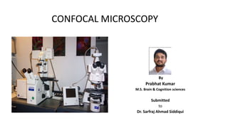
CONFOCAL MICROSCOPY
By
Prabhat Kumar
M.S. Brain & Cognition sciences
Submitted
to
Dr. Sarfraj Ahmad Siddiqui
CONFOCAL MICROSCOPY
 Introduction
 Principle
 Components
 Applications
 Advantage
 Disadvantage
 References
Introduction
 Marvin Minsky made the first confocal microscope in 1957, he was
postdoc at Harvard University.
 Also known as Confocal laser microscopy.
 This microscopy basically aim to solve the problem that is in different
microscopy techniques that is the lack of resolution.
 Confocal microscopy making balance between optical microscopy and Marvin Minsky
electron microscopy techniques in term of resolution. By using this
technique we get better resolution when we comparison other
optical microscopy.
 Uses pinhole to produce high resolution images because its exclude out of the focus light.
 So images have better contrast and are less hazy.
 Z-stack: A set of confocal images taken from the specimen so that the image area along the x- and
y-axis remains the same but the distance from the objective (z-axis) is different for each image. As
the distance between adjacent images is precisely controlled in the z-stack, it can be used to form
a 3-dimensional image.
 A series of thin slices of the specimen are assembled to generate a 3-dimensional image.(Optical
Sectioning )
 Fluorescence- emission of light by substance that has absorbed light & remission of light stop as
soon as incident light is cutoff. It occurs when electrons move
from their ground state to an excited state.
 These electrons keep the same spin as in the ground state,
but when they return to the ground state they emit energy.
 This energy has a longer wavelength than the originally
absorbed energy. If this longer wavelength is within the visible
spectrum, then we can see a glowing light.
 Example of fluorescence are :- DAPI, ALEXA , FITC , GFP etc.
Principle
 Similar to the wide field microscope, the confocal microscope
uses fluorescence optics.
 Instead of illuminating the whole sample at once, laser light is
focused onto a defined spot at a specific depth within the
sample.
 This leads to the emission of fluorescent light at exactly this
point.
 A pinhole inside the optical pathway cut off signals that are
out of focus, thus allowing only the fluorescence signals from
the illuminated spot to enter the light detector.
 3D objects can be visualized by scanning several optical planes
and stacking them using a suitable microscopy software (z-
stack).
Components
 Light Source Laser :- laser light can be chosen via
selection device and are matched with fluorophore
used in experiment.
 Pinhole :- Apertures placed near the light source and
detector enable the microscope to produce thin optical
sections of focal planes in the specimen. Its exclude the
out of focus light rays.
 Dichromic Mirror :- This is a filter which separates the
excitation from the emitted light in fluorescence beam
path of microscope, also called as beam splitter.
• Photomultiplier Detector :- Highly sensitive detectors
tube used in confocal microscopes to gain an amplified
electrical signal from a weak light source.
Applications
 Confocal microscopy has been adapted into numerous fields such as Cell biology, Life
sciences, Semiconductor inspection, Materials science, Neuroanatomy, and
Neurophysiology.
 Confocal Microscopy is a powerful tool for studying signaling mechanisms.
 Key applications include:
oDetail structure of specific part of cell
oLocalizations of proteins.
oImaging two or more fluorescence stains.
oAcquisition of high quality images for presentation.
Advantage
 It is non-invasive(no requirement of much effort ) microscopy techniques.
 We can do Qualitative as well as quantitative studies .
 We can do function and dynamic analysis.
 No requirement of vacuum .
 Examine live or fixed cell.
 We get 3-D image in confocal microscopy.
 Setup cost and maintenance cost are comparatively very low when we comparison with
electron microscopy.
 Precise and focus.
 Provides deep analysis of any part of specimen .
Disadvantage
 Damage to specimen in case of live cell.
 Decrees in Intensity of the incident light.
 The fluorophore should tagged properly to part of the specimen.
 Fluorophore should be sensitive enough for the given excitation wave length, so that they can
emit the light.
 Photo bleaching: photochemical alteration of a dye or a fluorophore molecule such that it
permanently is unable to fluorescence.
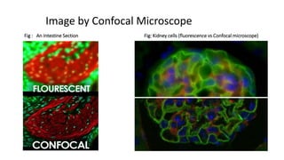
Image by Confocal Microscope
Fig : An Intestine Section Fig: Kidney cells (fluorescence vs Confocal microscope)
References
 Wilson and Walker's Principles and Techniques of Biochemistry and Molecular Biology.
 Confocal Microscopy Denis Semwogerere Eric R. Weeks, Emory University, Atlanta, Georgia, U.S.A.
 https://www.olympuslifescience.com/en/microscoperesource/primer/techniques/confocal/gloss
ary/
 http://www.physics.emory.edu/faculty/weeks//confocal/
 https://ibidi.com/content/216-confocal-microscopy
 http://microscopy.berkeley.edu/courses/tlm/clsm/index.html
Confocal microscopy by prabhat

More Related Content

What's hot

Electron microscope, principle and application
Electron microscope, principle and applicationElectron microscope, principle and application
Electron microscope, principle and application
KAUSHAL SAHU
 
Confocal microscope
Confocal microscopeConfocal microscope
Confocal microscope
SuganyaPaulraj
 
Bright field microscope
Bright field microscopeBright field microscope
Bright field microscope
Sivasangari Shanmugam
 
Principles and application of light, phase constrast and fluorescence microscope
Principles and application of light, phase constrast and fluorescence microscopePrinciples and application of light, phase constrast and fluorescence microscope
Principles and application of light, phase constrast and fluorescence microscope
MaitriThakor
 
Scanning and transmission electron microscopy
Scanning and transmission electron microscopyScanning and transmission electron microscopy
Scanning and transmission electron microscopy
gohil sanjay bhagvanji
 
Fluorescence microscopy
Fluorescence microscopyFluorescence microscopy
Fluorescence microscopy
Jyotsna Verma
 
Fluorescence and electron microscopy
Fluorescence and electron microscopyFluorescence and electron microscopy
Fluorescence and electron microscopy
Shmilona Jain
 
Fluorescence Microscopy
Fluorescence MicroscopyFluorescence Microscopy
Fluorescence Microscopy
VIVEK KUMAR SINGH
 
Transmission electron
Transmission        electronTransmission        electron
Transmission electron
Takeen Khurshid
 
Scanning Electron Microscope (SEM)
Scanning Electron Microscope (SEM)Scanning Electron Microscope (SEM)
Scanning Electron Microscope (SEM)
Amna Jalil
 
Confocal microscopy
Confocal microscopyConfocal microscopy
Confocal microscopy
akimiabdullah
 
5. Microsocope ELECTRON MICROSCOPE (TEM & SEM ) - Basics
5. Microsocope   ELECTRON MICROSCOPE (TEM & SEM ) - Basics5. Microsocope   ELECTRON MICROSCOPE (TEM & SEM ) - Basics
5. Microsocope ELECTRON MICROSCOPE (TEM & SEM ) - Basics
Nethravathi Siri
 
Transmission electron microscope
Transmission electron microscopeTransmission electron microscope
Transmission electron microscope
Senthil Arasan
 
PHASE MICROSCOPY
PHASE MICROSCOPYPHASE MICROSCOPY
PHASE MICROSCOPY
Rajpal Choudhary
 
Electron microscope ppt
Electron microscope pptElectron microscope ppt
Electron microscope ppt
NithyaNandapal
 
Electron Microscopy (SEM & TEM)
Electron Microscopy (SEM & TEM)Electron Microscopy (SEM & TEM)
Electron Microscopy (SEM & TEM)
Kalyan Acharjya
 
Microscopy
 Microscopy  Microscopy
Microscopy
Baljinder Gill
 
Confocal microscopy Likhith K
Confocal microscopy Likhith KConfocal microscopy Likhith K
Confocal microscopy Likhith K
LIKHITHK1
 
Phase contrast microscope
Phase contrast microscopePhase contrast microscope
Phase contrast microscope
SuganyaPaulraj
 
Electron microscope
Electron microscopeElectron microscope
Electron microscope
ISHRAK MAHADI
 

What's hot (20)

Electron microscope, principle and application
Electron microscope, principle and applicationElectron microscope, principle and application
Electron microscope, principle and application
 
Confocal microscope
Confocal microscopeConfocal microscope
Confocal microscope
 
Bright field microscope
Bright field microscopeBright field microscope
Bright field microscope
 
Principles and application of light, phase constrast and fluorescence microscope
Principles and application of light, phase constrast and fluorescence microscopePrinciples and application of light, phase constrast and fluorescence microscope
Principles and application of light, phase constrast and fluorescence microscope
 
Scanning and transmission electron microscopy
Scanning and transmission electron microscopyScanning and transmission electron microscopy
Scanning and transmission electron microscopy
 
Fluorescence microscopy
Fluorescence microscopyFluorescence microscopy
Fluorescence microscopy
 
Fluorescence and electron microscopy
Fluorescence and electron microscopyFluorescence and electron microscopy
Fluorescence and electron microscopy
 
Fluorescence Microscopy
Fluorescence MicroscopyFluorescence Microscopy
Fluorescence Microscopy
 
Transmission electron
Transmission        electronTransmission        electron
Transmission electron
 
Scanning Electron Microscope (SEM)
Scanning Electron Microscope (SEM)Scanning Electron Microscope (SEM)
Scanning Electron Microscope (SEM)
 
Confocal microscopy
Confocal microscopyConfocal microscopy
Confocal microscopy
 
5. Microsocope ELECTRON MICROSCOPE (TEM & SEM ) - Basics
5. Microsocope   ELECTRON MICROSCOPE (TEM & SEM ) - Basics5. Microsocope   ELECTRON MICROSCOPE (TEM & SEM ) - Basics
5. Microsocope ELECTRON MICROSCOPE (TEM & SEM ) - Basics
 
Transmission electron microscope
Transmission electron microscopeTransmission electron microscope
Transmission electron microscope
 
PHASE MICROSCOPY
PHASE MICROSCOPYPHASE MICROSCOPY
PHASE MICROSCOPY
 
Electron microscope ppt
Electron microscope pptElectron microscope ppt
Electron microscope ppt
 
Electron Microscopy (SEM & TEM)
Electron Microscopy (SEM & TEM)Electron Microscopy (SEM & TEM)
Electron Microscopy (SEM & TEM)
 
Microscopy
 Microscopy  Microscopy
Microscopy
 
Confocal microscopy Likhith K
Confocal microscopy Likhith KConfocal microscopy Likhith K
Confocal microscopy Likhith K
 
Phase contrast microscope
Phase contrast microscopePhase contrast microscope
Phase contrast microscope
 
Electron microscope
Electron microscopeElectron microscope
Electron microscope
 

Similar to Confocal microscopy by prabhat

Types of Light Microscopes used in Histological Studies.pptx
Types of Light Microscopes used in Histological Studies.pptxTypes of Light Microscopes used in Histological Studies.pptx
Types of Light Microscopes used in Histological Studies.pptx
ssuserab552f
 
Confocal Microscopy.pptx MSC BIOTECHNOLOGY
Confocal Microscopy.pptx MSC BIOTECHNOLOGYConfocal Microscopy.pptx MSC BIOTECHNOLOGY
Confocal Microscopy.pptx MSC BIOTECHNOLOGY
AnjaliKannaujiya1
 
Confocal Microscopy.pptx
Confocal Microscopy.pptxConfocal Microscopy.pptx
Confocal Microscopy.pptx
Rajadr
 
Confocal microscope ppt and their working mechanism
Confocal microscope ppt and their working mechanismConfocal microscope ppt and their working mechanism
Confocal microscope ppt and their working mechanism
PSobhangi
 
Introducation to Microscopy
Introducation to MicroscopyIntroducation to Microscopy
Introducation to Microscopy
Narottam Priyadarsahi
 
Microscope anwesha.pptx
Microscope anwesha.pptxMicroscope anwesha.pptx
Microscope anwesha.pptx
Anwesha Mandal
 
Confocal microscopy
Confocal microscopyConfocal microscopy
Confocal microscopy
DiwakarSharma71
 
2. Microscopy (Biochemistry)
2. Microscopy (Biochemistry)2. Microscopy (Biochemistry)
2. Microscopy (Biochemistry)
Jay Khaniya
 
2.0 - Types of Light Microscope.pptx ppt
2.0 - Types of Light Microscope.pptx ppt2.0 - Types of Light Microscope.pptx ppt
2.0 - Types of Light Microscope.pptx ppt
stephenopokuasante
 
2.0 - Types of Light Microscope wonder.pptx
2.0 - Types of Light Microscope wonder.pptx2.0 - Types of Light Microscope wonder.pptx
2.0 - Types of Light Microscope wonder.pptx
stephenopokuasante
 
2.0 - Types of Light Microscope.ppt microscope
2.0 - Types of Light Microscope.ppt microscope2.0 - Types of Light Microscope.ppt microscope
2.0 - Types of Light Microscope.ppt microscope
stephenopokuasante
 
Confocal laser scanning microscopy (clsm)
Confocal laser scanning microscopy (clsm)Confocal laser scanning microscopy (clsm)
Confocal laser scanning microscopy (clsm)
Nethravathi Siri
 
Application Of Biochemical Tools And Techniques.
Application Of Biochemical Tools And Techniques.Application Of Biochemical Tools And Techniques.
Application Of Biochemical Tools And Techniques.
Ashley Carter
 
MICROSCOPY.pptx
MICROSCOPY.pptxMICROSCOPY.pptx
MICROSCOPY.pptx
ArchanaBawari1
 
Microscopy - The Life of Bio science lab
Microscopy - The Life of Bio science labMicroscopy - The Life of Bio science lab
Microscopy - The Life of Bio science lab
Swami Viekananda Institute of Modern Sciences
 
ELECTRON MICROSCOPE-1.pptx
ELECTRON MICROSCOPE-1.pptxELECTRON MICROSCOPE-1.pptx
ELECTRON MICROSCOPE-1.pptx
OpenPhone
 
Microbiology Quick Learn microscope
Microbiology Quick Learn microscopeMicrobiology Quick Learn microscope
Microbiology Quick Learn microscope
Saajida Sultaana
 
Ppt f1
Ppt f1Ppt f1
Light microscope
Light microscopeLight microscope
Light microscope
khalilkazmi
 

Similar to Confocal microscopy by prabhat (20)

Types of Light Microscopes used in Histological Studies.pptx
Types of Light Microscopes used in Histological Studies.pptxTypes of Light Microscopes used in Histological Studies.pptx
Types of Light Microscopes used in Histological Studies.pptx
 
Confocal Microscopy.pptx MSC BIOTECHNOLOGY
Confocal Microscopy.pptx MSC BIOTECHNOLOGYConfocal Microscopy.pptx MSC BIOTECHNOLOGY
Confocal Microscopy.pptx MSC BIOTECHNOLOGY
 
Confocal Microscopy.pptx
Confocal Microscopy.pptxConfocal Microscopy.pptx
Confocal Microscopy.pptx
 
Confocal microscope ppt and their working mechanism
Confocal microscope ppt and their working mechanismConfocal microscope ppt and their working mechanism
Confocal microscope ppt and their working mechanism
 
Introducation to Microscopy
Introducation to MicroscopyIntroducation to Microscopy
Introducation to Microscopy
 
Microscope anwesha.pptx
Microscope anwesha.pptxMicroscope anwesha.pptx
Microscope anwesha.pptx
 
MPEF_Microscopy
MPEF_MicroscopyMPEF_Microscopy
MPEF_Microscopy
 
Confocal microscopy
Confocal microscopyConfocal microscopy
Confocal microscopy
 
2. Microscopy (Biochemistry)
2. Microscopy (Biochemistry)2. Microscopy (Biochemistry)
2. Microscopy (Biochemistry)
 
2.0 - Types of Light Microscope.pptx ppt
2.0 - Types of Light Microscope.pptx ppt2.0 - Types of Light Microscope.pptx ppt
2.0 - Types of Light Microscope.pptx ppt
 
2.0 - Types of Light Microscope wonder.pptx
2.0 - Types of Light Microscope wonder.pptx2.0 - Types of Light Microscope wonder.pptx
2.0 - Types of Light Microscope wonder.pptx
 
2.0 - Types of Light Microscope.ppt microscope
2.0 - Types of Light Microscope.ppt microscope2.0 - Types of Light Microscope.ppt microscope
2.0 - Types of Light Microscope.ppt microscope
 
Confocal laser scanning microscopy (clsm)
Confocal laser scanning microscopy (clsm)Confocal laser scanning microscopy (clsm)
Confocal laser scanning microscopy (clsm)
 
Application Of Biochemical Tools And Techniques.
Application Of Biochemical Tools And Techniques.Application Of Biochemical Tools And Techniques.
Application Of Biochemical Tools And Techniques.
 
MICROSCOPY.pptx
MICROSCOPY.pptxMICROSCOPY.pptx
MICROSCOPY.pptx
 
Microscopy - The Life of Bio science lab
Microscopy - The Life of Bio science labMicroscopy - The Life of Bio science lab
Microscopy - The Life of Bio science lab
 
ELECTRON MICROSCOPE-1.pptx
ELECTRON MICROSCOPE-1.pptxELECTRON MICROSCOPE-1.pptx
ELECTRON MICROSCOPE-1.pptx
 
Microbiology Quick Learn microscope
Microbiology Quick Learn microscopeMicrobiology Quick Learn microscope
Microbiology Quick Learn microscope
 
Ppt f1
Ppt f1Ppt f1
Ppt f1
 
Light microscope
Light microscopeLight microscope
Light microscope
 

Recently uploaded

BRACHYTHERAPY OVERVIEW AND APPLICATORS
BRACHYTHERAPY OVERVIEW  AND  APPLICATORSBRACHYTHERAPY OVERVIEW  AND  APPLICATORS
BRACHYTHERAPY OVERVIEW AND APPLICATORS
Krishan Murari
 
The Normal Electrocardiogram - Part I of II
The Normal Electrocardiogram - Part I of IIThe Normal Electrocardiogram - Part I of II
The Normal Electrocardiogram - Part I of II
MedicoseAcademics
 
MANAGEMENT OF ATRIOVENTRICULAR CONDUCTION BLOCK.pdf
MANAGEMENT OF ATRIOVENTRICULAR CONDUCTION BLOCK.pdfMANAGEMENT OF ATRIOVENTRICULAR CONDUCTION BLOCK.pdf
MANAGEMENT OF ATRIOVENTRICULAR CONDUCTION BLOCK.pdf
Jim Jacob Roy
 
Surat @ℂall @Girls ꧁❤8527049040❤꧂@ℂall @Girls Service Vip Top Model Safe
Surat @ℂall @Girls ꧁❤8527049040❤꧂@ℂall @Girls Service Vip Top Model SafeSurat @ℂall @Girls ꧁❤8527049040❤꧂@ℂall @Girls Service Vip Top Model Safe
Surat @ℂall @Girls ꧁❤8527049040❤꧂@ℂall @Girls Service Vip Top Model Safe
Savita Shen $i11
 
24 Upakrama.pptx class ppt useful in all
24 Upakrama.pptx class ppt useful in all24 Upakrama.pptx class ppt useful in all
24 Upakrama.pptx class ppt useful in all
DrSathishMS1
 
Ocular injury ppt Upendra pal optometrist upums saifai etawah
Ocular injury  ppt  Upendra pal  optometrist upums saifai etawahOcular injury  ppt  Upendra pal  optometrist upums saifai etawah
Ocular injury ppt Upendra pal optometrist upums saifai etawah
pal078100
 
Cervical & Brachial Plexus By Dr. RIG.pptx
Cervical & Brachial Plexus By Dr. RIG.pptxCervical & Brachial Plexus By Dr. RIG.pptx
Cervical & Brachial Plexus By Dr. RIG.pptx
Dr. Rabia Inam Gandapore
 
The POPPY STUDY (Preconception to post-partum cardiovascular function in prim...
The POPPY STUDY (Preconception to post-partum cardiovascular function in prim...The POPPY STUDY (Preconception to post-partum cardiovascular function in prim...
The POPPY STUDY (Preconception to post-partum cardiovascular function in prim...
Catherine Liao
 
Non-respiratory Functions of the Lungs.pdf
Non-respiratory Functions of the Lungs.pdfNon-respiratory Functions of the Lungs.pdf
Non-respiratory Functions of the Lungs.pdf
MedicoseAcademics
 
HOT NEW PRODUCT! BIG SALES FAST SHIPPING NOW FROM CHINA!! EU KU DB BK substit...
HOT NEW PRODUCT! BIG SALES FAST SHIPPING NOW FROM CHINA!! EU KU DB BK substit...HOT NEW PRODUCT! BIG SALES FAST SHIPPING NOW FROM CHINA!! EU KU DB BK substit...
HOT NEW PRODUCT! BIG SALES FAST SHIPPING NOW FROM CHINA!! EU KU DB BK substit...
GL Anaacs
 
Ophthalmology Clinical Tests for OSCE exam
Ophthalmology Clinical Tests for OSCE examOphthalmology Clinical Tests for OSCE exam
Ophthalmology Clinical Tests for OSCE exam
KafrELShiekh University
 
Surgical Site Infections, pathophysiology, and prevention.pptx
Surgical Site Infections, pathophysiology, and prevention.pptxSurgical Site Infections, pathophysiology, and prevention.pptx
Surgical Site Infections, pathophysiology, and prevention.pptx
jval Landero
 
POST OPERATIVE OLIGURIA and its management
POST OPERATIVE OLIGURIA and its managementPOST OPERATIVE OLIGURIA and its management
POST OPERATIVE OLIGURIA and its management
touseefaziz1
 
Couples presenting to the infertility clinic- Do they really have infertility...
Couples presenting to the infertility clinic- Do they really have infertility...Couples presenting to the infertility clinic- Do they really have infertility...
Couples presenting to the infertility clinic- Do they really have infertility...
Sujoy Dasgupta
 
heat stroke and heat exhaustion in children
heat stroke and heat exhaustion in childrenheat stroke and heat exhaustion in children
heat stroke and heat exhaustion in children
SumeraAhmad5
 
The hemodynamic and autonomic determinants of elevated blood pressure in obes...
The hemodynamic and autonomic determinants of elevated blood pressure in obes...The hemodynamic and autonomic determinants of elevated blood pressure in obes...
The hemodynamic and autonomic determinants of elevated blood pressure in obes...
Catherine Liao
 
THOA 2.ppt Human Organ Transplantation Act
THOA 2.ppt Human Organ Transplantation ActTHOA 2.ppt Human Organ Transplantation Act
THOA 2.ppt Human Organ Transplantation Act
DrSathishMS1
 
ARTHROLOGY PPT NCISM SYLLABUS AYURVEDA STUDENTS
ARTHROLOGY PPT NCISM SYLLABUS AYURVEDA STUDENTSARTHROLOGY PPT NCISM SYLLABUS AYURVEDA STUDENTS
ARTHROLOGY PPT NCISM SYLLABUS AYURVEDA STUDENTS
Dr. Vinay Pareek
 
Alcohol_Dr. Jeenal Mistry MD Pharmacology.pdf
Alcohol_Dr. Jeenal Mistry MD Pharmacology.pdfAlcohol_Dr. Jeenal Mistry MD Pharmacology.pdf
Alcohol_Dr. Jeenal Mistry MD Pharmacology.pdf
Dr Jeenal Mistry
 
How to Give Better Lectures: Some Tips for Doctors
How to Give Better Lectures: Some Tips for DoctorsHow to Give Better Lectures: Some Tips for Doctors
How to Give Better Lectures: Some Tips for Doctors
LanceCatedral
 

Recently uploaded (20)

BRACHYTHERAPY OVERVIEW AND APPLICATORS
BRACHYTHERAPY OVERVIEW  AND  APPLICATORSBRACHYTHERAPY OVERVIEW  AND  APPLICATORS
BRACHYTHERAPY OVERVIEW AND APPLICATORS
 
The Normal Electrocardiogram - Part I of II
The Normal Electrocardiogram - Part I of IIThe Normal Electrocardiogram - Part I of II
The Normal Electrocardiogram - Part I of II
 
MANAGEMENT OF ATRIOVENTRICULAR CONDUCTION BLOCK.pdf
MANAGEMENT OF ATRIOVENTRICULAR CONDUCTION BLOCK.pdfMANAGEMENT OF ATRIOVENTRICULAR CONDUCTION BLOCK.pdf
MANAGEMENT OF ATRIOVENTRICULAR CONDUCTION BLOCK.pdf
 
Surat @ℂall @Girls ꧁❤8527049040❤꧂@ℂall @Girls Service Vip Top Model Safe
Surat @ℂall @Girls ꧁❤8527049040❤꧂@ℂall @Girls Service Vip Top Model SafeSurat @ℂall @Girls ꧁❤8527049040❤꧂@ℂall @Girls Service Vip Top Model Safe
Surat @ℂall @Girls ꧁❤8527049040❤꧂@ℂall @Girls Service Vip Top Model Safe
 
24 Upakrama.pptx class ppt useful in all
24 Upakrama.pptx class ppt useful in all24 Upakrama.pptx class ppt useful in all
24 Upakrama.pptx class ppt useful in all
 
Ocular injury ppt Upendra pal optometrist upums saifai etawah
Ocular injury  ppt  Upendra pal  optometrist upums saifai etawahOcular injury  ppt  Upendra pal  optometrist upums saifai etawah
Ocular injury ppt Upendra pal optometrist upums saifai etawah
 
Cervical & Brachial Plexus By Dr. RIG.pptx
Cervical & Brachial Plexus By Dr. RIG.pptxCervical & Brachial Plexus By Dr. RIG.pptx
Cervical & Brachial Plexus By Dr. RIG.pptx
 
The POPPY STUDY (Preconception to post-partum cardiovascular function in prim...
The POPPY STUDY (Preconception to post-partum cardiovascular function in prim...The POPPY STUDY (Preconception to post-partum cardiovascular function in prim...
The POPPY STUDY (Preconception to post-partum cardiovascular function in prim...
 
Non-respiratory Functions of the Lungs.pdf
Non-respiratory Functions of the Lungs.pdfNon-respiratory Functions of the Lungs.pdf
Non-respiratory Functions of the Lungs.pdf
 
HOT NEW PRODUCT! BIG SALES FAST SHIPPING NOW FROM CHINA!! EU KU DB BK substit...
HOT NEW PRODUCT! BIG SALES FAST SHIPPING NOW FROM CHINA!! EU KU DB BK substit...HOT NEW PRODUCT! BIG SALES FAST SHIPPING NOW FROM CHINA!! EU KU DB BK substit...
HOT NEW PRODUCT! BIG SALES FAST SHIPPING NOW FROM CHINA!! EU KU DB BK substit...
 
Ophthalmology Clinical Tests for OSCE exam
Ophthalmology Clinical Tests for OSCE examOphthalmology Clinical Tests for OSCE exam
Ophthalmology Clinical Tests for OSCE exam
 
Surgical Site Infections, pathophysiology, and prevention.pptx
Surgical Site Infections, pathophysiology, and prevention.pptxSurgical Site Infections, pathophysiology, and prevention.pptx
Surgical Site Infections, pathophysiology, and prevention.pptx
 
POST OPERATIVE OLIGURIA and its management
POST OPERATIVE OLIGURIA and its managementPOST OPERATIVE OLIGURIA and its management
POST OPERATIVE OLIGURIA and its management
 
Couples presenting to the infertility clinic- Do they really have infertility...
Couples presenting to the infertility clinic- Do they really have infertility...Couples presenting to the infertility clinic- Do they really have infertility...
Couples presenting to the infertility clinic- Do they really have infertility...
 
heat stroke and heat exhaustion in children
heat stroke and heat exhaustion in childrenheat stroke and heat exhaustion in children
heat stroke and heat exhaustion in children
 
The hemodynamic and autonomic determinants of elevated blood pressure in obes...
The hemodynamic and autonomic determinants of elevated blood pressure in obes...The hemodynamic and autonomic determinants of elevated blood pressure in obes...
The hemodynamic and autonomic determinants of elevated blood pressure in obes...
 
THOA 2.ppt Human Organ Transplantation Act
THOA 2.ppt Human Organ Transplantation ActTHOA 2.ppt Human Organ Transplantation Act
THOA 2.ppt Human Organ Transplantation Act
 
ARTHROLOGY PPT NCISM SYLLABUS AYURVEDA STUDENTS
ARTHROLOGY PPT NCISM SYLLABUS AYURVEDA STUDENTSARTHROLOGY PPT NCISM SYLLABUS AYURVEDA STUDENTS
ARTHROLOGY PPT NCISM SYLLABUS AYURVEDA STUDENTS
 
Alcohol_Dr. Jeenal Mistry MD Pharmacology.pdf
Alcohol_Dr. Jeenal Mistry MD Pharmacology.pdfAlcohol_Dr. Jeenal Mistry MD Pharmacology.pdf
Alcohol_Dr. Jeenal Mistry MD Pharmacology.pdf
 
How to Give Better Lectures: Some Tips for Doctors
How to Give Better Lectures: Some Tips for DoctorsHow to Give Better Lectures: Some Tips for Doctors
How to Give Better Lectures: Some Tips for Doctors
 

Confocal microscopy by prabhat

  • 1. CONFOCAL MICROSCOPY By Prabhat Kumar M.S. Brain & Cognition sciences Submitted to Dr. Sarfraj Ahmad Siddiqui
  • 2. CONFOCAL MICROSCOPY  Introduction  Principle  Components  Applications  Advantage  Disadvantage  References
  • 3. Introduction  Marvin Minsky made the first confocal microscope in 1957, he was postdoc at Harvard University.  Also known as Confocal laser microscopy.  This microscopy basically aim to solve the problem that is in different microscopy techniques that is the lack of resolution.  Confocal microscopy making balance between optical microscopy and Marvin Minsky electron microscopy techniques in term of resolution. By using this technique we get better resolution when we comparison other optical microscopy.  Uses pinhole to produce high resolution images because its exclude out of the focus light.  So images have better contrast and are less hazy.
  • 4.  Z-stack: A set of confocal images taken from the specimen so that the image area along the x- and y-axis remains the same but the distance from the objective (z-axis) is different for each image. As the distance between adjacent images is precisely controlled in the z-stack, it can be used to form a 3-dimensional image.  A series of thin slices of the specimen are assembled to generate a 3-dimensional image.(Optical Sectioning )  Fluorescence- emission of light by substance that has absorbed light & remission of light stop as soon as incident light is cutoff. It occurs when electrons move from their ground state to an excited state.  These electrons keep the same spin as in the ground state, but when they return to the ground state they emit energy.  This energy has a longer wavelength than the originally absorbed energy. If this longer wavelength is within the visible spectrum, then we can see a glowing light.  Example of fluorescence are :- DAPI, ALEXA , FITC , GFP etc.
  • 5. Principle  Similar to the wide field microscope, the confocal microscope uses fluorescence optics.  Instead of illuminating the whole sample at once, laser light is focused onto a defined spot at a specific depth within the sample.  This leads to the emission of fluorescent light at exactly this point.  A pinhole inside the optical pathway cut off signals that are out of focus, thus allowing only the fluorescence signals from the illuminated spot to enter the light detector.  3D objects can be visualized by scanning several optical planes and stacking them using a suitable microscopy software (z- stack).
  • 6. Components  Light Source Laser :- laser light can be chosen via selection device and are matched with fluorophore used in experiment.  Pinhole :- Apertures placed near the light source and detector enable the microscope to produce thin optical sections of focal planes in the specimen. Its exclude the out of focus light rays.  Dichromic Mirror :- This is a filter which separates the excitation from the emitted light in fluorescence beam path of microscope, also called as beam splitter. • Photomultiplier Detector :- Highly sensitive detectors tube used in confocal microscopes to gain an amplified electrical signal from a weak light source.
  • 7. Applications  Confocal microscopy has been adapted into numerous fields such as Cell biology, Life sciences, Semiconductor inspection, Materials science, Neuroanatomy, and Neurophysiology.  Confocal Microscopy is a powerful tool for studying signaling mechanisms.  Key applications include: oDetail structure of specific part of cell oLocalizations of proteins. oImaging two or more fluorescence stains. oAcquisition of high quality images for presentation.
  • 8. Advantage  It is non-invasive(no requirement of much effort ) microscopy techniques.  We can do Qualitative as well as quantitative studies .  We can do function and dynamic analysis.  No requirement of vacuum .  Examine live or fixed cell.  We get 3-D image in confocal microscopy.  Setup cost and maintenance cost are comparatively very low when we comparison with electron microscopy.  Precise and focus.  Provides deep analysis of any part of specimen .
  • 9. Disadvantage  Damage to specimen in case of live cell.  Decrees in Intensity of the incident light.  The fluorophore should tagged properly to part of the specimen.  Fluorophore should be sensitive enough for the given excitation wave length, so that they can emit the light.  Photo bleaching: photochemical alteration of a dye or a fluorophore molecule such that it permanently is unable to fluorescence.
  • 10. Image by Confocal Microscope Fig : An Intestine Section Fig: Kidney cells (fluorescence vs Confocal microscope)
  • 11. References  Wilson and Walker's Principles and Techniques of Biochemistry and Molecular Biology.  Confocal Microscopy Denis Semwogerere Eric R. Weeks, Emory University, Atlanta, Georgia, U.S.A.  https://www.olympuslifescience.com/en/microscoperesource/primer/techniques/confocal/gloss ary/  http://www.physics.emory.edu/faculty/weeks//confocal/  https://ibidi.com/content/216-confocal-microscopy  http://microscopy.berkeley.edu/courses/tlm/clsm/index.html