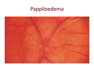
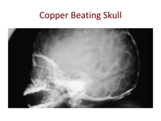
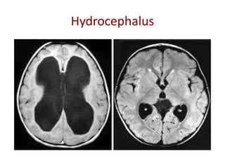
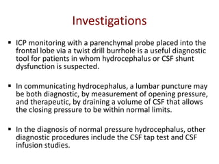
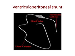
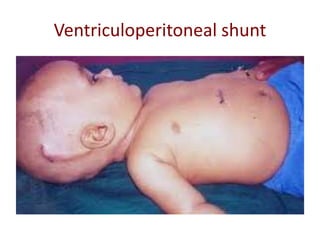
ELECTIVE NEUROSURGERY
Dr Tridip Dutta Baruah
Asst Prof, Dept Of Surgery
MGMCRI
Objectives
To understand and know:
 The physiology of CSF production and
understand the sign and symptoms of raised ICP
 The pathophysiology of hydrocephalus and its
treatment
Intracranial Pressure
 Intracranial pressure is the pressure inside the skull and
thus in the brain tissue and cerebrospinal fluid.
 Normal ICP varies from 5 to 15 mmHg in the adult at rest.
 ICP varies with venous pressure and is thus affected by
factors such as gravitational drainage an manoeuvres that
raise intrathoracic pressure .
 Normal values in small children and infants are lower
than for adults.
Raised intracranial pressure
 Results in reduced cerebral perfusion and
brain herniation
 The major causes of raised ICP are
haematomas, tumours and hydrocephalus
Pathophysiology Of Raised ICP
The Monroe Kellie Hypothesis: It is the pressure volume
relationship between ICP, volume of CSF, brain tissue and
Cerebral Perfusion Pressure.
 The hypothesis states that cranium is incompressible and
the volume inside the cranium is fixed.
 The addition of a new mass lesion can initially be
compensated for by the egress of CSF and venous blood
from the skull. During this compensation phase, there is
only a small increase in ICP.
 When compensation is maximal, there is then a rapid rise
in ICP. This increased pressure causes compression and
herniation of the brain
Raised ICP and Cerebrovascular Physiology
 The brain does not store much energy and is
unable to utilise anaerobic metabolism.
 Cerebral Perfusion Pressure=MAP – ICP
 In normal circumstances, cerebral blood flow is
maintained at a constant rate despite fluctuations
in mean arterial pressure (MAP) of between 50 and
150 mmHg via mechanisms termed cerebral
autoregulation.
 In the injured brain, cerebral autoregulation may
be impaired either locally or globally.
Cerebral Herniation
Sub-falcine herniation refers to shift of the cingulate gyrus of
one hemisphere under the falx cerebri .
Uncal herniation refers to shift of the medial temporal lobe
(uncus) medially towards the tentorial hiatus.
Tentorial herniation refers to a downwards shift of midbrain
structures through the tentorial hiatus. Tonsillar herniation
refers to a downwards shift of the cerebellar tonsils and
medulla through the foramen magnum. This type of
herniation is associated with death.
Cerebral Herniation
Symptoms Of Raised ICP
 Headache Early morning: Worse on lying
down
 Nausea and vomiting
 Visual blurring or double vision
 Drowsiness
Signs Of Raised ICP
Pappiloedema
Sunsetting Sign
Copper Beating Skull
Treatment Of Raised ICP
Appropriate treatment depends on identifying the cause.
Medical
i) Mannitol : osmotic diuretic
ii) High-dose steroids. Steroids reduce the permeability of the blood–
brain barrier.
iii) carbonic anhydrase inhibitor such as Acetazolamide.
Surgical
i) Craniotomy: in trauma, acute extradural
and subdural haematomas, intracerebral contusions and chronic
subdural haematomas.
ii) Occasionally, surgical control of ICP will involve a large
bony decompression (craniectomy), such as in traumatic brain
injury or extensive middle cerebral artery (MCA) infarction
Hydrocephalus
 Hydrocephalus is a condition in which there is
disequilibrium between CSF production and
absorption, leading to raised ICP, and is often
associated with dilated ventricles.
 Not all patients with ventriculomegaly have
hydrocephalus and not all patients with
hydrocephalus necessarily have enlarged
ventricles.
 It can be Obstructive or comunicating.
Cerebrospinal Fluid Physiology
 The total CSF volume in an adult is about 150 ml.
 CSF production occurs at a rate of approximately 0.33 ml per min
or 450 ml per day, resulting in a turnover of three volumes per day.
 CSF production is primarily by the choroid plexus of the ventricles
and is an active process independent of ICP.
 CSF flows from the lateral ventricles, through the foramen of
Munro, into the third ventricle and then into the cerebral aqueduct
and fourth ventricle before exiting into the subarachnoid space via
the midline foramen of Magendie and lateral foramina of Lushka.
 CSF absorption is a pressure-dependent passive process involving
filtration across the arachnoid villi, which are abundant along the
superior sagittal sinus into which the CSF is absorbed.
CSF Pathway
Aetiology
Hydrocephalus
Hydrocephalus
Hydrocephalus
Hydrocephalus
Investigations
 Lumbar puncture is contraindicated in obstructive hydrocephalus
because of the risk of causing tonsillar herniation and death.
 Ventricular size can be assessed with a computerised tomography
(CT) scan of the brain. The ventricles may be enlarged as a result of
generalised cerebral atrophy or localised neuronal cell loss (ex vacuo
dilatation) as well as by hydrocephalus.
 In children, chronic raised ICP can result in copperbeating of the
skull.
 A magnetic resonance imaging (MRI) scan of the brain can provide
better anatomical detail of lesions causing hydrocephalus and is
particularly useful in the diagnosis of aqueduct stenosis.
Investigations
 ICP monitoring with a parenchymal probe placed into the
frontal lobe via a twist drill burrhole is a useful diagnostic
tool for patients in whom hydrocephalus or CSF shunt
dysfunction is suspected.
 In communicating hydrocephalus, a lumbar puncture may
be both diagnostic, by measurement of opening pressure,
and therapeutic, by draining a volume of CSF that allows
the closing pressure to be within normal limits.
 In the diagnosis of normal pressure hydrocephalus, other
diagnostic procedures include the CSF tap test and CSF
infusion studies.
Management
Management of hydrocephalus will depend on
the underlying cause. Options include
 Removing a causative mass lesion
 Ventricular shunting or third ventriculostomy.
 Removing a causative mass lesion
External Drains
 External drains can be placed within the
ventricle (EVD) or the lumbar thecal sac
(lumbar drain).
 These are useful for temporary CSF drainage
and can be used to administer intrathecal
antibiotics to treat CSF infection.
Removing Causative Mass Lesion
 Intracranial mass lesions may present with
obstructive hydrocephalus.In some circumstances
it may be appropriate to treat the hydrocephalus
by tumour removal and decompression of the CSF
pathways.
 In other cases, such as a patient who presents
with an impaired conscious level secondary to
obstructive hydrocephalus, it may be appropriate
to treat the hydrocephalus with an EVD or
ventriculoperitoneal shunt and allow the patient
to recover before undertaking tumour surgery.
Ventriculoperitoneal shunt
 A ventriculoperitoneal shunt involves the insertion of
a catheter into the lateral ventricle (usually right
frontal or occipital).
 The catheter is then connected to a shunt valve under
the scalp and finally to a distal catheter, which is
tunnelled subcutaneously down to the abdomen and
inserted into the peritoneal cavity.
 If the CSF pressure exceeds the shunt valve pressure,
then CSF will flow out of the distal catheter and be
absorbed by the peritoneal lining.
Ventriculoperitoneal shunt
Complications Of VP Shunt
 Most common complications include shunt blockage and
infection. Approximately 15–20% of shunts are revised
withinthe first 3 yrs.
 Shunt blockage may affect the ventricular catheter, shunt
valve or distal catheter. Causes of blockage are choroid
plexus adhesion, blood, cellular debris or misplacement of
the distal catheter in the pre-peritoneal space.
 Shunt infection affects between 1% and 7% of shunt
insertions and is usually caused by skin commensals, such
as Staphylococcus epidermidis. Most infections become
apparent clinically by 6 weeks and over 90% are apparent
within 6 months. Treatment is by removal of the shunt,
external CSF drainage and treatment of infection prior to
Endoscopic Third Ventriculostomy
 ETV involves the insertion of a neuroendoscope into the frontal horn
of the lateral ventricle and then into the third ventricle through the
foramen of Munro. A stoma can be created in the floor of the third
ventricle in between the mamillary bodies and infundibular
(pituitary)recess.
 CSF can then communicate freely between the ventricular system
and interpeduncular subarachnoid space. It is useful when there is
obstruction of the CSF pathways below the third ventricle such
aqueduct stenosis or posterior fossa mass lesions.
 Advantage over shunting in that no tubing is left in the patient and
so infection rates are lower. ETVs may block off, however, with about
one-half of these patients ending up with a shunt. Rare, but serious,
complications include basilar artery rupture or memory impairment
from injury to the fornix.

Elective neurosurgery

  • 1.
    ELECTIVE NEUROSURGERY Dr TridipDutta Baruah Asst Prof, Dept Of Surgery MGMCRI
  • 2.
    Objectives To understand andknow:  The physiology of CSF production and understand the sign and symptoms of raised ICP  The pathophysiology of hydrocephalus and its treatment
  • 3.
    Intracranial Pressure  Intracranialpressure is the pressure inside the skull and thus in the brain tissue and cerebrospinal fluid.  Normal ICP varies from 5 to 15 mmHg in the adult at rest.  ICP varies with venous pressure and is thus affected by factors such as gravitational drainage an manoeuvres that raise intrathoracic pressure .  Normal values in small children and infants are lower than for adults.
  • 4.
    Raised intracranial pressure Results in reduced cerebral perfusion and brain herniation  The major causes of raised ICP are haematomas, tumours and hydrocephalus
  • 5.
    Pathophysiology Of RaisedICP The Monroe Kellie Hypothesis: It is the pressure volume relationship between ICP, volume of CSF, brain tissue and Cerebral Perfusion Pressure.  The hypothesis states that cranium is incompressible and the volume inside the cranium is fixed.  The addition of a new mass lesion can initially be compensated for by the egress of CSF and venous blood from the skull. During this compensation phase, there is only a small increase in ICP.  When compensation is maximal, there is then a rapid rise in ICP. This increased pressure causes compression and herniation of the brain
  • 6.
    Raised ICP andCerebrovascular Physiology  The brain does not store much energy and is unable to utilise anaerobic metabolism.  Cerebral Perfusion Pressure=MAP – ICP  In normal circumstances, cerebral blood flow is maintained at a constant rate despite fluctuations in mean arterial pressure (MAP) of between 50 and 150 mmHg via mechanisms termed cerebral autoregulation.  In the injured brain, cerebral autoregulation may be impaired either locally or globally.
  • 7.
    Cerebral Herniation Sub-falcine herniationrefers to shift of the cingulate gyrus of one hemisphere under the falx cerebri . Uncal herniation refers to shift of the medial temporal lobe (uncus) medially towards the tentorial hiatus. Tentorial herniation refers to a downwards shift of midbrain structures through the tentorial hiatus. Tonsillar herniation refers to a downwards shift of the cerebellar tonsils and medulla through the foramen magnum. This type of herniation is associated with death.
  • 8.
  • 9.
    Symptoms Of RaisedICP  Headache Early morning: Worse on lying down  Nausea and vomiting  Visual blurring or double vision  Drowsiness
  • 10.
  • 11.
  • 12.
  • 13.
  • 14.
    Treatment Of RaisedICP Appropriate treatment depends on identifying the cause. Medical i) Mannitol : osmotic diuretic ii) High-dose steroids. Steroids reduce the permeability of the blood– brain barrier. iii) carbonic anhydrase inhibitor such as Acetazolamide. Surgical i) Craniotomy: in trauma, acute extradural and subdural haematomas, intracerebral contusions and chronic subdural haematomas. ii) Occasionally, surgical control of ICP will involve a large bony decompression (craniectomy), such as in traumatic brain injury or extensive middle cerebral artery (MCA) infarction
  • 15.
    Hydrocephalus  Hydrocephalus isa condition in which there is disequilibrium between CSF production and absorption, leading to raised ICP, and is often associated with dilated ventricles.  Not all patients with ventriculomegaly have hydrocephalus and not all patients with hydrocephalus necessarily have enlarged ventricles.  It can be Obstructive or comunicating.
  • 16.
    Cerebrospinal Fluid Physiology The total CSF volume in an adult is about 150 ml.  CSF production occurs at a rate of approximately 0.33 ml per min or 450 ml per day, resulting in a turnover of three volumes per day.  CSF production is primarily by the choroid plexus of the ventricles and is an active process independent of ICP.  CSF flows from the lateral ventricles, through the foramen of Munro, into the third ventricle and then into the cerebral aqueduct and fourth ventricle before exiting into the subarachnoid space via the midline foramen of Magendie and lateral foramina of Lushka.  CSF absorption is a pressure-dependent passive process involving filtration across the arachnoid villi, which are abundant along the superior sagittal sinus into which the CSF is absorbed.
  • 17.
  • 18.
  • 19.
  • 20.
  • 21.
  • 22.
  • 23.
    Investigations  Lumbar punctureis contraindicated in obstructive hydrocephalus because of the risk of causing tonsillar herniation and death.  Ventricular size can be assessed with a computerised tomography (CT) scan of the brain. The ventricles may be enlarged as a result of generalised cerebral atrophy or localised neuronal cell loss (ex vacuo dilatation) as well as by hydrocephalus.  In children, chronic raised ICP can result in copperbeating of the skull.  A magnetic resonance imaging (MRI) scan of the brain can provide better anatomical detail of lesions causing hydrocephalus and is particularly useful in the diagnosis of aqueduct stenosis.
  • 24.
    Investigations  ICP monitoringwith a parenchymal probe placed into the frontal lobe via a twist drill burrhole is a useful diagnostic tool for patients in whom hydrocephalus or CSF shunt dysfunction is suspected.  In communicating hydrocephalus, a lumbar puncture may be both diagnostic, by measurement of opening pressure, and therapeutic, by draining a volume of CSF that allows the closing pressure to be within normal limits.  In the diagnosis of normal pressure hydrocephalus, other diagnostic procedures include the CSF tap test and CSF infusion studies.
  • 25.
    Management Management of hydrocephaluswill depend on the underlying cause. Options include  Removing a causative mass lesion  Ventricular shunting or third ventriculostomy.  Removing a causative mass lesion
  • 26.
    External Drains  Externaldrains can be placed within the ventricle (EVD) or the lumbar thecal sac (lumbar drain).  These are useful for temporary CSF drainage and can be used to administer intrathecal antibiotics to treat CSF infection.
  • 27.
    Removing Causative MassLesion  Intracranial mass lesions may present with obstructive hydrocephalus.In some circumstances it may be appropriate to treat the hydrocephalus by tumour removal and decompression of the CSF pathways.  In other cases, such as a patient who presents with an impaired conscious level secondary to obstructive hydrocephalus, it may be appropriate to treat the hydrocephalus with an EVD or ventriculoperitoneal shunt and allow the patient to recover before undertaking tumour surgery.
  • 28.
    Ventriculoperitoneal shunt  Aventriculoperitoneal shunt involves the insertion of a catheter into the lateral ventricle (usually right frontal or occipital).  The catheter is then connected to a shunt valve under the scalp and finally to a distal catheter, which is tunnelled subcutaneously down to the abdomen and inserted into the peritoneal cavity.  If the CSF pressure exceeds the shunt valve pressure, then CSF will flow out of the distal catheter and be absorbed by the peritoneal lining.
  • 29.
  • 30.
  • 31.
  • 32.
    Complications Of VPShunt  Most common complications include shunt blockage and infection. Approximately 15–20% of shunts are revised withinthe first 3 yrs.  Shunt blockage may affect the ventricular catheter, shunt valve or distal catheter. Causes of blockage are choroid plexus adhesion, blood, cellular debris or misplacement of the distal catheter in the pre-peritoneal space.  Shunt infection affects between 1% and 7% of shunt insertions and is usually caused by skin commensals, such as Staphylococcus epidermidis. Most infections become apparent clinically by 6 weeks and over 90% are apparent within 6 months. Treatment is by removal of the shunt, external CSF drainage and treatment of infection prior to
  • 33.
    Endoscopic Third Ventriculostomy ETV involves the insertion of a neuroendoscope into the frontal horn of the lateral ventricle and then into the third ventricle through the foramen of Munro. A stoma can be created in the floor of the third ventricle in between the mamillary bodies and infundibular (pituitary)recess.  CSF can then communicate freely between the ventricular system and interpeduncular subarachnoid space. It is useful when there is obstruction of the CSF pathways below the third ventricle such aqueduct stenosis or posterior fossa mass lesions.  Advantage over shunting in that no tubing is left in the patient and so infection rates are lower. ETVs may block off, however, with about one-half of these patients ending up with a shunt. Rare, but serious, complications include basilar artery rupture or memory impairment from injury to the fornix.