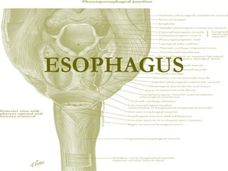
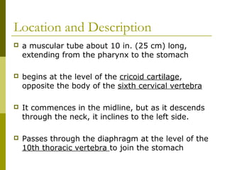
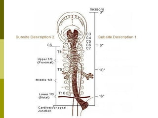
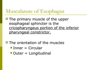
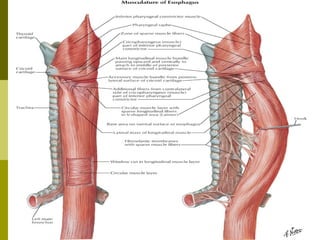
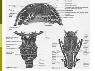
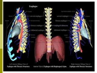
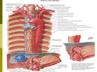
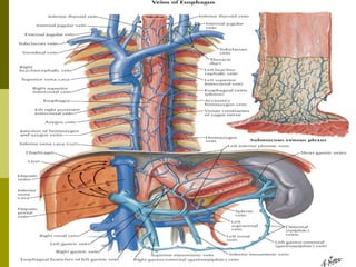
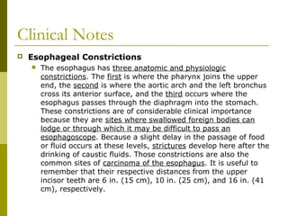
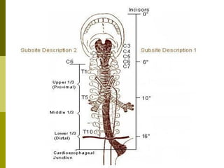
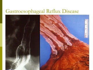
ESOPHAGUS
Location and Description
 a muscular tube about 10 in. (25 cm) long,
extending from the pharynx to the stomach
 begins at the level of the cricoid cartilage,
opposite the body of the sixth cervical vertebra
 It commences in the midline, but as it descends
through the neck, it inclines to the left side.
 Passes through the diaphragm at the level of the
10th thoracic vertebra to join the stomach
Musculature of Esophagus
 The primary muscle of the upper
esophageal sphincter is the
cricopharyngeus portion of the inferior
pharyngeal constrictor.
 The orientation of the muscles
 Inner = Circular
 Outer = Longitudinal
Location and Description
 Anteriorly
 The trachea
 the recurrent laryngeal nerves ascend one on
each side, in the groove between the trachea
and the esophagus
 Posteriorly
 The prevertebral layer of deep cervical fascia
 the longus colli
 the vertebral column
 Laterally
 On each side lie the lobe of the thyroid gland
and the carotid sheath
Blood Supply
Arterial Supply
1. upper third of the esophagus is supplied by the
inferior thyroid artery
1. middle third by branches from the descending
thoracic aorta
1. lower third by branches from the left gastric
artery
Blood Supply
Venous drainage
1. veins from the upper third drain into the inferior
thyroid veins
1. middle third into the azygos veins
1. the lower third into the left gastric vein, a
tributary of the portal vein
Lymph drainage
1. upper third of the esophagus drain into the deep
cervical nodes
1. middle third into the superior and posterior
mediastinal nodes
1. lower third into nodes along the left gastric
blood vessels and the celiac nodes
Nerve Supply
 The nerves are derived from the recurrent
laryngeal nerves and from the sympathetic
trunks
 In the lower part of its thoracic course, the
esophagus is surrounded by the
esophageal nerve plexus.
Clinical Notes
 Esophageal Constrictions
 The esophagus has three anatomic and physiologic
constrictions. The first is where the pharynx joins the upper
end, the second is where the aortic arch and the left bronchus
cross its anterior surface, and the third occurs where the
esophagus passes through the diaphragm into the stomach.
These constrictions are of considerable clinical importance
because they are sites where swallowed foreign bodies can
lodge or through which it may be difficult to pass an
esophagoscope. Because a slight delay in the passage of food
or fluid occurs at these levels, strictures develop here after the
drinking of caustic fluids. Those constrictions are also the
common sites of carcinoma of the esophagus. It is useful to
remember that their respective distances from the upper
incisor teeth are 6 in. (15 cm), 10 in. (25 cm), and 16 in. (41
cm), respectively.
Clinical Notes
 Achalasia or Cardiospasm
 The cause is unknown. The primary site of the
disorder maybe in the innervations of the
cardioesophageal sphincter by the vagus
nerve. Dysphagia (difficulty in swallowing) and
regurgitation are common symptoms.
Achalasia can be treated by surgically dilating
(enlarging) the esophagus.
Clinical Notes
 Gastroesophageal reflux disease (GERD)
 is defined as the movement of gastric contents into the
esophagus without vomiting. GERD occurs when gastric
contents irritate mucosal surfaces of the upper aerodigestive
tract. It is the most common esophageal disease. Besides the
typical presentation of heartburn and acid regurgitation, either
alone or in combination, GERD can cause atypical symptoms.
An estimated 20 to 60 percent of patients with GERD have
head and neck symptoms without any appreciable heartburn.
While the most common head and neck symptom is a globus
sensation (a lump in the throat), the head and neck
manifestations can be diverse and may be misleading in the
initial work-up. Thus, a high index of suspicion is required.
Gastroesophageal Reflux Disease
Clinical Notes
 Esophageal
atresia
 is a congenital
medical condition
which affects the
alimentary tract.
It causes the
esophagus to end
in a blind-ended
pouch rather than
connecting
normally to the
stomach.
Clinical Notes
Esophagitis
 Inflammation of the esophagus
 Causes include
 infection such as candida and herpes
simplex
 chemical injury by alkaline and acid
solutions
 fungi
Candida Esophagitis
Herpes Simplex Esophagitis
Chemical Esophagitis
Aspergillus Esophagitis
THANK YOU!! =)

Oesophagus 1

  • 1.
  • 2.
    Location and Description a muscular tube about 10 in. (25 cm) long, extending from the pharynx to the stomach  begins at the level of the cricoid cartilage, opposite the body of the sixth cervical vertebra  It commences in the midline, but as it descends through the neck, it inclines to the left side.  Passes through the diaphragm at the level of the 10th thoracic vertebra to join the stomach
  • 4.
    Musculature of Esophagus The primary muscle of the upper esophageal sphincter is the cricopharyngeus portion of the inferior pharyngeal constrictor.  The orientation of the muscles  Inner = Circular  Outer = Longitudinal
  • 6.
    Location and Description Anteriorly  The trachea  the recurrent laryngeal nerves ascend one on each side, in the groove between the trachea and the esophagus  Posteriorly  The prevertebral layer of deep cervical fascia  the longus colli  the vertebral column  Laterally  On each side lie the lobe of the thyroid gland and the carotid sheath
  • 9.
    Blood Supply Arterial Supply 1.upper third of the esophagus is supplied by the inferior thyroid artery 1. middle third by branches from the descending thoracic aorta 1. lower third by branches from the left gastric artery
  • 11.
    Blood Supply Venous drainage 1.veins from the upper third drain into the inferior thyroid veins 1. middle third into the azygos veins 1. the lower third into the left gastric vein, a tributary of the portal vein
  • 13.
    Lymph drainage 1. upperthird of the esophagus drain into the deep cervical nodes 1. middle third into the superior and posterior mediastinal nodes 1. lower third into nodes along the left gastric blood vessels and the celiac nodes
  • 15.
    Nerve Supply  Thenerves are derived from the recurrent laryngeal nerves and from the sympathetic trunks  In the lower part of its thoracic course, the esophagus is surrounded by the esophageal nerve plexus.
  • 17.
    Clinical Notes  EsophagealConstrictions  The esophagus has three anatomic and physiologic constrictions. The first is where the pharynx joins the upper end, the second is where the aortic arch and the left bronchus cross its anterior surface, and the third occurs where the esophagus passes through the diaphragm into the stomach. These constrictions are of considerable clinical importance because they are sites where swallowed foreign bodies can lodge or through which it may be difficult to pass an esophagoscope. Because a slight delay in the passage of food or fluid occurs at these levels, strictures develop here after the drinking of caustic fluids. Those constrictions are also the common sites of carcinoma of the esophagus. It is useful to remember that their respective distances from the upper incisor teeth are 6 in. (15 cm), 10 in. (25 cm), and 16 in. (41 cm), respectively.
  • 19.
    Clinical Notes  Achalasiaor Cardiospasm  The cause is unknown. The primary site of the disorder maybe in the innervations of the cardioesophageal sphincter by the vagus nerve. Dysphagia (difficulty in swallowing) and regurgitation are common symptoms. Achalasia can be treated by surgically dilating (enlarging) the esophagus.
  • 20.
    Clinical Notes  Gastroesophagealreflux disease (GERD)  is defined as the movement of gastric contents into the esophagus without vomiting. GERD occurs when gastric contents irritate mucosal surfaces of the upper aerodigestive tract. It is the most common esophageal disease. Besides the typical presentation of heartburn and acid regurgitation, either alone or in combination, GERD can cause atypical symptoms. An estimated 20 to 60 percent of patients with GERD have head and neck symptoms without any appreciable heartburn. While the most common head and neck symptom is a globus sensation (a lump in the throat), the head and neck manifestations can be diverse and may be misleading in the initial work-up. Thus, a high index of suspicion is required.
  • 21.
  • 22.
    Clinical Notes  Esophageal atresia is a congenital medical condition which affects the alimentary tract. It causes the esophagus to end in a blind-ended pouch rather than connecting normally to the stomach.
  • 23.
    Clinical Notes Esophagitis  Inflammationof the esophagus  Causes include  infection such as candida and herpes simplex  chemical injury by alkaline and acid solutions  fungi
  • 24.
  • 25.
  • 26.
  • 27.
  • 28.