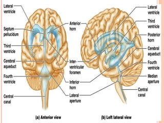
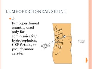
SEMINAR
ON
HYDROCEPHALUS
Presented by,
MR. Yogesh Dengale
MSc Nursing
CON LONI.
INTRODUCTION
 The term hydrocephalus is derived from the
Greek words "hydro" meaning water and
"cephalus" meaning head. As the name implies, it
is a condition in which the primary characteristic
is excessive accumulation of fluid in the brain.
DEFINITION
 Hydrocephalus is the abnormal accumulation of
cerebrospinal fluid in the intracranial spaces.
Or
 Hydrocephalus is an abnormal accumulation of
cerebrospinal fluid (CSF) in the ventricles and
cavities of brain. This causes increased
intracranial pressure inside the skull and may
cause progressive enlargement of the head
INCIDENCE:-
 it is found in 1-3 of every 1000 born children in
worldwide
CEREBROSPINAL FLUID
 The normal amount of CSF is about 150 ml
 CSF formed by choroid plexuses
 Absorbed by arachnoids villi
 Normally about 500 ml of CSF formed everyday
and an equal amount is absorbed
NORMAL CSF CIRCULATION
Reabsorbe
d in
venous
Sinuses
by
arachnoid
s villi
A. COMMUNICATING
HYDROCEPHALUS
B. NON- COMMUNICATING
HYDROCEPHALUS
COMMUNICATING
HYDROCEPHALUS
 Communicating hydrocephalus is a condition
that results when the arachnoid villi are unable
to adequately reabsorb CSF.
 Intraventricular or subarachnoid hemorrhage
 Infectious processes such as meningitis may also
render the arachnoid villi to be nonfunction.
 May also be due to the overproduction of CSF.
This is rare and is usually associated with a
choroid plexus papilloma or a choroid plexus
carcinoma.
NON- COMMUNICATING
HYDROCEPHALUS
 Noncommunicating hydrocephalus is a condition that
results when the ventricular system does not
communicate with the arachnoid villi due to some
obstruction in the normal pathways of CSF flow.
Consequently, CSF is produced in the ventricular
system but cannot flow to the arachnoid villa to be
reabsorbed.
 Such obstruction can occur when pathways
are blocked by a tumor, congenital abnormalities of the
brain, cysts, inflammation from infection, or any other
condition that interferes with the patency of these
pathways.
ETIOLOGY OF
HYDROCEPHALUS
Congenital
Intrauterine infections
Mainly in ruballa, toxoplasmosis,
cytomegalovirus.
CONT…
Congenital brain tumor
Intracranial hemorrhage.
Congenital malformation
Malformations of arachnoid villi
CONT…
Acquired
 Inflammation
 Trauma
 Neoplasm space occupying lesions like
tuberculoma, subdural hematoma or abscess,
gliomas, ependymoma, astrocytoma, choroid
plexus papilloma, pseudotomor cerebri.
 Chemical – hypervitaminosis ‘A’
CONT….
Connective tissue disorder- Hurler
syndrome, achondroplasia.
Degenerative atropy of brain
Arteriovenous malformations,
ruptured aneurysm, cavernous sinus
thrombosis.
CLINICAL FEATURES
CLINICAL FEATURES
CLINICAL MANIFESTATIONS
 An unusually large head
 A rapid increase in the size of the head
 A bulging or tense soft spot (fontanel) on the top
of the head
 Macewen’s sign
 Eyes fixed downward (sunsetting of the eyes)
 Shiny scalp with prominent scalp vein.
CONT….
 Poor feeding
 Seizures
 Deficits in muscle tone and strength,
responsiveness to touch, and expected growth
 Vomiting
 Sleepiness
 Irritability
DIAGNOSTIC EVALUATION
 Physical examination
 Positive transillumination of infant head
 Typical cracked-pot sound (Macewen’s sign)of
the skull bone
 Opthalmoscopy
 MRI
 CT scan
 Cranial ultrasonography and
 X-ray skull
LUMBAR PUNCTURE
Insertion of
spinal
needle into
subarachnoid
space
between the
lower
lumbar
vertebrae.
Modified sitting position for LP
CEREBRAL SPINAL FLUID
Normal CSF
 Clear odorless
 WBC’s 0 – 5
 Protein 15 to 45
 Glucose 50 – 80
 Pressure 50 to 180
Abnormal CSF
 Turbid, cloudy
 WBC’s 1000 – 2000
 Protein 100 – 500
 Glucose lower than
blood sugar
 Pressure 180 or
greater
MEDICAL MANAGEMENT
 Management of hydrocephalus directed
toward:-
I. Reducing intra cranial pressure
II. Prevention and Management of complication
III.Managing problems caused by pathology
 Medical Management include the use of osmotic
diuretics and loop diuretics to reduce CSF
production .medical management is temporary
relief but main management is surgery
SURGICAL MANAGEMENT
 A shunt is made up of radio plastic and has
ventricular cathetar, pressure valve, pumping
chamber, and distal catheter that directs the flow
of CSF from the ventricles to other areas of body
from where it is absorbed.
 Endoscopic Third Ventriculostomy
 Choroid plexectomy
 Ventriculo-peritoneal shunt (V-P shunt).
 Ventriculoatrial shunt
 Ventriculopleural shunt
 Ventriculogallbladder shunt.
ENDOSCOPIC THIRD
VENTRICULOSTOMY (ETV)
 An alternative procedure to shunt surgery is an
endoscopic third ventriculostomy (ETV). This
procedure involves making a hole in the floor of
the brain, allowing the trapped CSF to escape to
the surface of the brain where it can be absorbed,
instead of inserting a shunt.
VENTRICULO-PERITONEAL SHUNT (V-P
SHUNT).
 A VP shunt is a long, plastic tube that allows
fluid to drain from the brain to another part of
the body. This drainage prevents the increase in
pressure on the brain caused by hydrocephalus.
CONT…
 A VP shunt has at least three parts. The first
part is the ventricular catheter, which goes into
the brain. The second part is the valve, which
controls the pressure within the brain. The third
part is the distal catheter, which is underneath
the skin and connects the other parts of the VP
shunt to a space within the body, usually the
abdominal cavity (also called the peritoneal
cavity).
WARNING SIGNS OF A VP SHUNT
NOT WORKING PROPERLY
A headache that hurts more as time passes. Child
acts irritated ; this may be the only sign of a
headache in a child who is young or cannot
speak.
• Vomiting (throwing up) with little or no nausea
(feeling sick to his or her stomach)
• Changes in personality. For example, a child who
is easy to deal with becomes very hard to handle
all of a sudden or “ .. is just not acting right.”
CONT…
• Swelling of the skin that runs along the path of
the VP shunt
• A bulging soft spot on child’s head
• Vision problems (blurry or double vision, or loss
of vision)
• Loss of some mental and physical abilities that
he or she had already mastered (milestones that
fit your child’s age)
WHAT IS A SHUNT INFECTION?
 A shunt infection occurs when bacteria infect the
tissue around the VP shunt. When the tissue is
infected, it can cause the VP shunt to stop
working properly. A shunt that does not work
well can cause an increase in pressure within the
brain. This increase in pressure can damage the
brain or threaten child’s life.
CONT………..
Signs of a shunt infection
• A fever that is equal to or higher than 100.4
degrees F (38.0 degrees C)
• Redness or swelling of the skin that runs along
the path of the VP shunt
• Pain around the shunt or around the shunt
tubing
VENTRICULOATRIAL (VA)
 A ventriculoatrial (VA) shunt also is called
a "vascular shunt." It shunts the cerebral
ventricles through the jugular vein and
superior vena cava into the right cardiac
atrium. It is used when the patient has
abdominal abnormalities (eg, peritonitis,
morbid obesity, or after extensive
abdominal surgery). This shunt requires
repeated lengthening in a growing child.
VENTRICULO-PERITONEAL SHUNT (V-P
SHUNT). VENTRICULOATRIAL SHUNT
LUMBOPERITONEAL SHUNT
A
lumboperitoneal
shunt is used
only for
communicating
hydrocephalus,
CSF fistula, or
pseudotumor
cerebri.
VENTRICULOPLEURAL SHUNT
 A ventriculopleural
shunt is considered
second line. It is
used if other shunt
types are
contraindicated.
PROGNOSIS
 Prognosis depends on early diagnosis and prompt
therapy.
 With improved diagnostic and management
techniques, the prognosis is becoming
considerably better.
 Approximately two- thirds of patients will die at
an early age if they do not receive surgical
treatment.
COMPLICATIONS
 Seizure
 Herniation of brain
 Spontaneous arrest due to natural compensatory
mechanisms, persistent increased ICP and brain
herniation.
 Developmental delay
 Depression.
NURSING MANAGEMENT
 1. Explain the family about the
management required for the disorder.
 2. Provide preoperative nursing care
a. Assess head circumference, fontanelles,
cranial sutures, and LOC; check also for
irritability, altered feeding habits and a
high-pitched cry.
CONT….
b. Firmly support the head and neck when holding the child.
c. Provide skin care for the head to prevent breakdown.
d. Give small, frequent feedings to decrease the risk of
vomiting.
 3. Provide Postoperative nursing care
a. Assess for signs of increased ICP and check the
following; head circumference (daily), anterior fontanelle
for size.
CONT….
b. Administer prescribed medications which may include
antibiotics to prevent infection and analgesics for
pain.
c. Provide shunt care
1. Monitor for shunt infection and malfunction which
may be characterized by rapid onset of vomiting,
severe headache, irritability, lethargy, fever, redness
along the shunt tract, and fluid around the shunt
valve.
CONT…….
2. Prevent infection
3. Monitor for shunt overdrainage (headache, dizziness
and nausea).
CONT….
 4. Teach home care
a. Encourage the child to participate in age-
appropriate activities as tolerated. Encourage
the parents to provide as normal lifestyle as
possible.
CONT….
b. Explain how to recognize signs and symptoms of
increased ICP. Subtle signs include changes in
school performance, intermittent headache, and
mild behavior changes.
c. Arrange for the child to have frequent
developmental screenings and routine medical
checkups.
NEEDS IDENTIFIED
 PHYSICAL NEEDS:-
 Need to maintain personal hygiene.
 Prevention of skin breakdown
 Prevention or reduction of deformities.
 Maintenance of ideal weight.
 Change the position frequently.
 PHYSIOLOGICAL NEEDS:-
 Prevention of injury and infection of sac.
 Provision of adequate nutrition.
 Prevention of infection of urinary tract.
 Regulation of bladder and bowel function.
 PSYCHOLOGICAL NEEDS:-
 Relieve anxiety of parents.
 Maintain the psychology of the parents
NURSING DIAGNOSIS
 Ineffective cerebral tissue perfusion related to
increased ICP
 Imbalance nutrition less than body requirement
related to reduced oral intake and vomiting.
 Risk for impaired skin integrity related to
alterations in LOC and enlarged head
 Anxiety related to abnormal condition and
surgical intervention
 Risk of infection related to introduction of
infecting organism through the shunt.
CONT…
Nursing diagnosis
 Ineffective cerebral tissue perfusion related to
increased ICP .
 Interventions – maintaining cerebral perfusion
 Observe for evidence of increased ICP, and report
immediately.
 Assist with diagnostic procedures to determine cause of
hydrocephalus and administering treatment schedule
as indicated.
CONT….
 Imbalance nutrition less than body requirement
related to reduced oral intake and vomiting.
 Interventions – providing adequate nutrition
1. Be aware that feeding is frequently difficult because
the child may be listless, and prone to vomiting.
2. Complete nursing care and treatments before feeding
so the child will not be disturbed after feeding.
CONT…
3. Hold the infant in a semi-sitting position with
head well supported during feeding.
4. Offer small and frequent feedings.
5. Place the child on side with head elevated after
feeding to prevent aspiration.
CONT….
 Risk for impaired skin integrity related to
alterations in LOC and enlarged head.
 Interventions – maintain skin integrity
1. Prevent pressure sore
2. Keep the scalp clean and dry.
3. Turn the child head frequently.
4. Give range of motion exercise.
Research Study
 It is the belief of some that results of shunt surgery for the
treatment of hydrocephalus in children are improved if the surgery
is performed in high-volume centres. Currently in the UK
paediatric neurosurgery is undergoing a service review. As part of
this review a set of standards of care are being drafted which state
that 1-year failure rates and infection rates for de-novo ventriculo-
peritoneal shunts in children should be less than 40% and 10%,
respectively. Our de-novo shunt infection rate (4.3%) and our 1-
year failure rate (28.6%) are well within the standards set by this
process and comparable to published literature from much higher
volume centres.
SUMMARY
 Definition of hydrocephalus , CSF circulation,
types, etiology , clinical features, diagnostic
evaluation, management, prognosis,
complications, nursing management of
hydrocephalus.
REFERENCES
 Dorothy R. Marlow, Textbook Of Pediatric
Nursing, Fifth Edition, Page No 301-307
 Hoekelman, Primary Pediatric Care, secomd
edition, page no. 1286-1288.
 Lippincott , Manual of Nursing Practice, eighth
edition, page no. 1491-1495.
 Parul Dutta, Pediatric Nursing , Second Edition,
Page No. 406-409
 Internet searching on google.com
Hydrocephalus

Hydrocephalus

  • 2.
  • 3.
    INTRODUCTION  The termhydrocephalus is derived from the Greek words "hydro" meaning water and "cephalus" meaning head. As the name implies, it is a condition in which the primary characteristic is excessive accumulation of fluid in the brain.
  • 4.
    DEFINITION  Hydrocephalus isthe abnormal accumulation of cerebrospinal fluid in the intracranial spaces. Or  Hydrocephalus is an abnormal accumulation of cerebrospinal fluid (CSF) in the ventricles and cavities of brain. This causes increased intracranial pressure inside the skull and may cause progressive enlargement of the head
  • 5.
    INCIDENCE:-  it isfound in 1-3 of every 1000 born children in worldwide
  • 6.
    CEREBROSPINAL FLUID  Thenormal amount of CSF is about 150 ml  CSF formed by choroid plexuses  Absorbed by arachnoids villi  Normally about 500 ml of CSF formed everyday and an equal amount is absorbed
  • 8.
    NORMAL CSF CIRCULATION Reabsorbe din venous Sinuses by arachnoid s villi
  • 10.
    A. COMMUNICATING HYDROCEPHALUS B. NON-COMMUNICATING HYDROCEPHALUS
  • 11.
    COMMUNICATING HYDROCEPHALUS  Communicating hydrocephalusis a condition that results when the arachnoid villi are unable to adequately reabsorb CSF.  Intraventricular or subarachnoid hemorrhage  Infectious processes such as meningitis may also render the arachnoid villi to be nonfunction.  May also be due to the overproduction of CSF. This is rare and is usually associated with a choroid plexus papilloma or a choroid plexus carcinoma.
  • 12.
    NON- COMMUNICATING HYDROCEPHALUS  Noncommunicatinghydrocephalus is a condition that results when the ventricular system does not communicate with the arachnoid villi due to some obstruction in the normal pathways of CSF flow. Consequently, CSF is produced in the ventricular system but cannot flow to the arachnoid villa to be reabsorbed.  Such obstruction can occur when pathways are blocked by a tumor, congenital abnormalities of the brain, cysts, inflammation from infection, or any other condition that interferes with the patency of these pathways.
  • 13.
  • 14.
    CONT… Congenital brain tumor Intracranialhemorrhage. Congenital malformation Malformations of arachnoid villi
  • 15.
    CONT… Acquired  Inflammation  Trauma Neoplasm space occupying lesions like tuberculoma, subdural hematoma or abscess, gliomas, ependymoma, astrocytoma, choroid plexus papilloma, pseudotomor cerebri.  Chemical – hypervitaminosis ‘A’
  • 16.
    CONT…. Connective tissue disorder-Hurler syndrome, achondroplasia. Degenerative atropy of brain Arteriovenous malformations, ruptured aneurysm, cavernous sinus thrombosis.
  • 17.
  • 18.
  • 19.
    CLINICAL MANIFESTATIONS  Anunusually large head  A rapid increase in the size of the head  A bulging or tense soft spot (fontanel) on the top of the head  Macewen’s sign  Eyes fixed downward (sunsetting of the eyes)  Shiny scalp with prominent scalp vein.
  • 20.
    CONT….  Poor feeding Seizures  Deficits in muscle tone and strength, responsiveness to touch, and expected growth  Vomiting  Sleepiness  Irritability
  • 21.
    DIAGNOSTIC EVALUATION  Physicalexamination  Positive transillumination of infant head  Typical cracked-pot sound (Macewen’s sign)of the skull bone  Opthalmoscopy  MRI  CT scan  Cranial ultrasonography and  X-ray skull
  • 22.
    LUMBAR PUNCTURE Insertion of spinal needleinto subarachnoid space between the lower lumbar vertebrae. Modified sitting position for LP
  • 23.
    CEREBRAL SPINAL FLUID NormalCSF  Clear odorless  WBC’s 0 – 5  Protein 15 to 45  Glucose 50 – 80  Pressure 50 to 180 Abnormal CSF  Turbid, cloudy  WBC’s 1000 – 2000  Protein 100 – 500  Glucose lower than blood sugar  Pressure 180 or greater
  • 24.
    MEDICAL MANAGEMENT  Managementof hydrocephalus directed toward:- I. Reducing intra cranial pressure II. Prevention and Management of complication III.Managing problems caused by pathology  Medical Management include the use of osmotic diuretics and loop diuretics to reduce CSF production .medical management is temporary relief but main management is surgery
  • 25.
    SURGICAL MANAGEMENT  Ashunt is made up of radio plastic and has ventricular cathetar, pressure valve, pumping chamber, and distal catheter that directs the flow of CSF from the ventricles to other areas of body from where it is absorbed.
  • 26.
     Endoscopic ThirdVentriculostomy  Choroid plexectomy  Ventriculo-peritoneal shunt (V-P shunt).  Ventriculoatrial shunt  Ventriculopleural shunt  Ventriculogallbladder shunt.
  • 27.
    ENDOSCOPIC THIRD VENTRICULOSTOMY (ETV) An alternative procedure to shunt surgery is an endoscopic third ventriculostomy (ETV). This procedure involves making a hole in the floor of the brain, allowing the trapped CSF to escape to the surface of the brain where it can be absorbed, instead of inserting a shunt.
  • 28.
    VENTRICULO-PERITONEAL SHUNT (V-P SHUNT). A VP shunt is a long, plastic tube that allows fluid to drain from the brain to another part of the body. This drainage prevents the increase in pressure on the brain caused by hydrocephalus.
  • 30.
    CONT…  A VPshunt has at least three parts. The first part is the ventricular catheter, which goes into the brain. The second part is the valve, which controls the pressure within the brain. The third part is the distal catheter, which is underneath the skin and connects the other parts of the VP shunt to a space within the body, usually the abdominal cavity (also called the peritoneal cavity).
  • 31.
    WARNING SIGNS OFA VP SHUNT NOT WORKING PROPERLY A headache that hurts more as time passes. Child acts irritated ; this may be the only sign of a headache in a child who is young or cannot speak. • Vomiting (throwing up) with little or no nausea (feeling sick to his or her stomach) • Changes in personality. For example, a child who is easy to deal with becomes very hard to handle all of a sudden or “ .. is just not acting right.”
  • 32.
    CONT… • Swelling ofthe skin that runs along the path of the VP shunt • A bulging soft spot on child’s head • Vision problems (blurry or double vision, or loss of vision) • Loss of some mental and physical abilities that he or she had already mastered (milestones that fit your child’s age)
  • 33.
    WHAT IS ASHUNT INFECTION?  A shunt infection occurs when bacteria infect the tissue around the VP shunt. When the tissue is infected, it can cause the VP shunt to stop working properly. A shunt that does not work well can cause an increase in pressure within the brain. This increase in pressure can damage the brain or threaten child’s life.
  • 34.
    CONT……….. Signs of ashunt infection • A fever that is equal to or higher than 100.4 degrees F (38.0 degrees C) • Redness or swelling of the skin that runs along the path of the VP shunt • Pain around the shunt or around the shunt tubing
  • 35.
    VENTRICULOATRIAL (VA)  Aventriculoatrial (VA) shunt also is called a "vascular shunt." It shunts the cerebral ventricles through the jugular vein and superior vena cava into the right cardiac atrium. It is used when the patient has abdominal abnormalities (eg, peritonitis, morbid obesity, or after extensive abdominal surgery). This shunt requires repeated lengthening in a growing child.
  • 36.
  • 37.
    LUMBOPERITONEAL SHUNT A lumboperitoneal shunt isused only for communicating hydrocephalus, CSF fistula, or pseudotumor cerebri.
  • 38.
    VENTRICULOPLEURAL SHUNT  Aventriculopleural shunt is considered second line. It is used if other shunt types are contraindicated.
  • 39.
    PROGNOSIS  Prognosis dependson early diagnosis and prompt therapy.  With improved diagnostic and management techniques, the prognosis is becoming considerably better.  Approximately two- thirds of patients will die at an early age if they do not receive surgical treatment.
  • 40.
    COMPLICATIONS  Seizure  Herniationof brain  Spontaneous arrest due to natural compensatory mechanisms, persistent increased ICP and brain herniation.  Developmental delay  Depression.
  • 41.
    NURSING MANAGEMENT  1.Explain the family about the management required for the disorder.  2. Provide preoperative nursing care a. Assess head circumference, fontanelles, cranial sutures, and LOC; check also for irritability, altered feeding habits and a high-pitched cry.
  • 42.
    CONT…. b. Firmly supportthe head and neck when holding the child. c. Provide skin care for the head to prevent breakdown. d. Give small, frequent feedings to decrease the risk of vomiting.  3. Provide Postoperative nursing care a. Assess for signs of increased ICP and check the following; head circumference (daily), anterior fontanelle for size.
  • 43.
    CONT…. b. Administer prescribedmedications which may include antibiotics to prevent infection and analgesics for pain. c. Provide shunt care 1. Monitor for shunt infection and malfunction which may be characterized by rapid onset of vomiting, severe headache, irritability, lethargy, fever, redness along the shunt tract, and fluid around the shunt valve.
  • 44.
    CONT……. 2. Prevent infection 3.Monitor for shunt overdrainage (headache, dizziness and nausea).
  • 45.
    CONT….  4. Teachhome care a. Encourage the child to participate in age- appropriate activities as tolerated. Encourage the parents to provide as normal lifestyle as possible.
  • 46.
    CONT…. b. Explain howto recognize signs and symptoms of increased ICP. Subtle signs include changes in school performance, intermittent headache, and mild behavior changes. c. Arrange for the child to have frequent developmental screenings and routine medical checkups.
  • 47.
    NEEDS IDENTIFIED  PHYSICALNEEDS:-  Need to maintain personal hygiene.  Prevention of skin breakdown  Prevention or reduction of deformities.  Maintenance of ideal weight.  Change the position frequently.  PHYSIOLOGICAL NEEDS:-  Prevention of injury and infection of sac.  Provision of adequate nutrition.  Prevention of infection of urinary tract.  Regulation of bladder and bowel function.  PSYCHOLOGICAL NEEDS:-  Relieve anxiety of parents.  Maintain the psychology of the parents
  • 48.
    NURSING DIAGNOSIS  Ineffectivecerebral tissue perfusion related to increased ICP  Imbalance nutrition less than body requirement related to reduced oral intake and vomiting.  Risk for impaired skin integrity related to alterations in LOC and enlarged head  Anxiety related to abnormal condition and surgical intervention  Risk of infection related to introduction of infecting organism through the shunt.
  • 49.
    CONT… Nursing diagnosis  Ineffectivecerebral tissue perfusion related to increased ICP .  Interventions – maintaining cerebral perfusion  Observe for evidence of increased ICP, and report immediately.  Assist with diagnostic procedures to determine cause of hydrocephalus and administering treatment schedule as indicated.
  • 50.
    CONT….  Imbalance nutritionless than body requirement related to reduced oral intake and vomiting.  Interventions – providing adequate nutrition 1. Be aware that feeding is frequently difficult because the child may be listless, and prone to vomiting. 2. Complete nursing care and treatments before feeding so the child will not be disturbed after feeding.
  • 51.
    CONT… 3. Hold theinfant in a semi-sitting position with head well supported during feeding. 4. Offer small and frequent feedings. 5. Place the child on side with head elevated after feeding to prevent aspiration.
  • 52.
    CONT….  Risk forimpaired skin integrity related to alterations in LOC and enlarged head.  Interventions – maintain skin integrity 1. Prevent pressure sore 2. Keep the scalp clean and dry. 3. Turn the child head frequently. 4. Give range of motion exercise.
  • 53.
    Research Study  Itis the belief of some that results of shunt surgery for the treatment of hydrocephalus in children are improved if the surgery is performed in high-volume centres. Currently in the UK paediatric neurosurgery is undergoing a service review. As part of this review a set of standards of care are being drafted which state that 1-year failure rates and infection rates for de-novo ventriculo- peritoneal shunts in children should be less than 40% and 10%, respectively. Our de-novo shunt infection rate (4.3%) and our 1- year failure rate (28.6%) are well within the standards set by this process and comparable to published literature from much higher volume centres.
  • 54.
    SUMMARY  Definition ofhydrocephalus , CSF circulation, types, etiology , clinical features, diagnostic evaluation, management, prognosis, complications, nursing management of hydrocephalus.
  • 55.
    REFERENCES  Dorothy R.Marlow, Textbook Of Pediatric Nursing, Fifth Edition, Page No 301-307  Hoekelman, Primary Pediatric Care, secomd edition, page no. 1286-1288.  Lippincott , Manual of Nursing Practice, eighth edition, page no. 1491-1495.  Parul Dutta, Pediatric Nursing , Second Edition, Page No. 406-409  Internet searching on google.com