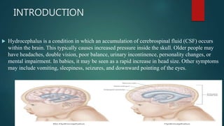
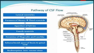
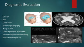
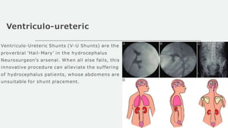
HYDROCEPHALUS
PROF. KOLANEEDI ANURDAHA
CHILD HEALTH NURSING
ASRAM NURSING COLLEGE
INTRODUCTION
 Hydrocephalus is a condition in which an accumulation of cerebrospinal fluid (CSF) occurs
within the brain. This typically causes increased pressure inside the skull. Older people may
have headaches, double vision, poor balance, urinary incontinence, personality changes, or
mental impairment. In babies, it may be seen as a rapid increase in head size. Other symptoms
may include vomiting, sleepiness, seizures, and downward pointing of the eyes.
DEFINATION
 Definition : Hydrocephalus is the
abnormal accumulation of
cerebrospinal fluid(CSF)in the
intracranial spaces. It occurs due to
imbalance between production or
absorption of CSF or due to
obstruction of the CSF pathways. Its
results in the dilatation of the
cerebral ventricles and enlargement
of head.
CSF circulation and pathway
 Cerebrospinal fluid is secreted at the choroid plexus within the cerebral ventricles by ultrafiltration
and active secretion .
 From the lateral ventricles CSF passes to the third ventricle through the foramina of monro.
 From the third ventricle, it passes through cerebral aqueduct to the fourth ventricle.
 From the fourth ventricle to CSF passes to the basal cisterns and subarachnoid spaces through the
foramina Luschka and magendie.
 CSF is absorbed via the arachnoid villi into the venous channel and sinuses.
Incidence
 The incidence of hydrocephalus varies depending on the geographic location.
Hydrocephalus associated with meningomyelocele is found in about 4 per
1000 live births in the parts of northern Ireland
In Japan is about 0.2 per 1000 live births.
Incidence equal in males and females
15-25% of neonates with open myelomeningocele(a form of spina bifida)
56-600 children and adolescents younger than age 18 years have a shunt in
place.
CAUSES
CONGENITAL
1. Intrauterineinfections:Rubella,Toxoplasmos
is, cytomegalovirus.
2. Congenital brain tumor obstructing the CSF
flow
3. Intracranial hemorrhage
4. Congenital malformations like aqueduct
stenosis, Arnold-chiari malformation
5. Malformations of arachnoid villi.
ACQUIRED
1. Inflammation : meningitis, Encephalitis
2. Trauma: Birth injury,head injury, intracranial
hemorrhage.
3. Neoplasm:Space occupying lesions like
tuberculosis,subdural hematoma or
abscess,gliomas,ependymoma,astrocytoma,choroid
plexus papilloma, pseudotumor cerebri
4. Chemical: Hypervitaminosis
5. Connective tissue disorder:Hurler syndrome
6. Degenerative atrophy of brain
7. Arteriovenous malformation:reptured aneurysm
TYPES OF HYDROCEPHALUS
1. COMMUNICATING
 In this type, there is no blockage
between ventricular system,
 The basal cisterns and the spinal
subarachnoid space
 There may be failure in the
absorption of CSF or excessive
production of CSF in choroid plexus
2. NON COMMUNICATING
 In this type of hydrocephalus, there
is obstruction at any level in the
ventricular system, commonly at
the level of aqueduct or at foramina
Luschka and or magendie.
The obstruction maybe
1. Partial
2. Intermittent
3. Complete.
pathophysiology
 COMMUNICATING HYDROCEPHALUS:
 Communication between the ventricular and Subarachnoid space
 There is an interference with the absorption of CSF caused by on occlusion of the subarachnoid cisterns
around the brain stem
 Interference with absorption of CSF
Path…..
Occlusion of the subarachnoid cisterns around the brain stem may be due to
Subarachnoid haemorrhage
Meningitis
Toxoplasmosis
cytomegaly virus infection
communicating hydrocephalus
Atrophy and convulsions can occur
Non communicating hydrocephalus
PATHOPHYSIOLOGY
Non communicating or obstructive hydrocephalus
Blockage between the ventricular and subarachnoid systems
Interference with the circulation of CSF
Stenosis of aqueduct of syvius
PATHO….
Inflammation and compression of aqueduct
Lesion can occur in brain stem resulting in
Aneurysm
Subdural haemorrhage
Atresia of the foramina
Obstructive hydrocephalus
Signs and symptoms
 The symptoms of hydrocephalus tend to vary greatly from person to person and across different age
groups. Infants and young children are more susceptible to symptoms from increased intracranial
pressure like vomiting and adults can experience loss of function like walking or thinking.
1. There is accumulation of CSF in ventricles leading enlargement of the skull
2. Sutures becomes of anterior fontanelle
3. Tense bulging fontanelle
4. A “cracked pot “ sound is heard on percussion of skull (macewen’s sign)
5. Scalp veins are prominent and scalp appears shiny
6. The eyes may have a wide bridge between them and visible sclera above the iris( sun-setting sign)
Signs and symptoms
 7.Neurologically, the infant may be fussy, restless,irritable, apathetic or have an altered or diminished
level of consciousness accompanied by sluggish pupillary response to light, posturing and spasticity of
lower limbs.
 8.Feeding difficulty and high pitch cry
 9. Increased intracranial pressure
 10.Physical and mental retardation
Infants symptoms
1. Unusually large head size
2. Rapidly increasing head circumference
3. Bulging and tense fontanelle or soft spot
4. Prominent scalp veins
5. Downward deviation of eyes or sunset sign
6. Vomiting
7. Sleepiness
8. Irritability
9. Seizures
Children and Adolescents
1. Nausea and vomiting
2. Swelling of the optic disc or papilledema
3. Blurred or double vision
4. Balance and gait abnormalities
5. Slowing or loss of developmental progress
6. Changes in personality
7. Inability to concentrate
8. Seizures
9. Poor appetite
10. Urinary incontinence
Adults
1. Headache
2. Nausea and vomiting
3. Difficulty walking or gait disturbances
4. Loss of balance or coordination
5. Lethargy
6. Bladder incontinence
7. Impaired vision
8. Impaired cognitive skills
9. Memory loss
10. Mild dementia
Diagnostic Evaluation
1. CT Scan
2. MRI
3. Ultrasound
4. Echoencephalography
5. X-ray of skull
6. Lumbar puncture (spinal tap)
7. Intracranial pressure monitoring
8. Isotope cisternography
COMPLICATIONS
 1. Visual changes
 2. Occlusion of posterior cerebral arteries secondary to downward trans tentorial herniation
 3. Chronic papilledema injuring the optic disc
 4. Dilatation of the third ventricle with compression of optic chiasm
 5. Cognitive dysfunction
 6. Incontinence
 7.Gait changes
MANAGEMENT OF HYDROCEPHALUS
1. Medical management
2. Surgical management
3. Nursing management
Medical management
1. Administer isosorbide pre operatively
2. Postoperatively administer: Acetazolamide dose 50mg/kg/day to diminishes CSF production
3. Frusemide
4. Antibiotics
5. Anticonvulsant
6. Oral glycerol: has also been used for the similar purpose
SURGICAL MANAGEMENT
 AIMS: Medicine provide temporary relief but the main management is surgery for removal of
any space –occupying lesion and insertion of a shunt.
1. Ventriculoperitoneal shunt
2. Ventriculoatrial shunt (from ventricles to left atrium)
3. Ventriculopleural shunt (from ventricles to the pleural cavity )
4. Ventriculoureteric shunt ( from ventricles to the ureter)
Ventriculoperitoneal shunt
Ventriculoperitoneal shunting
 Ventriculoperitoneal shunting is surgery to
treat excess cerebrospinal fluid (CSF) in the
cavities (ventricles) of the brain
(hydrocephalus).
 A tube (catheter) is passed from the cavities
of the head to the abdomen to drain the
excess cerebrospinal fluid (CSF).
 A pressure valve and an anti-syphon device
ensure that just the right amount of fluid is
drained
Ventriculoatrial shunt
 Ventricular shunts are used to drain
cerebrospinal fluid into extra-
cranial spaces. Ventriculoatrial (VA)
shunts are provided to transfer
cerebrospinal fluid from the
cerebral ventricle into the right
atrium of the heart.
Ventriculopleural shunt
 The peritoneum is the preferred distal
terminus of a cerebrospinal fluid
(CSF) shunt. However, other anatomic
locations must be considered when the
peritoneum is not suitable, such as in
patients with infection or enough
adhesions to prevent effective CSF
absorption. Other potential distal sites
include the pleural space, gallbladder,
and cardiac atrium.
Nursing management
 Pre-operative nursing care:
1. Measure the head circumference of the child daily
2. Palpate the fontanelle for evidence of increased intracranial pressure.
3. the anterior fontanelle is bulging and tense.
4. The sutures are widely separated
5. Assess the pupillary response and level of consciousness
6. Monitor vital signs regularly
7. Provide calm and quiet environment ,and provide adequate rest
8. Position the body with neck adequately supported
Continues…
 9.Provide water pillow or lamb’s wool may be used to keep head over it
 10.Change the infant’s position frequently
 11.The infant is prone to vomiting provide small ,frequent feeding with intermittent burping
 12.Keep the infant clean and dry.
Postoperative care
1. Place the infant in flat position to prevent rapid CSF drainage
2. Check the vital signs every 15-30 minutes
3. Assess the neurological status and level of consciousness frequently
4. Regularly assess head circumference
5. Monitor the intake and out put chart
6. Restrict the fluid intake for first 24 hours
7. Provide regular skin care
8. If the fontanelle become sunken, notify the physician immediately, Aslo
immediately lower the head end of the bed to decrease the outflow of CSF
through the shunt
Continues....
 9. Administer prophylactic antibiotic therapy to prevent infection
10.Home care :
While feeding positing the child
Recognizing the signs of increased intracranial pressure and
malfunctioning or blockage of shunt
Constipation can be prevented if the child has ventriculoperitoneal
shunt
Follow up care.
Nursing diagnosis
 1.Imbalance cerebral tissue perfusion related to increased ICP
 2.Imbalance nutrition, less than body requirement related to reduced oral intake
and vomiting
 3.Risk for impaired skin integrity related to enlarged head
 4.Anxiety related to the abnormal condition and surgical interventions
 5.Risk for infection related to introduction of infecting organism through the
shunt
 6.Risk for fluid volume deficit related to CSF drainage
 7.Ineffective family coping related to life threatening problem of infant
HYDROCEPHALUS

HYDROCEPHALUS

  • 1.
    HYDROCEPHALUS PROF. KOLANEEDI ANURDAHA CHILDHEALTH NURSING ASRAM NURSING COLLEGE
  • 2.
    INTRODUCTION  Hydrocephalus isa condition in which an accumulation of cerebrospinal fluid (CSF) occurs within the brain. This typically causes increased pressure inside the skull. Older people may have headaches, double vision, poor balance, urinary incontinence, personality changes, or mental impairment. In babies, it may be seen as a rapid increase in head size. Other symptoms may include vomiting, sleepiness, seizures, and downward pointing of the eyes.
  • 3.
    DEFINATION  Definition :Hydrocephalus is the abnormal accumulation of cerebrospinal fluid(CSF)in the intracranial spaces. It occurs due to imbalance between production or absorption of CSF or due to obstruction of the CSF pathways. Its results in the dilatation of the cerebral ventricles and enlargement of head.
  • 4.
    CSF circulation andpathway  Cerebrospinal fluid is secreted at the choroid plexus within the cerebral ventricles by ultrafiltration and active secretion .  From the lateral ventricles CSF passes to the third ventricle through the foramina of monro.  From the third ventricle, it passes through cerebral aqueduct to the fourth ventricle.  From the fourth ventricle to CSF passes to the basal cisterns and subarachnoid spaces through the foramina Luschka and magendie.  CSF is absorbed via the arachnoid villi into the venous channel and sinuses.
  • 6.
    Incidence  The incidenceof hydrocephalus varies depending on the geographic location. Hydrocephalus associated with meningomyelocele is found in about 4 per 1000 live births in the parts of northern Ireland In Japan is about 0.2 per 1000 live births. Incidence equal in males and females 15-25% of neonates with open myelomeningocele(a form of spina bifida) 56-600 children and adolescents younger than age 18 years have a shunt in place.
  • 7.
    CAUSES CONGENITAL 1. Intrauterineinfections:Rubella,Toxoplasmos is, cytomegalovirus. 2.Congenital brain tumor obstructing the CSF flow 3. Intracranial hemorrhage 4. Congenital malformations like aqueduct stenosis, Arnold-chiari malformation 5. Malformations of arachnoid villi. ACQUIRED 1. Inflammation : meningitis, Encephalitis 2. Trauma: Birth injury,head injury, intracranial hemorrhage. 3. Neoplasm:Space occupying lesions like tuberculosis,subdural hematoma or abscess,gliomas,ependymoma,astrocytoma,choroid plexus papilloma, pseudotumor cerebri 4. Chemical: Hypervitaminosis 5. Connective tissue disorder:Hurler syndrome 6. Degenerative atrophy of brain 7. Arteriovenous malformation:reptured aneurysm
  • 8.
    TYPES OF HYDROCEPHALUS 1.COMMUNICATING  In this type, there is no blockage between ventricular system,  The basal cisterns and the spinal subarachnoid space  There may be failure in the absorption of CSF or excessive production of CSF in choroid plexus 2. NON COMMUNICATING  In this type of hydrocephalus, there is obstruction at any level in the ventricular system, commonly at the level of aqueduct or at foramina Luschka and or magendie. The obstruction maybe 1. Partial 2. Intermittent 3. Complete.
  • 9.
    pathophysiology  COMMUNICATING HYDROCEPHALUS: Communication between the ventricular and Subarachnoid space  There is an interference with the absorption of CSF caused by on occlusion of the subarachnoid cisterns around the brain stem  Interference with absorption of CSF
  • 10.
    Path….. Occlusion of thesubarachnoid cisterns around the brain stem may be due to Subarachnoid haemorrhage Meningitis Toxoplasmosis cytomegaly virus infection communicating hydrocephalus Atrophy and convulsions can occur
  • 11.
    Non communicating hydrocephalus PATHOPHYSIOLOGY Noncommunicating or obstructive hydrocephalus Blockage between the ventricular and subarachnoid systems Interference with the circulation of CSF Stenosis of aqueduct of syvius
  • 12.
    PATHO…. Inflammation and compressionof aqueduct Lesion can occur in brain stem resulting in Aneurysm Subdural haemorrhage Atresia of the foramina Obstructive hydrocephalus
  • 13.
    Signs and symptoms The symptoms of hydrocephalus tend to vary greatly from person to person and across different age groups. Infants and young children are more susceptible to symptoms from increased intracranial pressure like vomiting and adults can experience loss of function like walking or thinking. 1. There is accumulation of CSF in ventricles leading enlargement of the skull 2. Sutures becomes of anterior fontanelle 3. Tense bulging fontanelle 4. A “cracked pot “ sound is heard on percussion of skull (macewen’s sign) 5. Scalp veins are prominent and scalp appears shiny 6. The eyes may have a wide bridge between them and visible sclera above the iris( sun-setting sign)
  • 14.
    Signs and symptoms 7.Neurologically, the infant may be fussy, restless,irritable, apathetic or have an altered or diminished level of consciousness accompanied by sluggish pupillary response to light, posturing and spasticity of lower limbs.  8.Feeding difficulty and high pitch cry  9. Increased intracranial pressure  10.Physical and mental retardation
  • 15.
    Infants symptoms 1. Unusuallylarge head size 2. Rapidly increasing head circumference 3. Bulging and tense fontanelle or soft spot 4. Prominent scalp veins 5. Downward deviation of eyes or sunset sign 6. Vomiting 7. Sleepiness 8. Irritability 9. Seizures
  • 16.
    Children and Adolescents 1.Nausea and vomiting 2. Swelling of the optic disc or papilledema 3. Blurred or double vision 4. Balance and gait abnormalities 5. Slowing or loss of developmental progress 6. Changes in personality 7. Inability to concentrate 8. Seizures 9. Poor appetite 10. Urinary incontinence
  • 18.
    Adults 1. Headache 2. Nauseaand vomiting 3. Difficulty walking or gait disturbances 4. Loss of balance or coordination 5. Lethargy 6. Bladder incontinence 7. Impaired vision 8. Impaired cognitive skills 9. Memory loss 10. Mild dementia
  • 19.
    Diagnostic Evaluation 1. CTScan 2. MRI 3. Ultrasound 4. Echoencephalography 5. X-ray of skull 6. Lumbar puncture (spinal tap) 7. Intracranial pressure monitoring 8. Isotope cisternography
  • 20.
    COMPLICATIONS  1. Visualchanges  2. Occlusion of posterior cerebral arteries secondary to downward trans tentorial herniation  3. Chronic papilledema injuring the optic disc  4. Dilatation of the third ventricle with compression of optic chiasm  5. Cognitive dysfunction  6. Incontinence  7.Gait changes
  • 21.
    MANAGEMENT OF HYDROCEPHALUS 1.Medical management 2. Surgical management 3. Nursing management
  • 22.
    Medical management 1. Administerisosorbide pre operatively 2. Postoperatively administer: Acetazolamide dose 50mg/kg/day to diminishes CSF production 3. Frusemide 4. Antibiotics 5. Anticonvulsant 6. Oral glycerol: has also been used for the similar purpose
  • 23.
    SURGICAL MANAGEMENT  AIMS:Medicine provide temporary relief but the main management is surgery for removal of any space –occupying lesion and insertion of a shunt. 1. Ventriculoperitoneal shunt 2. Ventriculoatrial shunt (from ventricles to left atrium) 3. Ventriculopleural shunt (from ventricles to the pleural cavity ) 4. Ventriculoureteric shunt ( from ventricles to the ureter)
  • 24.
  • 25.
    Ventriculoperitoneal shunting  Ventriculoperitonealshunting is surgery to treat excess cerebrospinal fluid (CSF) in the cavities (ventricles) of the brain (hydrocephalus).  A tube (catheter) is passed from the cavities of the head to the abdomen to drain the excess cerebrospinal fluid (CSF).  A pressure valve and an anti-syphon device ensure that just the right amount of fluid is drained
  • 26.
    Ventriculoatrial shunt  Ventricularshunts are used to drain cerebrospinal fluid into extra- cranial spaces. Ventriculoatrial (VA) shunts are provided to transfer cerebrospinal fluid from the cerebral ventricle into the right atrium of the heart.
  • 27.
    Ventriculopleural shunt  Theperitoneum is the preferred distal terminus of a cerebrospinal fluid (CSF) shunt. However, other anatomic locations must be considered when the peritoneum is not suitable, such as in patients with infection or enough adhesions to prevent effective CSF absorption. Other potential distal sites include the pleural space, gallbladder, and cardiac atrium.
  • 29.
    Nursing management  Pre-operativenursing care: 1. Measure the head circumference of the child daily 2. Palpate the fontanelle for evidence of increased intracranial pressure. 3. the anterior fontanelle is bulging and tense. 4. The sutures are widely separated 5. Assess the pupillary response and level of consciousness 6. Monitor vital signs regularly 7. Provide calm and quiet environment ,and provide adequate rest 8. Position the body with neck adequately supported
  • 30.
    Continues…  9.Provide waterpillow or lamb’s wool may be used to keep head over it  10.Change the infant’s position frequently  11.The infant is prone to vomiting provide small ,frequent feeding with intermittent burping  12.Keep the infant clean and dry.
  • 31.
    Postoperative care 1. Placethe infant in flat position to prevent rapid CSF drainage 2. Check the vital signs every 15-30 minutes 3. Assess the neurological status and level of consciousness frequently 4. Regularly assess head circumference 5. Monitor the intake and out put chart 6. Restrict the fluid intake for first 24 hours 7. Provide regular skin care 8. If the fontanelle become sunken, notify the physician immediately, Aslo immediately lower the head end of the bed to decrease the outflow of CSF through the shunt
  • 32.
    Continues....  9. Administerprophylactic antibiotic therapy to prevent infection 10.Home care : While feeding positing the child Recognizing the signs of increased intracranial pressure and malfunctioning or blockage of shunt Constipation can be prevented if the child has ventriculoperitoneal shunt Follow up care.
  • 33.
    Nursing diagnosis  1.Imbalancecerebral tissue perfusion related to increased ICP  2.Imbalance nutrition, less than body requirement related to reduced oral intake and vomiting  3.Risk for impaired skin integrity related to enlarged head  4.Anxiety related to the abnormal condition and surgical interventions  5.Risk for infection related to introduction of infecting organism through the shunt  6.Risk for fluid volume deficit related to CSF drainage  7.Ineffective family coping related to life threatening problem of infant