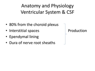
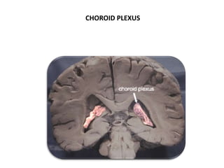
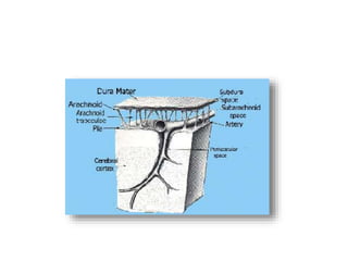
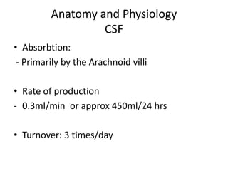
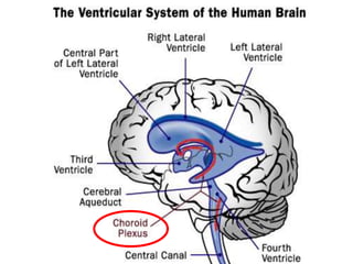
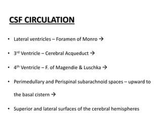
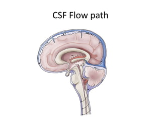
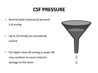
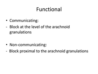
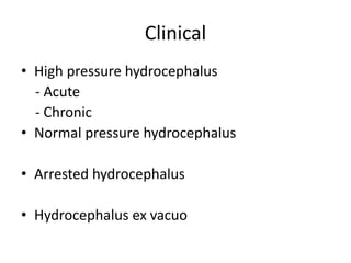
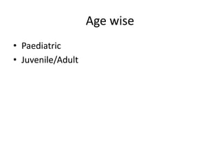
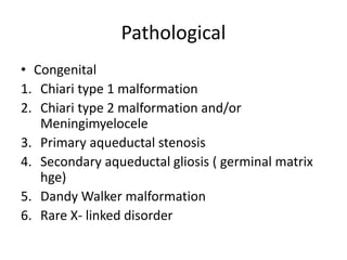
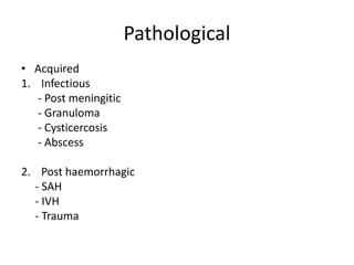
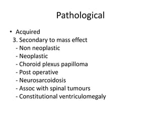
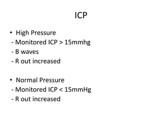
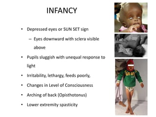
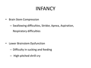
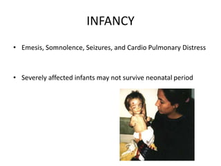
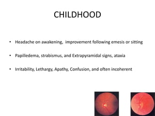
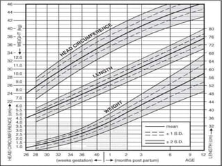
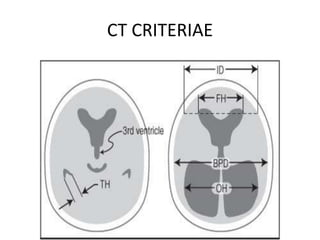
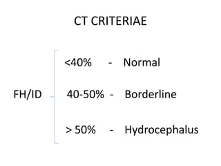
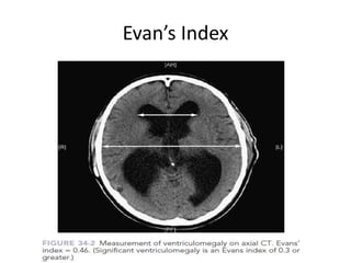
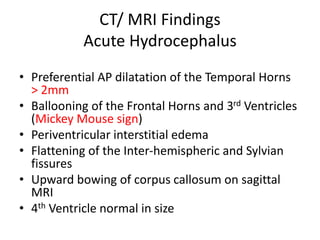
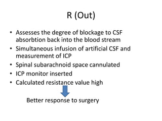
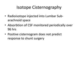
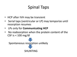
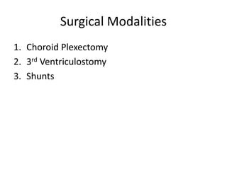
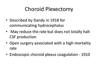
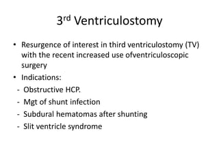
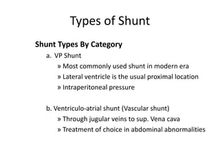
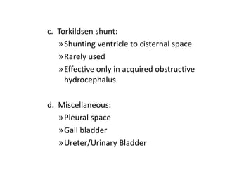
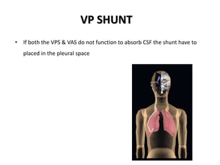
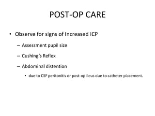
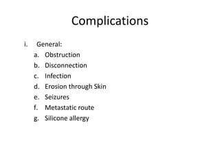
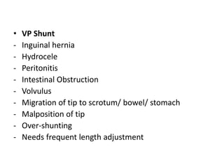
SlideShare a Scribd company logo
HYDROCEPHALUS
EVALUATION & MANAGEMENT
Anatomy and Physiology
Ventricular System & CSF
• 80% from the choroid plexus
• Interstitial spaces Production
• Ependymal lining
• Dura of nerve root sheaths
CHOROID PLEXUS
Anatomy and Physiology
CSF
• Absorbtion:
- Primarily by the Arachnoid villi
• Rate of production
- 0.3ml/min or approx 450ml/24 hrs
• Turnover: 3 times/day
CSF CIRCULATION
• Lateral ventricles – Foramen of Monro 
• 3rd Ventricle – Cerebral Acqueduct 
• 4th Ventricle – F. of Magendie & Luschka 
• Perimedullary and Perispinal subarachnoid spaces – upward to
the basal cistern 
• Superior and lateral surfaces of the cerebral hemispheres
CSF Flow path
CSF PRESSURE
• The CSF volume and pressure are
maintained every minute by the
systemic circulation
• CSF pressure is in equilibrium
with capillary pressure (arteriolar
tone)
• Hypoventilation 
– ↑ in blood PCO2 
– ↓ pH & ↓ arteriolar resistance 
– ↑ cerebral blood flow 
– ↑ CSF pressure
• Hyperventilation has the
opposite effect
CSF PRESSURE
• Normal adult intracranial pressure
2-8 mmHg
• Up to 16 mmHg are considered
normal
• ICP higher than 40 mmHg or lower BP
may combine to cause ischemic
damage to the brain
Definition
• An increase in CSF volume in an enlarged
ventricular system resulting
- primarily from decreased absorbtion
- rarely b’coz of increased production
• Prevalance: 1-1.5%
• Incidence: 0.3-3.5%
- Upto 20% after SAH
- 1% after meningitis
Definition
• Results in ventricular enlargement
• Lat ventricles
- frontal and occipital horns
• Volumes decrease in cerebral sulci, fissures
and cisterns
Classification
• Functional
• Clinical
• Age wise
• Pathological
• ICP/ R-out
• Special Types
Functional
• Communicating:
- Block at the level of the arachnoid
granulations
• Non-communicating:
- Block proximal to the arachnoid granulations
Clinical
• High pressure hydrocephalus
- Acute
- Chronic
• Normal pressure hydrocephalus
• Arrested hydrocephalus
• Hydrocephalus ex vacuo
Age wise
• Paediatric
• Juvenile/Adult
Pathological
• Congenital
1. Chiari type 1 malformation
2. Chiari type 2 malformation and/or
Meningimyelocele
3. Primary aqueductal stenosis
4. Secondary aqueductal gliosis ( germinal matrix
hge)
5. Dandy Walker malformation
6. Rare X- linked disorder
Pathological
• Acquired
1. Infectious
- Post meningitic
- Granuloma
- Cysticercosis
- Abscess
2. Post haemorrhagic
- SAH
- IVH
- Trauma
Pathological
• Acquired
3. Secondary to mass effect
- Non neoplastic
- Neoplastic
- Choroid plexus papilloma
- Post operative
- Neurosarcoidosis
- Assoc with spinal tumours
- Constitutional ventriculomegaly
ICP
• High Pressure
- Monitored ICP > 15mmhg
- B waves
- R out increased
• Normal Pressure
- Monitored ICP < 15mmHg
- R out increased
Special Types
HYYDROCEPHALUS EX VACUO
• enlargement of the ventricles due to loss of
cerebral tissue (cerebral atrophy)
• usually as a function of normal ageing
• Accelerated by Alzheimer's disease,
Creutzfeldt-Jakob, Alcoholism
Special Types
EXTERNAL HYDROCEPHALUS
• enlarged subarachnoid spaces over the frontal poles in the first year
of life
• ventricles are normal or minimally enlarged
• may be distinguished from subdural hematoma by the "cortical vein
sign"
• usually resolves spontaneously by 2 years of age
• Etiology :
• Unclear
• Defect in CSF resorption is postulated
• External hydrocephalus (EH) may be a variant of communicating
hydrocephalus
Special Types
ARRESTED HYDROCEPHALUS
• Compensated hydrocephalus interchangeably
• There is no progression or deleterious
sequelae requiring CSF shunting
• Criteriae in the absence of a CSF shunt:
- Near normal ventricular size
- Normal head growth curve
- Continued psychomotor development
Special Types
OTITIC HYDROCEPHALUS
• Obsolete term
• Describes the increased ICP in patients with
otitis media
Special Types
HYDRANENCEPHAL Y
• A post-neurulation defect
• Total or near-total absence ofthe cerebrum
• Intact cranial vault and meninges
• Intracranial cavity being filled with CSF
• There is usually progressive macrocrania
• Most commonly cited cause : B/L ICA infarcts
• Infection
- Congenital or neonatal herpes
- Toxoplasmosis
- Equine virus
Special Types
ENTRAPPED FOURTH VENTRICLE
• AKA isolated fourth ventricle,
• 3rd Ventricle X 4th ventricle X Foramina of
Luschka or Magendie
- Post-infectious hydrocephalus( fungal)
- Repeated shunt infections
• Choroid plexus of the 4th ventricle : produces
CSF which enlarges the ventricle
Special Types
NPH
• Classic triad:
- Dementia
- Gait disturbance
- Urinary incontinence
• Communicating hydrocephalus on CT or MRI
• Normal pressure on random LP
• Symptoms remediable with CSF shunting
NPH
• Etiology
- Post SAH
- Post-traumatic
- Post-meningitic
- Following posterior fossa surgery
- Tumors including carcinomatous meningitis
- Also seen in -15% of patients with Alzheimer's
disease
- Deficiency of the arachnoid granulations
- Aqueductal stenosis
CLINICAL FEATURES
INFANCY
• Head grows at alarming rate with hydrocephalus.
– First sign: Bulging pulsatile fontanelles
– Tense, non-pulsatile anterior fontanelle
– Dilated scalp veins
– Thin skull bones with separated sutures
• Cracked pot sounds on percussion : Mc Ewans
sign
INFANCY
• Depressed eyes or SUN SET sign
– Eyes downward with sclera visible
above
• Pupils sluggish with unequal response to
light
• Irritability, lethargy, feeds poorly,
• Changes in Level of Consciousness
• Arching of back (Opisthotonus)
• Lower extremity spasticity
INFANCY
• Brain Stem Compression
– Swallowing difficulties, Stridor, Apnea, Aspiration,
Respiratory difficulties
• Lower Brainstem Dysfunction
– Difficulty in sucking and feeding
– High-pitched shrill cry
INFANCY
• Emesis, Somnolence, Seizures, and Cardio Pulmonary Distress
• Severely affected infants may not survive neonatal period
CHILDHOOD
• Headache on awakening, improvement following emesis or sitting
• Papilledema, strabismus, and Extrapyramidal signs, ataxia
• Irritability, Lethargy, Apathy, Confusion, and often incoherent
SYMPTOMS AND SIGNS
• Irritability
• Poor feeding
• Headache
• Nausea, vomiting
• Diplopia
• Visual impairment
• Dementia
• Incontinence
• Gait disturbances
• Accelerated head growth
• Bulging fontanelles
• Forced down gaze
• Developmental delay
• Exotropia
• Papilledema
• Posturing
• Bradycardia
• Apnea / Death
Evaluation
• Clinical
• CT
• MRI
• ICP
• R (out)
• Isotope cisternography
Clinical
• Occipito Frontal Circumference
- OFC of a normal infant = Distance from Crown to
Rump
• Indicators:
- Crossing curves
- Head growth > 1.25cm/wk
- OFC approaching 2 SD above normal
- Out of proportion with body length or weight,
even if normal for age
CT CRITERIAE
CT CRITERIAE
<40% - Normal
FH/ID 40-50% - Borderline
> 50% - Hydrocephalus
Evan’s Index
CT/ MRI Findings
Acute Hydrocephalus
• Preferential AP dilatation of the Temporal Horns
> 2mm
• Ballooning of the Frontal Horns and 3rd Ventricles
(Mickey Mouse sign)
• Periventricular interstitial edema
• Flattening of the Inter-hemispheric and Sylvian
fissures
• Upward bowing of corpus callosum on sagittal
MRI
• 4th Ventricle normal in size
CT/ MRI Findings
Chronic Hydrocephalus
• Temporal horns may be less prominent
• 3rd ventricle may herniate into Sella Turcica
• Erosion of Sella
• Corpus callosum atrophy
• Irreversible white matter demyelination
R (Out)
• Assesses the degree of blockage to CSF
absorbtion back into the blood stream
• Simultaneous infusion of artificial CSF and
measurement of ICP
• Spinal subarachnoid space cannulated
• ICP monitor inserted
• Calculated resistance value high
Better response to surgery
Isotope Cisternography
• Radioisotope injected into Lumbar Sub-
arachnoid space
• Absorbtion of CSF monitored periodically over
96 hrs
• Positive cisternogram does not predict
response to shunt surgery
TREATMENT OF HYDROCEPHALUS
THERAPEUTIC MANAGEMENT
• Goals:
– Relieve hydrocephaly
– Treat complications
– Manage psychomotor problems
– Usually surgical
Drug Therapy
• The choroid plexus shares many ion pumps and enzyme
systems with renal tubular epithelium
– Acetazolamide:
 Start @ 25mg/kg/day PO TID
 Increase @ 25mg/kg/day to 100mg/kg/day
 Simultaneously start Frusemide @1mg/kg/day
Drug Therapy
 To counteract acidosis:
• tricitrate (Polycitra®) 4 ml/kg/day divided QID (each ml
is equivalent to 2 mEq of bicarbonate, and contains 1
mEq K+ and 1 mEq Na+)
• measure serial electrolytes, and adjust dosage to
maintain serum HC03 > 18 mEqIL .
• change to Polycitra-K® (2 mEq K+ per ml, no Na+)
ifserum potassium becomes low
• or to sodium bicarbonate if serum sodium becomes
low
Drug Therapy
• Watch for electrolyte imbalance and acetazolamide
side effects:
- Lethargy - tachypnea
- diarrhea - paresthesias
• Perform weekly CT scan and insert ventricular shunt
if progressive ventriculomegaly occurs.
• Otherwise, maintain therapy for a 6 month trial, then
taper dosage over 2-4 weeks
Spinal Taps
• HCP after IVH may be transient
• Serial taps (ventricular or LP) may temporize until
resorption resumes
• LPs only for Communicating HCP
• No reabsorption when the protein content of the
CSF is < 100 mg/dl
Spontaneous resorption unlikely
SHUNTING
Surgical Modalities
1. Choroid Plexectomy
2. 3rd Ventriculostomy
3. Shunts
Choroid Plexectomy
• Described by Dandy in 1918 for
communicating hydrocephalus
• May reduce the rate but does not totally halt
CSF production
• Open surgery associated with a high mortality
rate
• Endoscopic choroid plexus coagulation - 1910
3rd Ventriculostomy
• Resurgence of interest in third ventriculostomy (TV)
with the recent increased use ofventriculoscopic
surgery
• Indications:
- Obstructive HCP.
- Mgt of shunt infection
- Subdural hematomas after shunting
- Slit ventricle syndrome
3rd Ventriculostomy
• Contraindications:
- Communicating Hydrocepalus
- Tumor
- Previous shunt
- Previous SAH
- Previous whole brain radiation
- Significant adhesions visible when perforating
through the floor of the 3rd ventricle at the time
of performance of TV
3rd Ventriculostomy
• Complications
- Hypothalamic injury
- Transient 3rd and 6th nerve palsies
- Uncontrollable bleeding
- Cardiac arrest
- Traumatic basilar artery aneurysm
Shunts
Types of Shunt
Shunt Types By Category
a. VP Shunt
» Most commonly used shunt in modern era
» Lateral ventricle is the usual proximal location
» Intraperitoneal pressure
b. Ventriculo-atrial shunt (Vascular shunt)
» Through jugular veins to sup. Vena cava
» Treatment of choice in abdominal abnormalities
c. Torkildsen shunt:
»Shunting ventricle to cisternal space
»Rarely used
»Effective only in acquired obstructive
hydrocephalus
d. Miscellaneous:
»Pleural space
»Gall bladder
»Ureter/Urinary Bladder
e. Lumbo-peritoneal shunt:
»Only for communicating hydrocephalous
f. Cyst/Subdural-Peritoneal shunt:
»Draining arachnoid cyst/subdural
hygroma cavity
SHUNT MATERIALS
• Shunts are composed of Silastic material made from silicone.
VP SHUNT
• Shunt systems include three components:
– Ventricular catheter
– One way valve
– Distal catheter
• The ventricular catheter
– Straight piece of tube
– Closed on the proximal end
– With multiple holes upto 2cm for the entry of CSF
VA Shunt
• The VA shunt
– Must be accurately located
– Requires frequent revisions
– Distal end position to be maintained
– Infection may be more serious
VP SHUNT
• If both the VPS & VAS do not function to absorb CSF the shunt have to
placed in the pleural space
POST-OP CARE
• Observe for signs of Increased ICP
– Assessment pupil size
– Cushing’s Reflex
– Abdominal distention
• due to CSF peritonitis or post-op ileus due to catheter placement.
Complications
i. General:
a. Obstruction
b. Disconnection
c. Infection
d. Erosion through Skin
e. Seizures
f. Metastatic route
g. Silicone allergy
• VP Shunt
- Inguinal hernia
- Hydrocele
- Peritonitis
- Intestinal Obstruction
- Volvulus
- Migration of tip to scrotum/ bowel/ stomach
- Malposition of tip
- Over-shunting
- Needs frequent length adjustment
VA shunt:
– Requires repeated lengthening:
– High risk of infection/septicaemia:
– Risk of retrograde flow of blood: in case of valve
malfunction (rare)
– Shunt embolus
– Vascular complications: perforation,
thrombophlebitis, pulmonary micro-emboli
LP Shunt:
– Laminectomy incurs 15% chance of scoliosis
– Progressive cerebellar tonsillar herniation (up to 70%)
– Slit ventricle syndrome
– Overshunting is harder to control
– Difficult proximal end revision (if required:
– Lumber radiculopathy
– CSF leak
– Difficult pressure regulation
– Bilateral 6th, 7th, nerve dysfunction due to overshunting
– High incidence of arachnoiditis & adhesions

More Related Content

What's hot

Spinal cord injury (SCI)
Spinal cord injury (SCI)Spinal cord injury (SCI)
Spinal cord injury (SCI)
Sachin Dwivedi
 
Hydrocephalus disease
Hydrocephalus diseaseHydrocephalus disease
Hydrocephalus disease
Vipin Chandran
 
Extrapyramidal disorders
Extrapyramidal disordersExtrapyramidal disorders
Extrapyramidal disordersrahul arora
 
Hydrocephalus (1) (2)
Hydrocephalus (1) (2)Hydrocephalus (1) (2)
Hydrocephalus (1) (2)
Dr. Akshita Duha Juneja (PT)
 
Guillain-Barré syndrome (GBS)
Guillain-Barré syndrome (GBS)Guillain-Barré syndrome (GBS)
Guillain-Barré syndrome (GBS)
Prof Vijayraddi
 
Paraplegia ppt
Paraplegia pptParaplegia ppt
Paraplegia ppt
drsurajkanase7
 
Spina bifida
Spina bifidaSpina bifida
Spina bifida
HARSHITA
 
Subdural hematoma
Subdural hematomaSubdural hematoma
Subdural hematoma
Faizyab Ahmed
 
Neonatal seizures
Neonatal seizuresNeonatal seizures
Neonatal seizures
CSN Vittal
 
Craniotomy
CraniotomyCraniotomy
Craniotomy
maekrasniewic
 
Quadriplegia & Paraplegia
Quadriplegia & ParaplegiaQuadriplegia & Paraplegia
Quadriplegia & Paraplegia
zuni1412
 
Hydrocephalous, shunting & shunt systems
Hydrocephalous, shunting & shunt systemsHydrocephalous, shunting & shunt systems
Hydrocephalous, shunting & shunt systems
Mukhtar Khan
 
Transverse myelitis
Transverse myelitisTransverse myelitis
Transverse myelitis
Reyad Al_Faky
 
Tuberculous meningitis
Tuberculous meningitisTuberculous meningitis
Tuberculous meningitiszahid mehmood
 
Flail chest
Flail chest Flail chest
Flail chest
drbanuchanderpandians
 
Autonomic Dysreflexia
Autonomic DysreflexiaAutonomic Dysreflexia
Autonomic Dysreflexia
dsukumaran
 
Increased Intracranial Pressure
Increased Intracranial PressureIncreased Intracranial Pressure
Increased Intracranial PressureTosca Torres
 

What's hot (20)

Spinal cord injury (SCI)
Spinal cord injury (SCI)Spinal cord injury (SCI)
Spinal cord injury (SCI)
 
Hydrocephalus disease
Hydrocephalus diseaseHydrocephalus disease
Hydrocephalus disease
 
Club foot
Club footClub foot
Club foot
 
Extrapyramidal disorders
Extrapyramidal disordersExtrapyramidal disorders
Extrapyramidal disorders
 
Hydrocephalus (1) (2)
Hydrocephalus (1) (2)Hydrocephalus (1) (2)
Hydrocephalus (1) (2)
 
Guillain-Barré syndrome (GBS)
Guillain-Barré syndrome (GBS)Guillain-Barré syndrome (GBS)
Guillain-Barré syndrome (GBS)
 
Paraplegia ppt
Paraplegia pptParaplegia ppt
Paraplegia ppt
 
Peripheral Neuropathy
Peripheral NeuropathyPeripheral Neuropathy
Peripheral Neuropathy
 
Spina bifida
Spina bifidaSpina bifida
Spina bifida
 
Subdural hematoma
Subdural hematomaSubdural hematoma
Subdural hematoma
 
Neonatal seizures
Neonatal seizuresNeonatal seizures
Neonatal seizures
 
Craniotomy
CraniotomyCraniotomy
Craniotomy
 
Quadriplegia & Paraplegia
Quadriplegia & ParaplegiaQuadriplegia & Paraplegia
Quadriplegia & Paraplegia
 
Hydrocephalous, shunting & shunt systems
Hydrocephalous, shunting & shunt systemsHydrocephalous, shunting & shunt systems
Hydrocephalous, shunting & shunt systems
 
Transverse myelitis
Transverse myelitisTransverse myelitis
Transverse myelitis
 
Tuberculous meningitis
Tuberculous meningitisTuberculous meningitis
Tuberculous meningitis
 
Spinal Bifida
Spinal BifidaSpinal Bifida
Spinal Bifida
 
Flail chest
Flail chest Flail chest
Flail chest
 
Autonomic Dysreflexia
Autonomic DysreflexiaAutonomic Dysreflexia
Autonomic Dysreflexia
 
Increased Intracranial Pressure
Increased Intracranial PressureIncreased Intracranial Pressure
Increased Intracranial Pressure
 

Viewers also liked

Hydrocephalus
HydrocephalusHydrocephalus
Hydrocephalus
Mohammad Ihmeidan
 
Hydrocephalus
HydrocephalusHydrocephalus
Hydrocephalus
clinicalstudio
 
Hydrocephalus
HydrocephalusHydrocephalus
Hydrocephalus
Kuotho Nyuwi
 
Hydrocephalus with fetal hydrops
Hydrocephalus with fetal hydropsHydrocephalus with fetal hydrops
Hydrocephalus with fetal hydrops
Patrick Adu-poku
 
Hydrocephalus presentation
Hydrocephalus presentationHydrocephalus presentation
Hydrocephalus presentation
Hussain Karimi
 
Csf analysis presentation
Csf analysis presentationCsf analysis presentation
Csf analysis presentation
Bijo Augustine
 
Surgical management of NPH
Surgical management of NPHSurgical management of NPH
Surgical management of NPH
shashank_smg
 
infrequently performed investigations in nuclear medicine
infrequently performed investigations in nuclear medicineinfrequently performed investigations in nuclear medicine
infrequently performed investigations in nuclear medicineGanesh Kumar
 
Assessing Shunt patency in Neuclear Medicine
Assessing Shunt patency in Neuclear MedicineAssessing Shunt patency in Neuclear Medicine
Assessing Shunt patency in Neuclear Medicine
Yasser Asiri
 
Hydrocephalus
HydrocephalusHydrocephalus
Hydrocephalus
Mano Ranjitha Kumari
 
CSF Shunt Infection: Diagnosis and Treatment
CSF Shunt Infection: Diagnosis and TreatmentCSF Shunt Infection: Diagnosis and Treatment
CSF Shunt Infection: Diagnosis and Treatment
Liew Boon Seng
 
Metabolisme vitamin E
Metabolisme vitamin EMetabolisme vitamin E
Metabolisme vitamin E
Muhammad Luthfan
 
Human resources managenent
Human resources managenentHuman resources managenent
Human resources managenentnavee7nvn
 
Normal Pressure Hydrocephalus
Normal Pressure HydrocephalusNormal Pressure Hydrocephalus
Normal Pressure Hydrocephalus
joemdas
 
Pediatric neurologic nurs 3340 spring 2017
Pediatric neurologic nurs 3340 spring 2017Pediatric neurologic nurs 3340 spring 2017
Pediatric neurologic nurs 3340 spring 2017
Shepard Joy
 
CPR( Cardiopulmonary Resuscitation
CPR( Cardiopulmonary Resuscitation CPR( Cardiopulmonary Resuscitation
CPR( Cardiopulmonary Resuscitation
clinicalstudio
 
Otitis media intracranial complications
Otitis media intracranial complicationsOtitis media intracranial complications
Otitis media intracranial complications
Surendra Poudel
 
Ppts on hydrocephalus
Ppts on hydrocephalusPpts on hydrocephalus
Ppts on hydrocephalus
anhadwalia
 

Viewers also liked (20)

Hydrocephalus
HydrocephalusHydrocephalus
Hydrocephalus
 
Hydrocephalus
HydrocephalusHydrocephalus
Hydrocephalus
 
Hydrocephalus
HydrocephalusHydrocephalus
Hydrocephalus
 
Hydrocephalus
HydrocephalusHydrocephalus
Hydrocephalus
 
Hydrocephalus with fetal hydrops
Hydrocephalus with fetal hydropsHydrocephalus with fetal hydrops
Hydrocephalus with fetal hydrops
 
Hydrocephalus presentation
Hydrocephalus presentationHydrocephalus presentation
Hydrocephalus presentation
 
Csf analysis presentation
Csf analysis presentationCsf analysis presentation
Csf analysis presentation
 
Hydrocephalus
HydrocephalusHydrocephalus
Hydrocephalus
 
Surgical management of NPH
Surgical management of NPHSurgical management of NPH
Surgical management of NPH
 
infrequently performed investigations in nuclear medicine
infrequently performed investigations in nuclear medicineinfrequently performed investigations in nuclear medicine
infrequently performed investigations in nuclear medicine
 
Assessing Shunt patency in Neuclear Medicine
Assessing Shunt patency in Neuclear MedicineAssessing Shunt patency in Neuclear Medicine
Assessing Shunt patency in Neuclear Medicine
 
Hydrocephalus
HydrocephalusHydrocephalus
Hydrocephalus
 
CSF Shunt Infection: Diagnosis and Treatment
CSF Shunt Infection: Diagnosis and TreatmentCSF Shunt Infection: Diagnosis and Treatment
CSF Shunt Infection: Diagnosis and Treatment
 
Metabolisme vitamin E
Metabolisme vitamin EMetabolisme vitamin E
Metabolisme vitamin E
 
Human resources managenent
Human resources managenentHuman resources managenent
Human resources managenent
 
Normal Pressure Hydrocephalus
Normal Pressure HydrocephalusNormal Pressure Hydrocephalus
Normal Pressure Hydrocephalus
 
Pediatric neurologic nurs 3340 spring 2017
Pediatric neurologic nurs 3340 spring 2017Pediatric neurologic nurs 3340 spring 2017
Pediatric neurologic nurs 3340 spring 2017
 
CPR( Cardiopulmonary Resuscitation
CPR( Cardiopulmonary Resuscitation CPR( Cardiopulmonary Resuscitation
CPR( Cardiopulmonary Resuscitation
 
Otitis media intracranial complications
Otitis media intracranial complicationsOtitis media intracranial complications
Otitis media intracranial complications
 
Ppts on hydrocephalus
Ppts on hydrocephalusPpts on hydrocephalus
Ppts on hydrocephalus
 

Similar to Hydrocephalus diagnosis and management

Hydrocephalus Detailed Neurosurgery
Hydrocephalus Detailed NeurosurgeryHydrocephalus Detailed Neurosurgery
Hydrocephalus Detailed Neurosurgery
BlackOrchid2
 
Hydrocephalus
HydrocephalusHydrocephalus
Hydrocephalus
Dr,Kaushik Barot
 
Hydrocephalus.pptx
Hydrocephalus.pptxHydrocephalus.pptx
Hydrocephalus.pptx
Dr. Adamu Ibrahim
 
Neuro oncological emergency
Neuro oncological emergencyNeuro oncological emergency
Neuro oncological emergencyLiew Boon Seng
 
Pathology of Stroke & CVA
Pathology of Stroke & CVAPathology of Stroke & CVA
Pathology of Stroke & CVA
Shashidhar Venkatesh Murthy
 
Hydrocephalus
HydrocephalusHydrocephalus
Hydrocephalus
Nabaz Mohammed
 
Hydrocephalus
HydrocephalusHydrocephalus
CSF. Anaesthesia
CSF. Anaesthesia CSF. Anaesthesia
CSF. Anaesthesia
Soumya Nath Maiti
 
8--Intracranial Hemorrhage of the Newborn{8}.ppt
8--Intracranial Hemorrhage of the Newborn{8}.ppt8--Intracranial Hemorrhage of the Newborn{8}.ppt
8--Intracranial Hemorrhage of the Newborn{8}.ppt
ShamiPokhrel2
 
8--Intracranial Hemorrhage of the Newborn{8}.ppt
8--Intracranial Hemorrhage of the Newborn{8}.ppt8--Intracranial Hemorrhage of the Newborn{8}.ppt
8--Intracranial Hemorrhage of the Newborn{8}.ppt
ShamiPokhrel2
 
Pediatric Hydrocephalus
Pediatric HydrocephalusPediatric Hydrocephalus
Pediatric Hydrocephalus
MOHAMED HASSANEIN
 
Congenital Heart Disease for Anaesthesia
Congenital Heart Disease for AnaesthesiaCongenital Heart Disease for Anaesthesia
Congenital Heart Disease for Anaesthesia
AnmolSrivastava74
 
034 Clinical evaluation of adult hydrocephalus
034 Clinical evaluation of adult hydrocephalus034 Clinical evaluation of adult hydrocephalus
034 Clinical evaluation of adult hydrocephalus
Neurosurgery Vajira
 
Cisternography sujan
Cisternography sujanCisternography sujan
Cisternography sujan
SUJAN KARKI
 
Increased intracranial pressure
Increased intracranial pressureIncreased intracranial pressure
Increased intracranial pressure
Shweta Sharma
 
Cardiac Transplantation
Cardiac TransplantationCardiac Transplantation
Cardiac Transplantation
Usman Shams
 
SAH for Neurology Residents
SAH for Neurology ResidentsSAH for Neurology Residents
SAH for Neurology Residents
Dhaval Shukla
 
Valvular heart disease
Valvular heart diseaseValvular heart disease
Valvular heart disease
MR. JAGDISH SAMBAD
 
valvularheartdisease-180524125726 (1).pptx
valvularheartdisease-180524125726 (1).pptxvalvularheartdisease-180524125726 (1).pptx
valvularheartdisease-180524125726 (1).pptx
ArpitaHalder8
 

Similar to Hydrocephalus diagnosis and management (20)

Hydrocephalus Detailed Neurosurgery
Hydrocephalus Detailed NeurosurgeryHydrocephalus Detailed Neurosurgery
Hydrocephalus Detailed Neurosurgery
 
Hydrocephalus
HydrocephalusHydrocephalus
Hydrocephalus
 
Hydrocephalus.pptx
Hydrocephalus.pptxHydrocephalus.pptx
Hydrocephalus.pptx
 
Neuro oncological emergency
Neuro oncological emergencyNeuro oncological emergency
Neuro oncological emergency
 
Pathology of Stroke & CVA
Pathology of Stroke & CVAPathology of Stroke & CVA
Pathology of Stroke & CVA
 
Hydrocephalus
HydrocephalusHydrocephalus
Hydrocephalus
 
Hydrocephalus
HydrocephalusHydrocephalus
Hydrocephalus
 
CSF. Anaesthesia
CSF. Anaesthesia CSF. Anaesthesia
CSF. Anaesthesia
 
8--Intracranial Hemorrhage of the Newborn{8}.ppt
8--Intracranial Hemorrhage of the Newborn{8}.ppt8--Intracranial Hemorrhage of the Newborn{8}.ppt
8--Intracranial Hemorrhage of the Newborn{8}.ppt
 
8--Intracranial Hemorrhage of the Newborn{8}.ppt
8--Intracranial Hemorrhage of the Newborn{8}.ppt8--Intracranial Hemorrhage of the Newborn{8}.ppt
8--Intracranial Hemorrhage of the Newborn{8}.ppt
 
Myocardial infarction
Myocardial infarctionMyocardial infarction
Myocardial infarction
 
Pediatric Hydrocephalus
Pediatric HydrocephalusPediatric Hydrocephalus
Pediatric Hydrocephalus
 
Congenital Heart Disease for Anaesthesia
Congenital Heart Disease for AnaesthesiaCongenital Heart Disease for Anaesthesia
Congenital Heart Disease for Anaesthesia
 
034 Clinical evaluation of adult hydrocephalus
034 Clinical evaluation of adult hydrocephalus034 Clinical evaluation of adult hydrocephalus
034 Clinical evaluation of adult hydrocephalus
 
Cisternography sujan
Cisternography sujanCisternography sujan
Cisternography sujan
 
Increased intracranial pressure
Increased intracranial pressureIncreased intracranial pressure
Increased intracranial pressure
 
Cardiac Transplantation
Cardiac TransplantationCardiac Transplantation
Cardiac Transplantation
 
SAH for Neurology Residents
SAH for Neurology ResidentsSAH for Neurology Residents
SAH for Neurology Residents
 
Valvular heart disease
Valvular heart diseaseValvular heart disease
Valvular heart disease
 
valvularheartdisease-180524125726 (1).pptx
valvularheartdisease-180524125726 (1).pptxvalvularheartdisease-180524125726 (1).pptx
valvularheartdisease-180524125726 (1).pptx
 

More from sanyal1981

Ascending Aortic Pseudoaneurysm: Post Aortic Valve Replacement
Ascending Aortic Pseudoaneurysm: Post Aortic Valve ReplacementAscending Aortic Pseudoaneurysm: Post Aortic Valve Replacement
Ascending Aortic Pseudoaneurysm: Post Aortic Valve Replacement
sanyal1981
 
Procurement and packaging of Donor Heart
Procurement and packaging of Donor HeartProcurement and packaging of Donor Heart
Procurement and packaging of Donor Heart
sanyal1981
 
Haemodialysis access surgeries
Haemodialysis access surgeriesHaemodialysis access surgeries
Haemodialysis access surgeries
sanyal1981
 
Chronic mesesnteric ischaemia
Chronic mesesnteric ischaemiaChronic mesesnteric ischaemia
Chronic mesesnteric ischaemia
sanyal1981
 
Esophagus diagnostic: modalities
Esophagus diagnostic: modalitiesEsophagus diagnostic: modalities
Esophagus diagnostic: modalities
sanyal1981
 
ICMR guidelines on antimicrobial use in Sepsis and SSI's in India
ICMR guidelines on antimicrobial use in Sepsis and SSI's in IndiaICMR guidelines on antimicrobial use in Sepsis and SSI's in India
ICMR guidelines on antimicrobial use in Sepsis and SSI's in India
sanyal1981
 
Median nerve injuries and mangement
Median nerve injuries and mangementMedian nerve injuries and mangement
Median nerve injuries and mangement
sanyal1981
 
gastric resection, reconstruction and post gastrectomy syndromes
gastric resection, reconstruction and post gastrectomy syndromesgastric resection, reconstruction and post gastrectomy syndromes
gastric resection, reconstruction and post gastrectomy syndromes
sanyal1981
 
Surg in ac pancreatitis
Surg in ac pancreatitisSurg in ac pancreatitis
Surg in ac pancreatitis
sanyal1981
 
Melanoma clinical features, pathology and management
Melanoma clinical features, pathology and managementMelanoma clinical features, pathology and management
Melanoma clinical features, pathology and management
sanyal1981
 
Vesicouretric reflux
Vesicouretric refluxVesicouretric reflux
Vesicouretric reflux
sanyal1981
 
Management of-wilms-tumors-9306 - copy
Management of-wilms-tumors-9306 - copyManagement of-wilms-tumors-9306 - copy
Management of-wilms-tumors-9306 - copy
sanyal1981
 
Levels of evidence, recommendations &amp; phases of
Levels of evidence, recommendations &amp; phases ofLevels of evidence, recommendations &amp; phases of
Levels of evidence, recommendations &amp; phases of
sanyal1981
 
cleft lip and palate
cleft lip and palatecleft lip and palate
cleft lip and palate
sanyal1981
 
Sugical anatomy of hand and its infections
Sugical anatomy of hand and its infectionsSugical anatomy of hand and its infections
Sugical anatomy of hand and its infections
sanyal1981
 
Acute appendicitis
Acute appendicitisAcute appendicitis
Acute appendicitis
sanyal1981
 

More from sanyal1981 (16)

Ascending Aortic Pseudoaneurysm: Post Aortic Valve Replacement
Ascending Aortic Pseudoaneurysm: Post Aortic Valve ReplacementAscending Aortic Pseudoaneurysm: Post Aortic Valve Replacement
Ascending Aortic Pseudoaneurysm: Post Aortic Valve Replacement
 
Procurement and packaging of Donor Heart
Procurement and packaging of Donor HeartProcurement and packaging of Donor Heart
Procurement and packaging of Donor Heart
 
Haemodialysis access surgeries
Haemodialysis access surgeriesHaemodialysis access surgeries
Haemodialysis access surgeries
 
Chronic mesesnteric ischaemia
Chronic mesesnteric ischaemiaChronic mesesnteric ischaemia
Chronic mesesnteric ischaemia
 
Esophagus diagnostic: modalities
Esophagus diagnostic: modalitiesEsophagus diagnostic: modalities
Esophagus diagnostic: modalities
 
ICMR guidelines on antimicrobial use in Sepsis and SSI's in India
ICMR guidelines on antimicrobial use in Sepsis and SSI's in IndiaICMR guidelines on antimicrobial use in Sepsis and SSI's in India
ICMR guidelines on antimicrobial use in Sepsis and SSI's in India
 
Median nerve injuries and mangement
Median nerve injuries and mangementMedian nerve injuries and mangement
Median nerve injuries and mangement
 
gastric resection, reconstruction and post gastrectomy syndromes
gastric resection, reconstruction and post gastrectomy syndromesgastric resection, reconstruction and post gastrectomy syndromes
gastric resection, reconstruction and post gastrectomy syndromes
 
Surg in ac pancreatitis
Surg in ac pancreatitisSurg in ac pancreatitis
Surg in ac pancreatitis
 
Melanoma clinical features, pathology and management
Melanoma clinical features, pathology and managementMelanoma clinical features, pathology and management
Melanoma clinical features, pathology and management
 
Vesicouretric reflux
Vesicouretric refluxVesicouretric reflux
Vesicouretric reflux
 
Management of-wilms-tumors-9306 - copy
Management of-wilms-tumors-9306 - copyManagement of-wilms-tumors-9306 - copy
Management of-wilms-tumors-9306 - copy
 
Levels of evidence, recommendations &amp; phases of
Levels of evidence, recommendations &amp; phases ofLevels of evidence, recommendations &amp; phases of
Levels of evidence, recommendations &amp; phases of
 
cleft lip and palate
cleft lip and palatecleft lip and palate
cleft lip and palate
 
Sugical anatomy of hand and its infections
Sugical anatomy of hand and its infectionsSugical anatomy of hand and its infections
Sugical anatomy of hand and its infections
 
Acute appendicitis
Acute appendicitisAcute appendicitis
Acute appendicitis
 

Recently uploaded

A Classical Text Review on Basavarajeeyam
A Classical Text Review on BasavarajeeyamA Classical Text Review on Basavarajeeyam
A Classical Text Review on Basavarajeeyam
Dr. Jyothirmai Paindla
 
Knee anatomy and clinical tests 2024.pdf
Knee anatomy and clinical tests 2024.pdfKnee anatomy and clinical tests 2024.pdf
Knee anatomy and clinical tests 2024.pdf
vimalpl1234
 
Ozempic: Preoperative Management of Patients on GLP-1 Receptor Agonists
Ozempic: Preoperative Management of Patients on GLP-1 Receptor Agonists  Ozempic: Preoperative Management of Patients on GLP-1 Receptor Agonists
Ozempic: Preoperative Management of Patients on GLP-1 Receptor Agonists
Saeid Safari
 
Tom Selleck Health: A Comprehensive Look at the Iconic Actor’s Wellness Journey
Tom Selleck Health: A Comprehensive Look at the Iconic Actor’s Wellness JourneyTom Selleck Health: A Comprehensive Look at the Iconic Actor’s Wellness Journey
Tom Selleck Health: A Comprehensive Look at the Iconic Actor’s Wellness Journey
greendigital
 
SURGICAL ANATOMY OF THE RETROPERITONEUM, ADRENALS, KIDNEYS AND URETERS.pptx
SURGICAL ANATOMY OF THE RETROPERITONEUM, ADRENALS, KIDNEYS AND URETERS.pptxSURGICAL ANATOMY OF THE RETROPERITONEUM, ADRENALS, KIDNEYS AND URETERS.pptx
SURGICAL ANATOMY OF THE RETROPERITONEUM, ADRENALS, KIDNEYS AND URETERS.pptx
Bright Chipili
 
Top 10 Best Ayurvedic Kidney Stone Syrups in India
Top 10 Best Ayurvedic Kidney Stone Syrups in IndiaTop 10 Best Ayurvedic Kidney Stone Syrups in India
Top 10 Best Ayurvedic Kidney Stone Syrups in India
SwastikAyurveda
 
Non-respiratory Functions of the Lungs.pdf
Non-respiratory Functions of the Lungs.pdfNon-respiratory Functions of the Lungs.pdf
Non-respiratory Functions of the Lungs.pdf
MedicoseAcademics
 
Cervical & Brachial Plexus By Dr. RIG.pptx
Cervical & Brachial Plexus By Dr. RIG.pptxCervical & Brachial Plexus By Dr. RIG.pptx
Cervical & Brachial Plexus By Dr. RIG.pptx
Dr. Rabia Inam Gandapore
 
NVBDCP.pptx Nation vector borne disease control program
NVBDCP.pptx Nation vector borne disease control programNVBDCP.pptx Nation vector borne disease control program
NVBDCP.pptx Nation vector borne disease control program
Sapna Thakur
 
Ophthalmology Clinical Tests for OSCE exam
Ophthalmology Clinical Tests for OSCE examOphthalmology Clinical Tests for OSCE exam
Ophthalmology Clinical Tests for OSCE exam
KafrELShiekh University
 
Effective-Soaps-for-Fungal-Skin-Infections.pptx
Effective-Soaps-for-Fungal-Skin-Infections.pptxEffective-Soaps-for-Fungal-Skin-Infections.pptx
Effective-Soaps-for-Fungal-Skin-Infections.pptx
SwisschemDerma
 
Flu Vaccine Alert in Bangalore Karnataka
Flu Vaccine Alert in Bangalore KarnatakaFlu Vaccine Alert in Bangalore Karnataka
Flu Vaccine Alert in Bangalore Karnataka
addon Scans
 
ANATOMY AND PHYSIOLOGY OF URINARY SYSTEM.pptx
ANATOMY AND PHYSIOLOGY OF URINARY SYSTEM.pptxANATOMY AND PHYSIOLOGY OF URINARY SYSTEM.pptx
ANATOMY AND PHYSIOLOGY OF URINARY SYSTEM.pptx
Swetaba Besh
 
BRACHYTHERAPY OVERVIEW AND APPLICATORS
BRACHYTHERAPY OVERVIEW  AND  APPLICATORSBRACHYTHERAPY OVERVIEW  AND  APPLICATORS
BRACHYTHERAPY OVERVIEW AND APPLICATORS
Krishan Murari
 
Triangles of Neck and Clinical Correlation by Dr. RIG.pptx
Triangles of Neck and Clinical Correlation by Dr. RIG.pptxTriangles of Neck and Clinical Correlation by Dr. RIG.pptx
Triangles of Neck and Clinical Correlation by Dr. RIG.pptx
Dr. Rabia Inam Gandapore
 
Dehradun #ℂall #gIRLS Oyo Hotel 9719300533 #ℂall #gIRL in Dehradun
Dehradun #ℂall #gIRLS Oyo Hotel 9719300533 #ℂall #gIRL in DehradunDehradun #ℂall #gIRLS Oyo Hotel 9719300533 #ℂall #gIRL in Dehradun
Dehradun #ℂall #gIRLS Oyo Hotel 9719300533 #ℂall #gIRL in Dehradun
chandankumarsmartiso
 
Role of Mukta Pishti in the Management of Hyperthyroidism
Role of Mukta Pishti in the Management of HyperthyroidismRole of Mukta Pishti in the Management of Hyperthyroidism
Role of Mukta Pishti in the Management of Hyperthyroidism
Dr. Jyothirmai Paindla
 
Cardiac Assessment for B.sc Nursing Student.pdf
Cardiac Assessment for B.sc Nursing Student.pdfCardiac Assessment for B.sc Nursing Student.pdf
Cardiac Assessment for B.sc Nursing Student.pdf
shivalingatalekar1
 
263778731218 Abortion Clinic /Pills In Harare ,
263778731218 Abortion Clinic /Pills In Harare ,263778731218 Abortion Clinic /Pills In Harare ,
263778731218 Abortion Clinic /Pills In Harare ,
sisternakatoto
 
Hemodialysis: Chapter 4, Dialysate Circuit - Dr.Gawad
Hemodialysis: Chapter 4, Dialysate Circuit - Dr.GawadHemodialysis: Chapter 4, Dialysate Circuit - Dr.Gawad
Hemodialysis: Chapter 4, Dialysate Circuit - Dr.Gawad
NephroTube - Dr.Gawad
 

Recently uploaded (20)

A Classical Text Review on Basavarajeeyam
A Classical Text Review on BasavarajeeyamA Classical Text Review on Basavarajeeyam
A Classical Text Review on Basavarajeeyam
 
Knee anatomy and clinical tests 2024.pdf
Knee anatomy and clinical tests 2024.pdfKnee anatomy and clinical tests 2024.pdf
Knee anatomy and clinical tests 2024.pdf
 
Ozempic: Preoperative Management of Patients on GLP-1 Receptor Agonists
Ozempic: Preoperative Management of Patients on GLP-1 Receptor Agonists  Ozempic: Preoperative Management of Patients on GLP-1 Receptor Agonists
Ozempic: Preoperative Management of Patients on GLP-1 Receptor Agonists
 
Tom Selleck Health: A Comprehensive Look at the Iconic Actor’s Wellness Journey
Tom Selleck Health: A Comprehensive Look at the Iconic Actor’s Wellness JourneyTom Selleck Health: A Comprehensive Look at the Iconic Actor’s Wellness Journey
Tom Selleck Health: A Comprehensive Look at the Iconic Actor’s Wellness Journey
 
SURGICAL ANATOMY OF THE RETROPERITONEUM, ADRENALS, KIDNEYS AND URETERS.pptx
SURGICAL ANATOMY OF THE RETROPERITONEUM, ADRENALS, KIDNEYS AND URETERS.pptxSURGICAL ANATOMY OF THE RETROPERITONEUM, ADRENALS, KIDNEYS AND URETERS.pptx
SURGICAL ANATOMY OF THE RETROPERITONEUM, ADRENALS, KIDNEYS AND URETERS.pptx
 
Top 10 Best Ayurvedic Kidney Stone Syrups in India
Top 10 Best Ayurvedic Kidney Stone Syrups in IndiaTop 10 Best Ayurvedic Kidney Stone Syrups in India
Top 10 Best Ayurvedic Kidney Stone Syrups in India
 
Non-respiratory Functions of the Lungs.pdf
Non-respiratory Functions of the Lungs.pdfNon-respiratory Functions of the Lungs.pdf
Non-respiratory Functions of the Lungs.pdf
 
Cervical & Brachial Plexus By Dr. RIG.pptx
Cervical & Brachial Plexus By Dr. RIG.pptxCervical & Brachial Plexus By Dr. RIG.pptx
Cervical & Brachial Plexus By Dr. RIG.pptx
 
NVBDCP.pptx Nation vector borne disease control program
NVBDCP.pptx Nation vector borne disease control programNVBDCP.pptx Nation vector borne disease control program
NVBDCP.pptx Nation vector borne disease control program
 
Ophthalmology Clinical Tests for OSCE exam
Ophthalmology Clinical Tests for OSCE examOphthalmology Clinical Tests for OSCE exam
Ophthalmology Clinical Tests for OSCE exam
 
Effective-Soaps-for-Fungal-Skin-Infections.pptx
Effective-Soaps-for-Fungal-Skin-Infections.pptxEffective-Soaps-for-Fungal-Skin-Infections.pptx
Effective-Soaps-for-Fungal-Skin-Infections.pptx
 
Flu Vaccine Alert in Bangalore Karnataka
Flu Vaccine Alert in Bangalore KarnatakaFlu Vaccine Alert in Bangalore Karnataka
Flu Vaccine Alert in Bangalore Karnataka
 
ANATOMY AND PHYSIOLOGY OF URINARY SYSTEM.pptx
ANATOMY AND PHYSIOLOGY OF URINARY SYSTEM.pptxANATOMY AND PHYSIOLOGY OF URINARY SYSTEM.pptx
ANATOMY AND PHYSIOLOGY OF URINARY SYSTEM.pptx
 
BRACHYTHERAPY OVERVIEW AND APPLICATORS
BRACHYTHERAPY OVERVIEW  AND  APPLICATORSBRACHYTHERAPY OVERVIEW  AND  APPLICATORS
BRACHYTHERAPY OVERVIEW AND APPLICATORS
 
Triangles of Neck and Clinical Correlation by Dr. RIG.pptx
Triangles of Neck and Clinical Correlation by Dr. RIG.pptxTriangles of Neck and Clinical Correlation by Dr. RIG.pptx
Triangles of Neck and Clinical Correlation by Dr. RIG.pptx
 
Dehradun #ℂall #gIRLS Oyo Hotel 9719300533 #ℂall #gIRL in Dehradun
Dehradun #ℂall #gIRLS Oyo Hotel 9719300533 #ℂall #gIRL in DehradunDehradun #ℂall #gIRLS Oyo Hotel 9719300533 #ℂall #gIRL in Dehradun
Dehradun #ℂall #gIRLS Oyo Hotel 9719300533 #ℂall #gIRL in Dehradun
 
Role of Mukta Pishti in the Management of Hyperthyroidism
Role of Mukta Pishti in the Management of HyperthyroidismRole of Mukta Pishti in the Management of Hyperthyroidism
Role of Mukta Pishti in the Management of Hyperthyroidism
 
Cardiac Assessment for B.sc Nursing Student.pdf
Cardiac Assessment for B.sc Nursing Student.pdfCardiac Assessment for B.sc Nursing Student.pdf
Cardiac Assessment for B.sc Nursing Student.pdf
 
263778731218 Abortion Clinic /Pills In Harare ,
263778731218 Abortion Clinic /Pills In Harare ,263778731218 Abortion Clinic /Pills In Harare ,
263778731218 Abortion Clinic /Pills In Harare ,
 
Hemodialysis: Chapter 4, Dialysate Circuit - Dr.Gawad
Hemodialysis: Chapter 4, Dialysate Circuit - Dr.GawadHemodialysis: Chapter 4, Dialysate Circuit - Dr.Gawad
Hemodialysis: Chapter 4, Dialysate Circuit - Dr.Gawad
 

Hydrocephalus diagnosis and management

  • 2. Anatomy and Physiology Ventricular System & CSF • 80% from the choroid plexus • Interstitial spaces Production • Ependymal lining • Dura of nerve root sheaths
  • 4.
  • 5. Anatomy and Physiology CSF • Absorbtion: - Primarily by the Arachnoid villi • Rate of production - 0.3ml/min or approx 450ml/24 hrs • Turnover: 3 times/day
  • 6.
  • 7. CSF CIRCULATION • Lateral ventricles – Foramen of Monro  • 3rd Ventricle – Cerebral Acqueduct  • 4th Ventricle – F. of Magendie & Luschka  • Perimedullary and Perispinal subarachnoid spaces – upward to the basal cistern  • Superior and lateral surfaces of the cerebral hemispheres
  • 9. CSF PRESSURE • The CSF volume and pressure are maintained every minute by the systemic circulation • CSF pressure is in equilibrium with capillary pressure (arteriolar tone) • Hypoventilation  – ↑ in blood PCO2  – ↓ pH & ↓ arteriolar resistance  – ↑ cerebral blood flow  – ↑ CSF pressure • Hyperventilation has the opposite effect
  • 10. CSF PRESSURE • Normal adult intracranial pressure 2-8 mmHg • Up to 16 mmHg are considered normal • ICP higher than 40 mmHg or lower BP may combine to cause ischemic damage to the brain
  • 11. Definition • An increase in CSF volume in an enlarged ventricular system resulting - primarily from decreased absorbtion - rarely b’coz of increased production • Prevalance: 1-1.5% • Incidence: 0.3-3.5% - Upto 20% after SAH - 1% after meningitis
  • 12. Definition • Results in ventricular enlargement • Lat ventricles - frontal and occipital horns • Volumes decrease in cerebral sulci, fissures and cisterns
  • 13. Classification • Functional • Clinical • Age wise • Pathological • ICP/ R-out • Special Types
  • 14. Functional • Communicating: - Block at the level of the arachnoid granulations • Non-communicating: - Block proximal to the arachnoid granulations
  • 15. Clinical • High pressure hydrocephalus - Acute - Chronic • Normal pressure hydrocephalus • Arrested hydrocephalus • Hydrocephalus ex vacuo
  • 16. Age wise • Paediatric • Juvenile/Adult
  • 17. Pathological • Congenital 1. Chiari type 1 malformation 2. Chiari type 2 malformation and/or Meningimyelocele 3. Primary aqueductal stenosis 4. Secondary aqueductal gliosis ( germinal matrix hge) 5. Dandy Walker malformation 6. Rare X- linked disorder
  • 18. Pathological • Acquired 1. Infectious - Post meningitic - Granuloma - Cysticercosis - Abscess 2. Post haemorrhagic - SAH - IVH - Trauma
  • 19. Pathological • Acquired 3. Secondary to mass effect - Non neoplastic - Neoplastic - Choroid plexus papilloma - Post operative - Neurosarcoidosis - Assoc with spinal tumours - Constitutional ventriculomegaly
  • 20. ICP • High Pressure - Monitored ICP > 15mmhg - B waves - R out increased • Normal Pressure - Monitored ICP < 15mmHg - R out increased
  • 21. Special Types HYYDROCEPHALUS EX VACUO • enlargement of the ventricles due to loss of cerebral tissue (cerebral atrophy) • usually as a function of normal ageing • Accelerated by Alzheimer's disease, Creutzfeldt-Jakob, Alcoholism
  • 22. Special Types EXTERNAL HYDROCEPHALUS • enlarged subarachnoid spaces over the frontal poles in the first year of life • ventricles are normal or minimally enlarged • may be distinguished from subdural hematoma by the "cortical vein sign" • usually resolves spontaneously by 2 years of age • Etiology : • Unclear • Defect in CSF resorption is postulated • External hydrocephalus (EH) may be a variant of communicating hydrocephalus
  • 23. Special Types ARRESTED HYDROCEPHALUS • Compensated hydrocephalus interchangeably • There is no progression or deleterious sequelae requiring CSF shunting • Criteriae in the absence of a CSF shunt: - Near normal ventricular size - Normal head growth curve - Continued psychomotor development
  • 24. Special Types OTITIC HYDROCEPHALUS • Obsolete term • Describes the increased ICP in patients with otitis media
  • 25. Special Types HYDRANENCEPHAL Y • A post-neurulation defect • Total or near-total absence ofthe cerebrum • Intact cranial vault and meninges • Intracranial cavity being filled with CSF • There is usually progressive macrocrania • Most commonly cited cause : B/L ICA infarcts • Infection - Congenital or neonatal herpes - Toxoplasmosis - Equine virus
  • 26. Special Types ENTRAPPED FOURTH VENTRICLE • AKA isolated fourth ventricle, • 3rd Ventricle X 4th ventricle X Foramina of Luschka or Magendie - Post-infectious hydrocephalus( fungal) - Repeated shunt infections • Choroid plexus of the 4th ventricle : produces CSF which enlarges the ventricle
  • 27. Special Types NPH • Classic triad: - Dementia - Gait disturbance - Urinary incontinence • Communicating hydrocephalus on CT or MRI • Normal pressure on random LP • Symptoms remediable with CSF shunting
  • 28. NPH • Etiology - Post SAH - Post-traumatic - Post-meningitic - Following posterior fossa surgery - Tumors including carcinomatous meningitis - Also seen in -15% of patients with Alzheimer's disease - Deficiency of the arachnoid granulations - Aqueductal stenosis
  • 30. INFANCY • Head grows at alarming rate with hydrocephalus. – First sign: Bulging pulsatile fontanelles – Tense, non-pulsatile anterior fontanelle – Dilated scalp veins – Thin skull bones with separated sutures • Cracked pot sounds on percussion : Mc Ewans sign
  • 31. INFANCY • Depressed eyes or SUN SET sign – Eyes downward with sclera visible above • Pupils sluggish with unequal response to light • Irritability, lethargy, feeds poorly, • Changes in Level of Consciousness • Arching of back (Opisthotonus) • Lower extremity spasticity
  • 32. INFANCY • Brain Stem Compression – Swallowing difficulties, Stridor, Apnea, Aspiration, Respiratory difficulties • Lower Brainstem Dysfunction – Difficulty in sucking and feeding – High-pitched shrill cry
  • 33. INFANCY • Emesis, Somnolence, Seizures, and Cardio Pulmonary Distress • Severely affected infants may not survive neonatal period
  • 34. CHILDHOOD • Headache on awakening, improvement following emesis or sitting • Papilledema, strabismus, and Extrapyramidal signs, ataxia • Irritability, Lethargy, Apathy, Confusion, and often incoherent
  • 35. SYMPTOMS AND SIGNS • Irritability • Poor feeding • Headache • Nausea, vomiting • Diplopia • Visual impairment • Dementia • Incontinence • Gait disturbances • Accelerated head growth • Bulging fontanelles • Forced down gaze • Developmental delay • Exotropia • Papilledema • Posturing • Bradycardia • Apnea / Death
  • 36. Evaluation • Clinical • CT • MRI • ICP • R (out) • Isotope cisternography
  • 37. Clinical • Occipito Frontal Circumference - OFC of a normal infant = Distance from Crown to Rump • Indicators: - Crossing curves - Head growth > 1.25cm/wk - OFC approaching 2 SD above normal - Out of proportion with body length or weight, even if normal for age
  • 38.
  • 39.
  • 41. CT CRITERIAE <40% - Normal FH/ID 40-50% - Borderline > 50% - Hydrocephalus
  • 43. CT/ MRI Findings Acute Hydrocephalus • Preferential AP dilatation of the Temporal Horns > 2mm • Ballooning of the Frontal Horns and 3rd Ventricles (Mickey Mouse sign) • Periventricular interstitial edema • Flattening of the Inter-hemispheric and Sylvian fissures • Upward bowing of corpus callosum on sagittal MRI • 4th Ventricle normal in size
  • 44. CT/ MRI Findings Chronic Hydrocephalus • Temporal horns may be less prominent • 3rd ventricle may herniate into Sella Turcica • Erosion of Sella • Corpus callosum atrophy • Irreversible white matter demyelination
  • 45. R (Out) • Assesses the degree of blockage to CSF absorbtion back into the blood stream • Simultaneous infusion of artificial CSF and measurement of ICP • Spinal subarachnoid space cannulated • ICP monitor inserted • Calculated resistance value high Better response to surgery
  • 46. Isotope Cisternography • Radioisotope injected into Lumbar Sub- arachnoid space • Absorbtion of CSF monitored periodically over 96 hrs • Positive cisternogram does not predict response to shunt surgery
  • 48. THERAPEUTIC MANAGEMENT • Goals: – Relieve hydrocephaly – Treat complications – Manage psychomotor problems – Usually surgical
  • 49. Drug Therapy • The choroid plexus shares many ion pumps and enzyme systems with renal tubular epithelium – Acetazolamide:  Start @ 25mg/kg/day PO TID  Increase @ 25mg/kg/day to 100mg/kg/day  Simultaneously start Frusemide @1mg/kg/day
  • 50. Drug Therapy  To counteract acidosis: • tricitrate (Polycitra®) 4 ml/kg/day divided QID (each ml is equivalent to 2 mEq of bicarbonate, and contains 1 mEq K+ and 1 mEq Na+) • measure serial electrolytes, and adjust dosage to maintain serum HC03 > 18 mEqIL . • change to Polycitra-K® (2 mEq K+ per ml, no Na+) ifserum potassium becomes low • or to sodium bicarbonate if serum sodium becomes low
  • 51. Drug Therapy • Watch for electrolyte imbalance and acetazolamide side effects: - Lethargy - tachypnea - diarrhea - paresthesias • Perform weekly CT scan and insert ventricular shunt if progressive ventriculomegaly occurs. • Otherwise, maintain therapy for a 6 month trial, then taper dosage over 2-4 weeks
  • 52. Spinal Taps • HCP after IVH may be transient • Serial taps (ventricular or LP) may temporize until resorption resumes • LPs only for Communicating HCP • No reabsorption when the protein content of the CSF is < 100 mg/dl Spontaneous resorption unlikely SHUNTING
  • 53. Surgical Modalities 1. Choroid Plexectomy 2. 3rd Ventriculostomy 3. Shunts
  • 54. Choroid Plexectomy • Described by Dandy in 1918 for communicating hydrocephalus • May reduce the rate but does not totally halt CSF production • Open surgery associated with a high mortality rate • Endoscopic choroid plexus coagulation - 1910
  • 55. 3rd Ventriculostomy • Resurgence of interest in third ventriculostomy (TV) with the recent increased use ofventriculoscopic surgery • Indications: - Obstructive HCP. - Mgt of shunt infection - Subdural hematomas after shunting - Slit ventricle syndrome
  • 56. 3rd Ventriculostomy • Contraindications: - Communicating Hydrocepalus - Tumor - Previous shunt - Previous SAH - Previous whole brain radiation - Significant adhesions visible when perforating through the floor of the 3rd ventricle at the time of performance of TV
  • 57. 3rd Ventriculostomy • Complications - Hypothalamic injury - Transient 3rd and 6th nerve palsies - Uncontrollable bleeding - Cardiac arrest - Traumatic basilar artery aneurysm
  • 59. Types of Shunt Shunt Types By Category a. VP Shunt » Most commonly used shunt in modern era » Lateral ventricle is the usual proximal location » Intraperitoneal pressure b. Ventriculo-atrial shunt (Vascular shunt) » Through jugular veins to sup. Vena cava » Treatment of choice in abdominal abnormalities
  • 60. c. Torkildsen shunt: »Shunting ventricle to cisternal space »Rarely used »Effective only in acquired obstructive hydrocephalus d. Miscellaneous: »Pleural space »Gall bladder »Ureter/Urinary Bladder
  • 61. e. Lumbo-peritoneal shunt: »Only for communicating hydrocephalous f. Cyst/Subdural-Peritoneal shunt: »Draining arachnoid cyst/subdural hygroma cavity
  • 62. SHUNT MATERIALS • Shunts are composed of Silastic material made from silicone.
  • 63. VP SHUNT • Shunt systems include three components: – Ventricular catheter – One way valve – Distal catheter • The ventricular catheter – Straight piece of tube – Closed on the proximal end – With multiple holes upto 2cm for the entry of CSF
  • 64. VA Shunt • The VA shunt – Must be accurately located – Requires frequent revisions – Distal end position to be maintained – Infection may be more serious
  • 65. VP SHUNT • If both the VPS & VAS do not function to absorb CSF the shunt have to placed in the pleural space
  • 66. POST-OP CARE • Observe for signs of Increased ICP – Assessment pupil size – Cushing’s Reflex – Abdominal distention • due to CSF peritonitis or post-op ileus due to catheter placement.
  • 67. Complications i. General: a. Obstruction b. Disconnection c. Infection d. Erosion through Skin e. Seizures f. Metastatic route g. Silicone allergy
  • 68. • VP Shunt - Inguinal hernia - Hydrocele - Peritonitis - Intestinal Obstruction - Volvulus - Migration of tip to scrotum/ bowel/ stomach - Malposition of tip - Over-shunting - Needs frequent length adjustment
  • 69. VA shunt: – Requires repeated lengthening: – High risk of infection/septicaemia: – Risk of retrograde flow of blood: in case of valve malfunction (rare) – Shunt embolus – Vascular complications: perforation, thrombophlebitis, pulmonary micro-emboli
  • 70. LP Shunt: – Laminectomy incurs 15% chance of scoliosis – Progressive cerebellar tonsillar herniation (up to 70%) – Slit ventricle syndrome – Overshunting is harder to control – Difficult proximal end revision (if required: – Lumber radiculopathy – CSF leak – Difficult pressure regulation – Bilateral 6th, 7th, nerve dysfunction due to overshunting – High incidence of arachnoiditis & adhesions

Editor's Notes

  1. vascular malformations, arachnoid cysts Medulloblastomas, suprsellar & pituitary tumours Post op: Post fossa tumours
  2. Shunts TYPES OF SHUNTS SHUNT TYPE BY CATEGORY 1. ventriculoperitoneal (VP) shunt: A. most commonly used shunt in modern era B. lateral ventricle is the usual proximal location C. intraperitoneal pressure: normal is near atmospheric 2. ventriculo-atrial (VA) shunt (“vascular shunt”): A. shunts ventricles through jugular vein to superior vena cava, so-called “ventriculo-atrial” shunt because it shunts the cerebral ventricles to the vascular system with the catheter tip in the region of the right cardiac atrium) B. treatment of choice when abdominal abnormalities are present (extensive abdominal surgery, peritonitis, morbid obesity, in preemies who have had NEC and may not tolerate VP shunt…) C. shorter length of tubing results in lower distal pressure and less siphon effect than VP shunt, however pulsatile pressures may alter CSF hydrodynamics 3.
  3. 3: Torkildsen shunt: A. shunts ventricle to cisternal space B. rarely used C. effective only in acquired obstructive HCP, as patients with congenital HCP frequently do not develop normal subarachnoid CSF pathways 4. miscellaneous: various distal projections used historically or in patients who have had significant problems with traditional shunt locations (e.g. peritonitis with VP shunt, SBE with vascular shunts): A. pleural space (ventriculopleural shunt): not a first choice, but a viable alternative if the peritoneum is not available. To avoid symptomatic hydrothorax necessitating relocating distal end, it is recommended only for patients > 7 yrs age. Pressure in pleural space is less than atmospheric B. gall bladder C. ureter or bladder: causes electrolyte imbalances due to losses through urine 5. lumboperitoneal (LP) shunt A. only for communicating HCP: primarily pseudotumor cerebri or CSF fistula37. Useful in situations with small ventricles B. over age 2 yrs, percutaneous insertion with Tuohy needle is preferred 6. cyst or subdural shunt: from arachnoid cyst or subdural hygroma cavity, usually to peritoneum
  4. Disadvantages/complications of various shunts 1. those that may occur with any shunt: A. obstruction: the most common cause of shunt malfunction proximal: ventricular catheter (the most common site) valve mechanism distal: reported incidence of 12-34%. Occurs in peritoneal catheter in VP shunt (see below), in atrial catheter in VA shunt B. disconnection at a junction, or break at any point C. infection D. hardware erosion through skin, usually only in debilitated patients (especially preemies with enlarged heads and thin scalp from chronic HCP, who lay on one side of head due to elongated cranium). May also indicate silicone allergy (see below) E. seizures (ventricular shunts only): there is ≈ 5.5% risk of seizures in the first year after placement of a shunt which drops to ≈ 1.1% after the 3rd year39 (NB: this does not mean that the shunt was the cause of all of these seizures). Seizure risk is questionably higher with frontal catheters than with parieto-occipital F. act as a conduit for extraneural metastases of certain tumours (e.g. medulloblastoma). This is probably a relatively low risk40 G. silicone allergy41: rare (if it occurs at all). May resemble shunt infection with skin breakdown and fungating granulomas. CSF is initially sterile but later infections may occur. May require fabrication of a custom silicone-free device (e.g. polyurethane)
  5. . VA shunt: A. requires repeated lengthening in growing child B. higher risk of infection, septicemia C. possible retrograde flow of blood into ventricles if valve malfunctions (rare) D. shunt embolus E. vascular complications: perforation, thrombophlebitis, pulmonary micro-emboli may cause pulmonary hypertension (incidence ≈ 0.3%)
  6. 4. LP shunt: A. if at all possible, should not be used in growing child unless ventricular access is unavailable (e.g. due to slit ventricles) because of: laminectomy in children causes scoliosis in 14%; risk of progressive cerebellar tonsillar herniation (Chiari I malformation) in up to 70% of cases B. overshunting harder to control when it occurs (a special horizontal-vertical (H-V) valve increases resistance when upright, see below) C. difficult access to proximal end for revision or assessment of patency (see Lumboperitoneal (LP) shunt evaluation, page 214) D. lumbar nerve root irritation (radiculopathy) E. leakage of CSF around catheter F. pressure regulation is difficult G. bilateral 6th and even 7th cranial nerve dysfunction from overshunting H. high incidence of arachnoiditis and adhesions