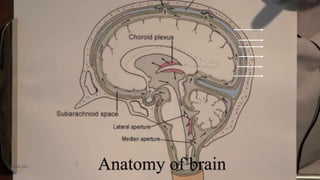
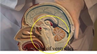
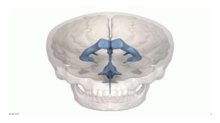
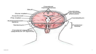
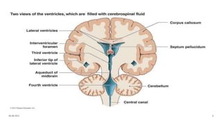
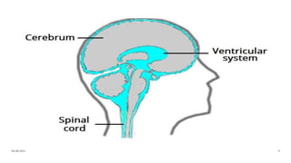
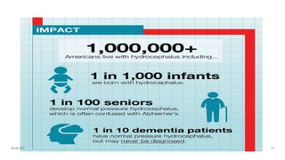
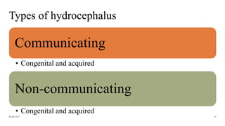
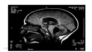
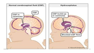
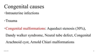
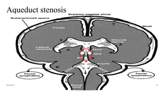
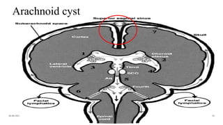
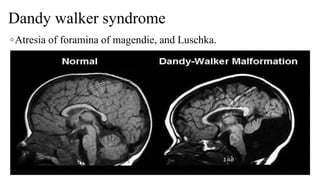
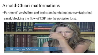
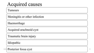
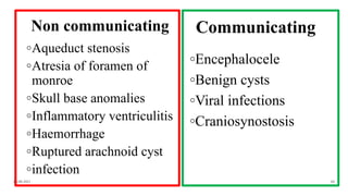
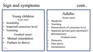
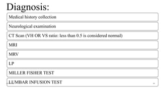
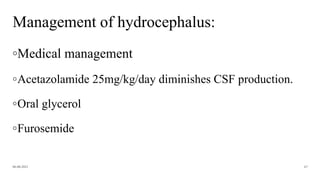
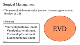
HYDROCEPHALUS
Evaluator: Mr L Anand Presenter: Shruti Shirke
[Asso professor, CON AIIMS BBSR] M.Sc Neuroscience Nursing
06-08-2021
1
Introduction
06-08-2021 2
06-08-2021 3
Anatomy of brain
06-08-2021 4
Anatomy of ventricles
06-08-2021 5
06-08-2021 6
06-08-2021 7
06-08-2021 8
06-08-2021 9
06-08-2021 10
06-08-2021 11
About choroid plexus
◦Choroid plexus is present in each ventricles
◦In the lateral ventricles it is only present in the inferior horn
◦Choroid plexus is also located in the interventricular foramina,
the channels between the lateral and the third ventricles. The
choroid plexus in the fourth ventricle is beneath the
cerebellum.
06-08-2021 12
06-08-2021 13
06-08-2021 14
Functions of choroid plexus
Production of CSF as plasma is filtered
from the blood through the epithelial cells.
Choroid plexus is also a major source of
transferrin secretion that plays a part in iron
homeostasis in the brain.
06-08-2021 15
Production of CSF
06-08-2021 16
About Cerebrospinal fluid
◦Production: Choroid plexus
◦Absorption: Arachnoid villi
◦Rate of production: Approximate 0.3-0.5ml/min or
20ml/hr and 450ml/day
06-08-2021 17
About CSF Cont..
◦Turn over: 3 times a day
◦CSF volume: 125-150 ml
◦ICP (adult): 2-8mmhg up to 16mmhg (70-
180mmH2O) is considered normal.
06-08-2021 18
06-08-2021 19
ⱺThe CSF volume and pressure are
maintained every minute by the
systemic circulation.
ⱺICP higher than 40mmhg or lower
bp may combine to cause ischemic
damage to the brain.
06-08-2021 20
Function of CSF
To keep the
brain tissue
buoyant
Vehicle for
delivering
nutrients to
brain
Removal of
waste
Compensate
for changes
intracranial
blood volume
06-08-2021 21
06-08-2021 22
CSF
Circulation
06-08-2021 23
06-08-2021 24
06-08-2021 25
Definition:
◦“Hydrocephalus is a condition caused by an
imbalance in the production and absorption of
CSF in the ventricular system”.
06-08-2021 26
Incidence:
◦1-2 of every 1000 babies are born with hydrocephalus.
◦Approximately 200,000 cases each year all over world and
12,000 cases of congenital hydrocephalus in India every
year.
06-08-2021 27
Incidence: cont..
◦According to hydrocephalus association
◦It is estimated that up to 5% of all cases of Alzheimer’s,
Parkinson’s, and dementia are actually undiagnosed cases of
NPH.
◦In US currently over 1million individuals are living with
hydrocephalus.
06-08-2021 28
Incidence: cont..
◦Prevalence – over 55 years old 5.5 patient per 100000 of
people, At any age, both man and women, more often in
elderly population between 60-70 (NPH)
◦Congenital hydrocephalus 0.2-0.5/1000 live birth, A higher
incidence is reported in elderly primiparous mother.
06-08-2021 29
06-08-2021 30
06-08-2021 31
Types of hydrocephalus
Communicating
• Congenital and acquired
Non-communicating
• Congenital and acquired
06-08-2021 32
06-08-2021 33
06-08-2021 34
Congenital
hydrocephalus
Acquired
hydrocephalus
Normal pressure
hydrocephalus
Pseudotumor
cerebri
06-08-2021 35
06-08-2021 36
Types of NPH
Idiopathic/
Primary
Secondary/
Hydrocephalus
ex-vacuo
06-08-2021 37
Hydrocephalus ex-vacuo - Subarachnoid
haemorrhage, head trauma, tumours, infections
in CNS, complication if cranial surgery.
Normal pressure hydrocephalus
◦It describes a condition that rarely occurs in patients younger
than 60 years.
◦Enlarged ventricles and normal pressure at lumbar
puncture(LP).
◦It is a type of communicating hydrocephalus.
06-08-2021 38
Normal pressure hydrocephalus
◦Intermittent intracranial
hypertension has been noted during
monitoring of patients in whom
NPH is suspected, usually at night.
◦Headache is NOT a typical
symptom in NPH.
06-08-2021 39
Sign and symptoms of NPH
Gait apraxia
Incontinence
Dementia
06-08-2021 40
The classic hakim’
triad
Gait disturbance – same as Parkinson’s
disease.
Dementia – apathy, forgetfulness, inertia, inattention,
disturbed manipulation of acquired knowledge. (same
as Alzheimer’s )
Incontinence – frontal lobe compression.
06-08-2021 41
Pseudotumor cerebri
06-08-2021 42
Mostly seen in women (obese, childbearing)
It is also known as idiopathic
intracranial hypertension.
pseudotumor cerebri is a
condition in which the
pressure around brain
increases, causing headache
and vision loss
Pseudotumor cerebri
Increased intracranial pressure / intracranial
hypertension
No mass / tumor
#Obstruction of venous drainage & impaired
reabsorption of CSF
06-08-2021 43
06-08-2021 44
Pseudotumor cerebri (sign and symptoms)
06-08-2021 45
Headache
Nausea
and
vomiting
Vision
problems
Papilledema
Diagnosis
MRI: Normal
MRV: (Magnetic resonance
venography) Stenosis of
transverse sinuses
06-08-2021 46
Etiology:
Congenital Acquired
06-08-2021 47
Congenital causes
◦Intrauterine infections
◦Trauma
◦Congenital malformations: Aqueduct stenosis (30%),
Dandy walker syndrome, Neural tube defect, Congenital
Arachnoid cyst, Arnold Chiari malformations
06-08-2021 48
Aqueduct stenosis
06-08-2021 49
Arachnoid cyst
06-08-2021 50
06-08-2021 51
Dandy walker syndrome
◦Atresia of foramina of magendie, and Luschka.
06-08-2021 52
Arnold-Chiari malformations
◦Portion of cerebellum and brainstem herniating into cervical spinal
canal, blocking the flow of CSF into the posterior fossa.
06-08-2021 53
06-08-2021 54
06-08-2021 55
06-08-2021 56
06-08-2021 57
Atresia of foramen of monro
Myelodysplasia (MDS)
Craniosynostosis
06-08-2021 58
Acquired causes
Tumours
Meningitis or other infection
Haemorrhage
Acquired arachnoid cyst
Traumatic brain injury
Idiopathic
Posterior fossa cyst
06-08-2021 59
Non communicating
◦Aqueduct stenosis
◦Atresia of foramen of
monroe
◦Skull base anomalies
◦Inflammatory ventriculitis
◦Haemorrhage
◦Ruptured arachnoid cyst
◦infection
Communicating
◦Encephalocele
◦Benign cysts
◦Viral infections
◦Craniosynostosis
06-08-2021 60
Pathophysiology
Following subarachnoid hemorrhage, blood in
the subarachnoid space makes it harder for the
CSF to reach the arachnoid villi and slows
down or prevents its absorption.
Following intraventricular hemorrhage, blood
in the ventricles can enter the subarachnoid
space and impair CSF absorption.
06-08-2021 61
Pathophysiology cont..
Exudate from infection (such as meningitis or
encephalitis) can block the cerebral aqueduct and
therefore obstruct CSF flow.
Genetic disorders, such as: aqueduct stenosis
(abnormally narrow cerebral aqueduct); Dandy-Walker
malformation (several abnormal brain structures
including a dilated.
06-08-2021 62
Pathophysiology cont..
Tumors of the choroid plexus, which are
rare, can cause overproduction of CSF.
06-08-2021 63
Tumors near the third and fourth ventricles
can obstruct CSF flow.
Sign and symptoms
06-08-2021 64
• Head grows to abnormal rate.
• Anterior fontanel is tense, often
bulging, non pulsatile.
• Scalp veins are dilated.
• Macewen’s sign- cracked pot
sound on the percussion of the
skull.
• Frontal bossing with depressed
eyes
Sign and symptoms cont..
06-08-2021 65
Young children:
Acute onset:
• Irritability
• Impaired conscious level
• Vomiting
Gradual onset:
• Mental retardation
• Failure to thrive
Adults:
Acute onset
• Headache
• Vomiting
• Deterioration of conscious level
• Impaired upward gaze (perinaud’s
phenomenon)
Gradual onset
• Dementia
• Gait ataxia
• Incontinence
Diagnosis:
Medical history collection
Neurological examination
CT Scan (VH OR VS ratio: less than 0.5 is considered normal)
MRI
MRV
LP
MILLER FISHER TEST
LUMBAR INFUSION TEST
06-08-2021 66
Management of hydrocephalus:
◦Medical management
◦Acetazolamide 25mg/kg/day diminishes CSF production.
◦Oral glycerol
◦Furosemide
06-08-2021 67
Surgical Management
◦The removal of the obstruction (tumour, haemorrhage or cyst) to
the flow of CSF
◦Shunting
06-08-2021 68
Ventriculoperitoneal shunt.
Ventriculoarterial shunt.
Ventriculopleural shunt.
Lumboperitoneal shunt.
EVD
Shunting
◦High pressure: 8-10 cm h2o
◦Medium pressure: 4-7 cm h2o
◦Low pressure: 2-4 cm h2o
06-08-2021 69
MPVP Shunt
06-08-2021 70
Lumboperotoneal shunt
06-08-2021 71
Endoscopic third ventricle
◦Puncture third ventricles to cistern
06-08-2021 72
EVD/ Ventriculostomy
◦Hydrocephalus is temporarily treated by insertion of an EVD.
Also known as an external ventriculostomy, the EVD is a small
soft catheter inserted directly into one of the lateral ventricles
usually of the right hemisphere, to drain excess CSF. The right
hemisphere is the non-dominant hemisphere for language, so
insertion into the right lateral ventricle reduces the risk of
language dysfunction.
06-08-2021 73
Clinical indications for EVD insertion
Monitoring intracranial pressure
Correcting intracranial hypertension.
Administering medication for intraventricular haemorrhage or
ventriculitis, Diverting infected or bloodstained cerebrospinal fluid,
preventing its absorption by the arachnoid villi
Treating hydrocephalus secondary to aneurysmal subarachnoid
hemorrhage or a tumour
06-08-2021 74
Care of the patient with EVD
06-08-2021 75
◦ Set at zero level - zero corresponds to the pressure where the catheter enters the ventricle, and should always be
horizontally level with the tragus of the patient’s ear.
◦ When the patient is lying on one side, this anatomical reference point becomes the bridge of the nose.
◦ It is a key nursing responsibility to ensure that zero on the pressure scale is level with the patient’s tragus at all times.
◦ The prescribed pressure level must be documented.
◦ The collection chamber must be checked frequently to ensure it is neither too high nor too low.
◦ Touching EVD components, such as the stopcock or drainage bag, must be an aseptic procedure and handling must be
kept to a minimum.
◦ Dressing should only be changed if it becomes soiled or loose.
◦ The drainage bag should be changed when it is three-quarters full, as too much weight could disrupt drainage,
◦ The integrity of the entire EVD system must be checked at a minimum of every four hours, and damage or
disconnection of any of the components reported as an emergency.
06-08-2021 76
◦ Patients must also be checked every four hours for early signs of infection such as an increase in temperature, pulse
and status of their neurological and vital observations.
◦ Drainage should be turned off while suctioning, walking, physiotherapy and repositioning in bed.
06-08-2021 77
◦ Humphrey E (2018) Caring for neurosurgical patients with external ventricular drains. Nursing Times
[online]; 114: 4, 52-56.
06-08-2021 78
06-08-2021 79
Hydrocephalus is temporarily treated by insertion of
an EVD. Also known as an external ventriculostomy
(Hammer et al, 2016), the EVD is a small soft
catheter
inserted directly into one of the lateral ventricles
(Hickey, 2009), usually of the right hemisphere, to
drain excess CSF (Fig 2). The right hemisphere is
the
non-dominant hemisphere for language (Grandhi et
al, 2015), so insertion into the right lateral ventricle
reduces the risk of language dysfunction. Box 1 lists
the clinical indications for EVD insertion

Hydrocephalus

  • 1.
    HYDROCEPHALUS Evaluator: Mr LAnand Presenter: Shruti Shirke [Asso professor, CON AIIMS BBSR] M.Sc Neuroscience Nursing 06-08-2021 1
  • 2.
  • 3.
  • 4.
  • 5.
  • 6.
  • 7.
  • 8.
  • 9.
  • 10.
  • 11.
  • 12.
    About choroid plexus ◦Choroidplexus is present in each ventricles ◦In the lateral ventricles it is only present in the inferior horn ◦Choroid plexus is also located in the interventricular foramina, the channels between the lateral and the third ventricles. The choroid plexus in the fourth ventricle is beneath the cerebellum. 06-08-2021 12
  • 13.
  • 14.
  • 15.
    Functions of choroidplexus Production of CSF as plasma is filtered from the blood through the epithelial cells. Choroid plexus is also a major source of transferrin secretion that plays a part in iron homeostasis in the brain. 06-08-2021 15
  • 16.
  • 17.
    About Cerebrospinal fluid ◦Production:Choroid plexus ◦Absorption: Arachnoid villi ◦Rate of production: Approximate 0.3-0.5ml/min or 20ml/hr and 450ml/day 06-08-2021 17
  • 18.
    About CSF Cont.. ◦Turnover: 3 times a day ◦CSF volume: 125-150 ml ◦ICP (adult): 2-8mmhg up to 16mmhg (70- 180mmH2O) is considered normal. 06-08-2021 18
  • 19.
  • 20.
    ⱺThe CSF volumeand pressure are maintained every minute by the systemic circulation. ⱺICP higher than 40mmhg or lower bp may combine to cause ischemic damage to the brain. 06-08-2021 20
  • 21.
    Function of CSF Tokeep the brain tissue buoyant Vehicle for delivering nutrients to brain Removal of waste Compensate for changes intracranial blood volume 06-08-2021 21
  • 22.
  • 23.
  • 24.
  • 25.
  • 26.
    Definition: ◦“Hydrocephalus is acondition caused by an imbalance in the production and absorption of CSF in the ventricular system”. 06-08-2021 26
  • 27.
    Incidence: ◦1-2 of every1000 babies are born with hydrocephalus. ◦Approximately 200,000 cases each year all over world and 12,000 cases of congenital hydrocephalus in India every year. 06-08-2021 27
  • 28.
    Incidence: cont.. ◦According tohydrocephalus association ◦It is estimated that up to 5% of all cases of Alzheimer’s, Parkinson’s, and dementia are actually undiagnosed cases of NPH. ◦In US currently over 1million individuals are living with hydrocephalus. 06-08-2021 28
  • 29.
    Incidence: cont.. ◦Prevalence –over 55 years old 5.5 patient per 100000 of people, At any age, both man and women, more often in elderly population between 60-70 (NPH) ◦Congenital hydrocephalus 0.2-0.5/1000 live birth, A higher incidence is reported in elderly primiparous mother. 06-08-2021 29
  • 30.
  • 31.
  • 32.
    Types of hydrocephalus Communicating •Congenital and acquired Non-communicating • Congenital and acquired 06-08-2021 32
  • 33.
  • 34.
  • 35.
  • 36.
  • 37.
    Types of NPH Idiopathic/ Primary Secondary/ Hydrocephalus ex-vacuo 06-08-202137 Hydrocephalus ex-vacuo - Subarachnoid haemorrhage, head trauma, tumours, infections in CNS, complication if cranial surgery.
  • 38.
    Normal pressure hydrocephalus ◦Itdescribes a condition that rarely occurs in patients younger than 60 years. ◦Enlarged ventricles and normal pressure at lumbar puncture(LP). ◦It is a type of communicating hydrocephalus. 06-08-2021 38
  • 39.
    Normal pressure hydrocephalus ◦Intermittentintracranial hypertension has been noted during monitoring of patients in whom NPH is suspected, usually at night. ◦Headache is NOT a typical symptom in NPH. 06-08-2021 39
  • 40.
    Sign and symptomsof NPH Gait apraxia Incontinence Dementia 06-08-2021 40 The classic hakim’ triad
  • 41.
    Gait disturbance –same as Parkinson’s disease. Dementia – apathy, forgetfulness, inertia, inattention, disturbed manipulation of acquired knowledge. (same as Alzheimer’s ) Incontinence – frontal lobe compression. 06-08-2021 41
  • 42.
    Pseudotumor cerebri 06-08-2021 42 Mostlyseen in women (obese, childbearing) It is also known as idiopathic intracranial hypertension. pseudotumor cerebri is a condition in which the pressure around brain increases, causing headache and vision loss
  • 43.
    Pseudotumor cerebri Increased intracranialpressure / intracranial hypertension No mass / tumor #Obstruction of venous drainage & impaired reabsorption of CSF 06-08-2021 43
  • 44.
  • 45.
    Pseudotumor cerebri (signand symptoms) 06-08-2021 45 Headache Nausea and vomiting Vision problems Papilledema Diagnosis MRI: Normal MRV: (Magnetic resonance venography) Stenosis of transverse sinuses
  • 46.
  • 47.
  • 48.
    Congenital causes ◦Intrauterine infections ◦Trauma ◦Congenitalmalformations: Aqueduct stenosis (30%), Dandy walker syndrome, Neural tube defect, Congenital Arachnoid cyst, Arnold Chiari malformations 06-08-2021 48
  • 49.
  • 50.
  • 51.
  • 52.
    Dandy walker syndrome ◦Atresiaof foramina of magendie, and Luschka. 06-08-2021 52
  • 53.
    Arnold-Chiari malformations ◦Portion ofcerebellum and brainstem herniating into cervical spinal canal, blocking the flow of CSF into the posterior fossa. 06-08-2021 53
  • 54.
  • 55.
  • 56.
  • 57.
  • 58.
    Atresia of foramenof monro Myelodysplasia (MDS) Craniosynostosis 06-08-2021 58
  • 59.
    Acquired causes Tumours Meningitis orother infection Haemorrhage Acquired arachnoid cyst Traumatic brain injury Idiopathic Posterior fossa cyst 06-08-2021 59
  • 60.
    Non communicating ◦Aqueduct stenosis ◦Atresiaof foramen of monroe ◦Skull base anomalies ◦Inflammatory ventriculitis ◦Haemorrhage ◦Ruptured arachnoid cyst ◦infection Communicating ◦Encephalocele ◦Benign cysts ◦Viral infections ◦Craniosynostosis 06-08-2021 60
  • 61.
    Pathophysiology Following subarachnoid hemorrhage,blood in the subarachnoid space makes it harder for the CSF to reach the arachnoid villi and slows down or prevents its absorption. Following intraventricular hemorrhage, blood in the ventricles can enter the subarachnoid space and impair CSF absorption. 06-08-2021 61
  • 62.
    Pathophysiology cont.. Exudate frominfection (such as meningitis or encephalitis) can block the cerebral aqueduct and therefore obstruct CSF flow. Genetic disorders, such as: aqueduct stenosis (abnormally narrow cerebral aqueduct); Dandy-Walker malformation (several abnormal brain structures including a dilated. 06-08-2021 62
  • 63.
    Pathophysiology cont.. Tumors ofthe choroid plexus, which are rare, can cause overproduction of CSF. 06-08-2021 63 Tumors near the third and fourth ventricles can obstruct CSF flow.
  • 64.
    Sign and symptoms 06-08-202164 • Head grows to abnormal rate. • Anterior fontanel is tense, often bulging, non pulsatile. • Scalp veins are dilated. • Macewen’s sign- cracked pot sound on the percussion of the skull. • Frontal bossing with depressed eyes
  • 65.
    Sign and symptomscont.. 06-08-2021 65 Young children: Acute onset: • Irritability • Impaired conscious level • Vomiting Gradual onset: • Mental retardation • Failure to thrive Adults: Acute onset • Headache • Vomiting • Deterioration of conscious level • Impaired upward gaze (perinaud’s phenomenon) Gradual onset • Dementia • Gait ataxia • Incontinence
  • 66.
    Diagnosis: Medical history collection Neurologicalexamination CT Scan (VH OR VS ratio: less than 0.5 is considered normal) MRI MRV LP MILLER FISHER TEST LUMBAR INFUSION TEST 06-08-2021 66
  • 67.
    Management of hydrocephalus: ◦Medicalmanagement ◦Acetazolamide 25mg/kg/day diminishes CSF production. ◦Oral glycerol ◦Furosemide 06-08-2021 67
  • 68.
    Surgical Management ◦The removalof the obstruction (tumour, haemorrhage or cyst) to the flow of CSF ◦Shunting 06-08-2021 68 Ventriculoperitoneal shunt. Ventriculoarterial shunt. Ventriculopleural shunt. Lumboperitoneal shunt. EVD
  • 69.
    Shunting ◦High pressure: 8-10cm h2o ◦Medium pressure: 4-7 cm h2o ◦Low pressure: 2-4 cm h2o 06-08-2021 69
  • 70.
  • 71.
  • 72.
    Endoscopic third ventricle ◦Puncturethird ventricles to cistern 06-08-2021 72
  • 73.
    EVD/ Ventriculostomy ◦Hydrocephalus istemporarily treated by insertion of an EVD. Also known as an external ventriculostomy, the EVD is a small soft catheter inserted directly into one of the lateral ventricles usually of the right hemisphere, to drain excess CSF. The right hemisphere is the non-dominant hemisphere for language, so insertion into the right lateral ventricle reduces the risk of language dysfunction. 06-08-2021 73
  • 74.
    Clinical indications forEVD insertion Monitoring intracranial pressure Correcting intracranial hypertension. Administering medication for intraventricular haemorrhage or ventriculitis, Diverting infected or bloodstained cerebrospinal fluid, preventing its absorption by the arachnoid villi Treating hydrocephalus secondary to aneurysmal subarachnoid hemorrhage or a tumour 06-08-2021 74
  • 75.
    Care of thepatient with EVD 06-08-2021 75 ◦ Set at zero level - zero corresponds to the pressure where the catheter enters the ventricle, and should always be horizontally level with the tragus of the patient’s ear. ◦ When the patient is lying on one side, this anatomical reference point becomes the bridge of the nose. ◦ It is a key nursing responsibility to ensure that zero on the pressure scale is level with the patient’s tragus at all times. ◦ The prescribed pressure level must be documented. ◦ The collection chamber must be checked frequently to ensure it is neither too high nor too low. ◦ Touching EVD components, such as the stopcock or drainage bag, must be an aseptic procedure and handling must be kept to a minimum. ◦ Dressing should only be changed if it becomes soiled or loose. ◦ The drainage bag should be changed when it is three-quarters full, as too much weight could disrupt drainage, ◦ The integrity of the entire EVD system must be checked at a minimum of every four hours, and damage or disconnection of any of the components reported as an emergency.
  • 76.
    06-08-2021 76 ◦ Patientsmust also be checked every four hours for early signs of infection such as an increase in temperature, pulse and status of their neurological and vital observations. ◦ Drainage should be turned off while suctioning, walking, physiotherapy and repositioning in bed.
  • 77.
  • 78.
    ◦ Humphrey E(2018) Caring for neurosurgical patients with external ventricular drains. Nursing Times [online]; 114: 4, 52-56. 06-08-2021 78
  • 79.
    06-08-2021 79 Hydrocephalus istemporarily treated by insertion of an EVD. Also known as an external ventriculostomy (Hammer et al, 2016), the EVD is a small soft catheter inserted directly into one of the lateral ventricles (Hickey, 2009), usually of the right hemisphere, to drain excess CSF (Fig 2). The right hemisphere is the non-dominant hemisphere for language (Grandhi et al, 2015), so insertion into the right lateral ventricle reduces the risk of language dysfunction. Box 1 lists the clinical indications for EVD insertion