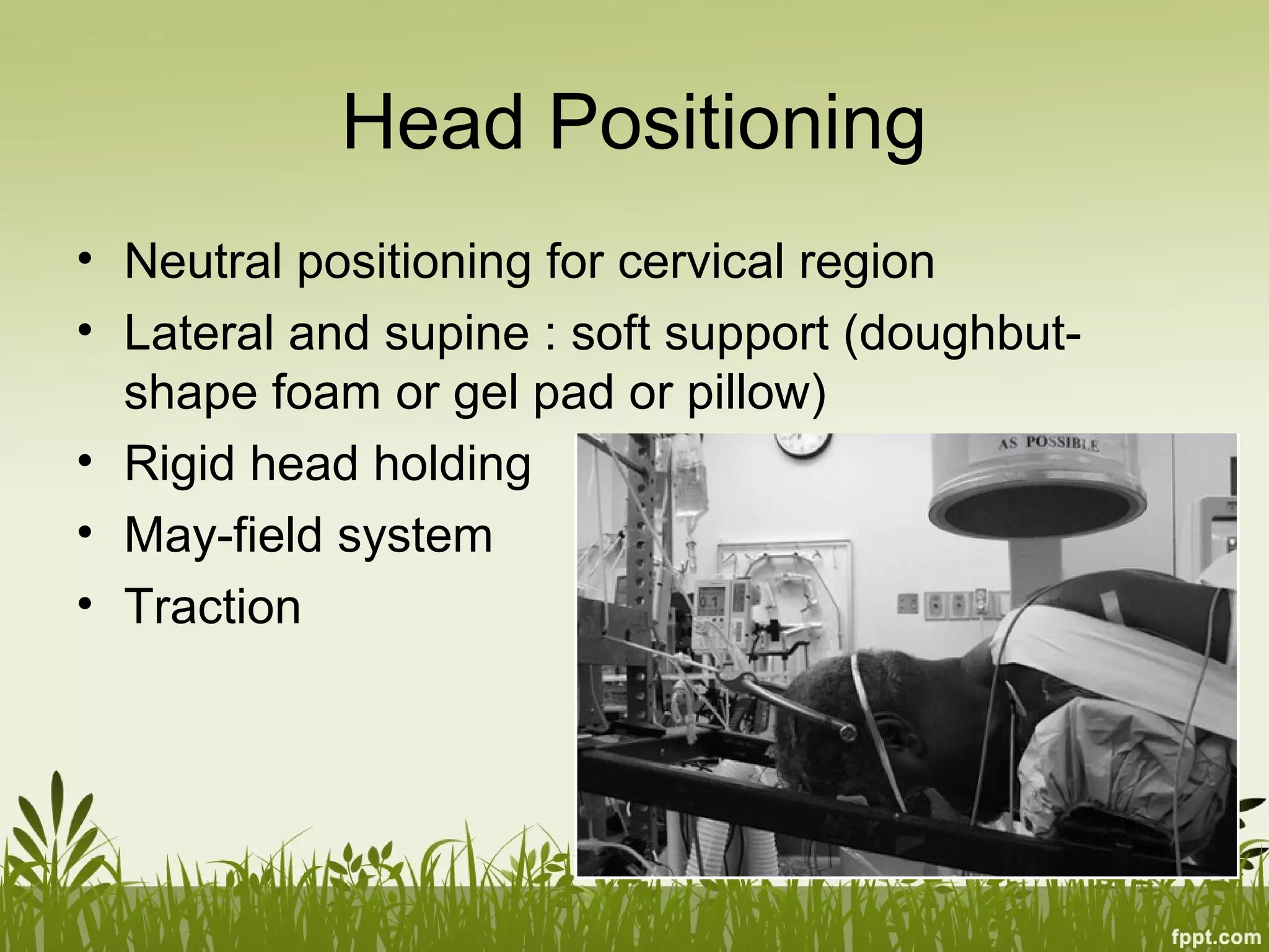
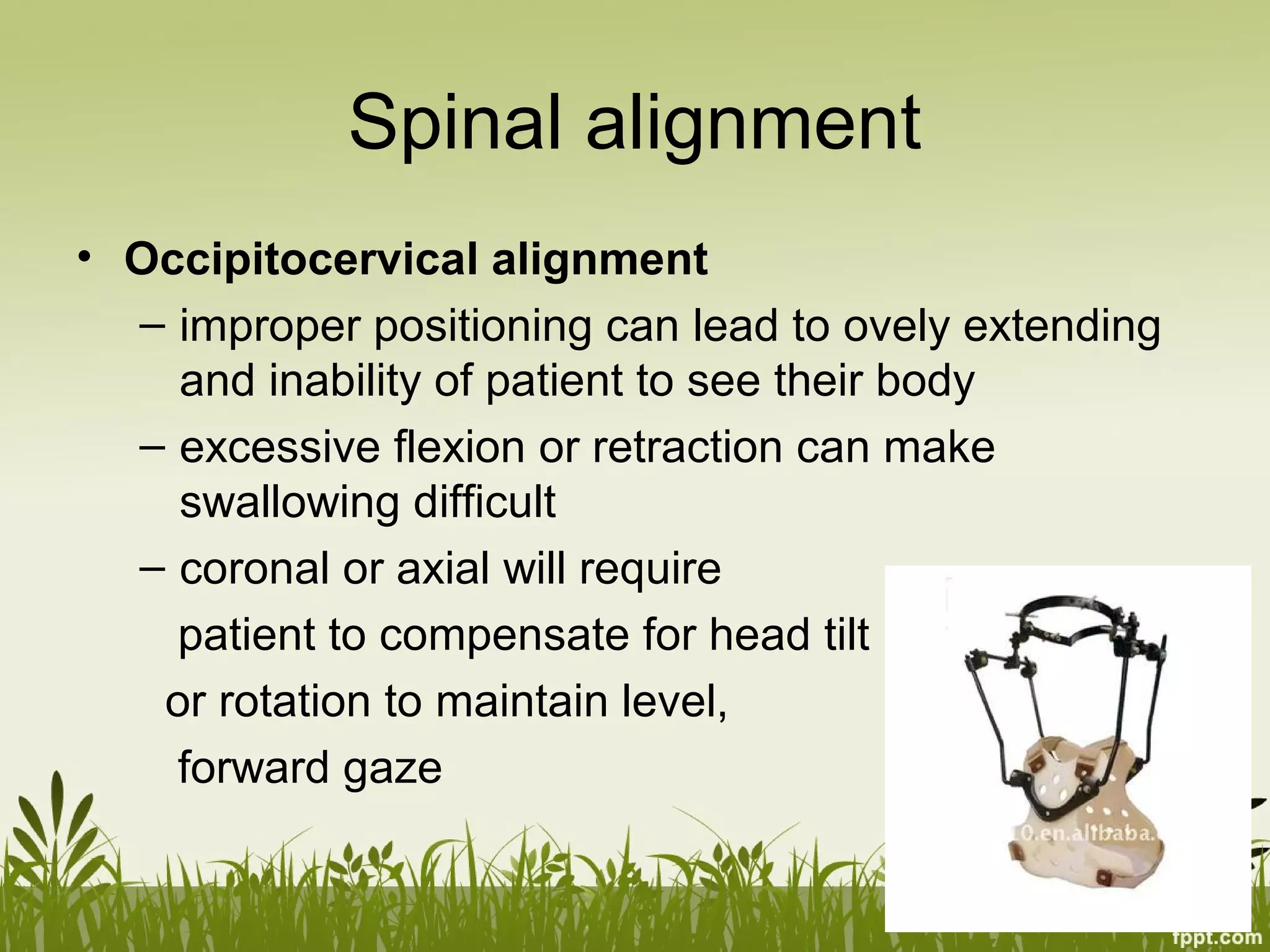
Patient Positioning for
Spinal Surgery
Youmans Chapter 27
Outline
• Equipment
• Principles of positioning
• Specific procedures
Equipment
Tables
Tables
• Operations in the supine position
– anterior cervical procedures
– anterior lumbar fusions in the distal lumbar spine
(L3-S1)
• A lateral approach for thoracic, thoracolumbar,
and lumbar procedure
• Thoracoabdominal and retroperitoneal flank
approaches, it is often helpful to place the level
of pathology at the table break and flex the
patient laterally.
Jackson spinal table
• Radiolucent
• Greater length of
height
• Full 360 clearance
• Use of multiple pad
Head Holders
• Cutouts for the eyes and endotracheal tube are
the main safety features
• Other
– Bean bag : lateral position for thoracotomy or
retroperitoneum flank approach
– Armrests
– Foam pad
– Disposable heating blanket
Principles of positioning
• Surgical access : allow the surgeon to achieve
the surgeon objective
• Patient safety and Protection
– Neuropathies and prevention
– Soft tissue injuries
– Head Positioning
– Visual loss and Its prevention
– Air embolism
• Spinal alignment
• Surgeon Ergonomics
Neuropathies and prevention
• Ulnar neuropathy : most common
postoperative neuropathies
• it is thought to be related to intraneural capillary
ischemia resulting from nerve overstretch or
compression, perhaps exacerbated by
prolonged intraoperative hypotension
• Time of onset of ulnar nerve symptom : after
surgery to 3 day postoperative
• Duration : day to year
• Risk factor : diabete, old age, male gender
Neuropathies and prevention
• Superficial condyle
groove
• Elbow flexion>110
• External
compression
Neuropathies and prevention
• Supine position : direct
pressure on ulnar n. at
the elbow is significantly
higher if both arm are
pronated than if they are
neutral position and
supinate
Neuropathies and prevention
• Brachial plexus neuropathy : shoulder pain,
scapular winging, and shoulder weakness
• Incidence during posterior spinal surgery : 3.6-
15 %
• Duration : persistent at late 1-3 yrs
• Upper trunk in supine position
• Lower trunk in prone position
• Pt congenital anomaly : cervical rib, shoulder
contracture
Neuropathies and prevention
• Somatosensory evoked potential (SSEP)
monitoring as a way to detect impending nerve
injury
• Lower extremity neuropathies : common
peroneal n. injury(superficial location as it
transverse the head of fibula)
Neuropathies and prevention
• Peroneal neuropathy : complete plegia of
dorsiflexion and eversion without significant pain
complain
• L5 radiculopathy : dermatomal pain, sensory
deficit, weakness of dorsiflexion, toe extension,
and foot inversion
Soft tissue injuries
• Prolong pressure leads to local ischemia, tissue
necrosis
• Not bear significant structure
• No EKG leads, IV line connector on supporting
pad
• Lateral or prone position : abdomen should be
free as possible  decrease intra-abdominal
pressure, decrease pressure in the valveless
epidural venous plexus (reduce epidural
bleeding)
Head Positioning
• Neutral positioning for cervical region
• Lateral and supine : soft support (doughbut-
shape foam or gel pad or pillow)
• Rigid head holding
• May-field system
• Traction
Visual loss and Its prevention
• POVL : post operative visual loss
• Most common cause : ischemic optic
neuropathy from compromised blood
flow(increase venous pressure and interstitial
edema), unilateral more than bilateral, prone
position
• May be attribute to central renal artery occlusion
• Associated with prolong anesthetic
operation(>6hr), significant blood loss(>1 lit)
Air embolism
• Sitting position,Cervical osteotomies
• Operative field above the heart  air may be entrained
into open  uncoaulated venous channel  air
embolism
• Precordial Doppler probe for diagnose an air embolism
• Long venous line used in attempt to aspirate air
• If air embolism is suspected during surgery : the field
should be flooded with sterile irrigation and position
change to bring the head close to the level of heart
Spinal alignment
• For procedure with no arthrodesis is performed :
lumbar microdiskectomy or cervical foraminotomy :
optimized to facilitate safe, thorough neural
decompression
Spinal alignment
• Occipitocervical alignment
– improper positioning can lead to ovely extending
and inability of patient to see their body
– excessive flexion or retraction can make
swallowing difficult
– coronal or axial will require
patient to compensate for head tilt
or rotation to maintain level,
forward gaze
Spinal alignment
• Lumbar decompressive : lumbar flexion
create
• This position would not be used if an
arthrodesis were also to be performed
• Hipextension enhances lumbar
lordosis, thereby resulting in
optimal spinal alignment for
instrumented arthrodesis
Surgeon ergonomic
• Optomize the working environment for surgeon
• Operative field heigt should be comfortable for
surgeon
• Horizontal plane as possible
• Lower cervical or cervicothoracic : reverse
trendelenberg
• Operating microscope
Specific procedure
• Anterior cervical
• Posterior occipitocervical, cervical,
cervicothoracic
• Posterior Thoracolumbar Arthrodesis
• Anterolateral, Retropleural Thoracic, Lateral
lumbar
• Anterior lumbar
• Intraoperative Repositioning
Anterior cervical
• Maintain gentle cervical extension(lordosis), maintain
head and cervical spine are neutrally aligned in the axial
plane
• Hypoextension: kyphosis
• Hyperextension : cervical spinal stenosis, neurological
risk intraoperative
• Small padded roll is placed underneath the patient and
extended transversely to about the T2 level,
• Foam doughnut is placed under the occiput
• Paper tape extending from one side to the other and
adherent to the forehead is adequate to maintain neutral
alignment
Posterior occipitocervical,
cervical, cervicothoracic
• First, unobstructed anteroposterior and lateral
radiographs or fluoroscopy can be obtained
• Second, the tabletop can be set up in a
moderate reverse Trendelenburg position
without raising the head unit
• Third, the modular pads can accommodate a
wide variety of body types.
• Finally, the dual-vector traction is easily set up
and manipulated.
Posterior Thoracolumbar Arthrodesis
• Position to maintain or enhance lumbar lordosis
• All contact point,particulary the knees, are
padded carefully
• It is also important to flex the knees and to
ensure that the feet are in a relaxed,neutral
position and not in forced plantar flexion
Anterolateral, lateral lumbar
Retropleural Thoracic,
• Lateral positioning follow the same principle as
for the common anterior and posterior approach
• Soft tissue or pheripheral n. injury secondary to
focal pressure
• Dependent axilla : soft roll to prevent excessive
shoulder abduction
• Dependent arm : externally rotate, elbox flex 90,
upper part gently flex, pillow
• Dependent leg : flex hip, flex knee
Anterior lumbar
• The arm may be abductes to allow access for
anesthesiologist
• Heel support
Intraoperative repositioning
• Supine to prone, prone to supine
• Two standard electric operating table

027 Patient posioning for spine surgery

  • 1.
    Patient Positioning for SpinalSurgery Youmans Chapter 27
  • 2.
    Outline • Equipment • Principlesof positioning • Specific procedures
  • 3.
  • 4.
  • 5.
    Tables • Operations inthe supine position – anterior cervical procedures – anterior lumbar fusions in the distal lumbar spine (L3-S1) • A lateral approach for thoracic, thoracolumbar, and lumbar procedure • Thoracoabdominal and retroperitoneal flank approaches, it is often helpful to place the level of pathology at the table break and flex the patient laterally.
  • 6.
    Jackson spinal table •Radiolucent • Greater length of height • Full 360 clearance • Use of multiple pad
  • 7.
    Head Holders • Cutoutsfor the eyes and endotracheal tube are the main safety features • Other – Bean bag : lateral position for thoracotomy or retroperitoneum flank approach – Armrests – Foam pad – Disposable heating blanket
  • 8.
    Principles of positioning •Surgical access : allow the surgeon to achieve the surgeon objective • Patient safety and Protection – Neuropathies and prevention – Soft tissue injuries – Head Positioning – Visual loss and Its prevention – Air embolism • Spinal alignment • Surgeon Ergonomics
  • 9.
    Neuropathies and prevention •Ulnar neuropathy : most common postoperative neuropathies • it is thought to be related to intraneural capillary ischemia resulting from nerve overstretch or compression, perhaps exacerbated by prolonged intraoperative hypotension • Time of onset of ulnar nerve symptom : after surgery to 3 day postoperative • Duration : day to year • Risk factor : diabete, old age, male gender
  • 10.
    Neuropathies and prevention •Superficial condyle groove • Elbow flexion>110 • External compression
  • 11.
    Neuropathies and prevention •Supine position : direct pressure on ulnar n. at the elbow is significantly higher if both arm are pronated than if they are neutral position and supinate
  • 12.
    Neuropathies and prevention •Brachial plexus neuropathy : shoulder pain, scapular winging, and shoulder weakness • Incidence during posterior spinal surgery : 3.6- 15 % • Duration : persistent at late 1-3 yrs • Upper trunk in supine position • Lower trunk in prone position • Pt congenital anomaly : cervical rib, shoulder contracture
  • 13.
    Neuropathies and prevention •Somatosensory evoked potential (SSEP) monitoring as a way to detect impending nerve injury • Lower extremity neuropathies : common peroneal n. injury(superficial location as it transverse the head of fibula)
  • 14.
    Neuropathies and prevention •Peroneal neuropathy : complete plegia of dorsiflexion and eversion without significant pain complain • L5 radiculopathy : dermatomal pain, sensory deficit, weakness of dorsiflexion, toe extension, and foot inversion
  • 15.
    Soft tissue injuries •Prolong pressure leads to local ischemia, tissue necrosis • Not bear significant structure • No EKG leads, IV line connector on supporting pad • Lateral or prone position : abdomen should be free as possible  decrease intra-abdominal pressure, decrease pressure in the valveless epidural venous plexus (reduce epidural bleeding)
  • 16.
    Head Positioning • Neutralpositioning for cervical region • Lateral and supine : soft support (doughbut- shape foam or gel pad or pillow) • Rigid head holding • May-field system • Traction
  • 17.
    Visual loss andIts prevention • POVL : post operative visual loss • Most common cause : ischemic optic neuropathy from compromised blood flow(increase venous pressure and interstitial edema), unilateral more than bilateral, prone position • May be attribute to central renal artery occlusion • Associated with prolong anesthetic operation(>6hr), significant blood loss(>1 lit)
  • 18.
    Air embolism • Sittingposition,Cervical osteotomies • Operative field above the heart  air may be entrained into open  uncoaulated venous channel  air embolism • Precordial Doppler probe for diagnose an air embolism • Long venous line used in attempt to aspirate air • If air embolism is suspected during surgery : the field should be flooded with sterile irrigation and position change to bring the head close to the level of heart
  • 19.
    Spinal alignment • Forprocedure with no arthrodesis is performed : lumbar microdiskectomy or cervical foraminotomy : optimized to facilitate safe, thorough neural decompression
  • 20.
    Spinal alignment • Occipitocervicalalignment – improper positioning can lead to ovely extending and inability of patient to see their body – excessive flexion or retraction can make swallowing difficult – coronal or axial will require patient to compensate for head tilt or rotation to maintain level, forward gaze
  • 21.
    Spinal alignment • Lumbardecompressive : lumbar flexion create • This position would not be used if an arthrodesis were also to be performed • Hipextension enhances lumbar lordosis, thereby resulting in optimal spinal alignment for instrumented arthrodesis
  • 22.
    Surgeon ergonomic • Optomizethe working environment for surgeon • Operative field heigt should be comfortable for surgeon • Horizontal plane as possible • Lower cervical or cervicothoracic : reverse trendelenberg • Operating microscope
  • 23.
    Specific procedure • Anteriorcervical • Posterior occipitocervical, cervical, cervicothoracic • Posterior Thoracolumbar Arthrodesis • Anterolateral, Retropleural Thoracic, Lateral lumbar • Anterior lumbar • Intraoperative Repositioning
  • 24.
    Anterior cervical • Maintaingentle cervical extension(lordosis), maintain head and cervical spine are neutrally aligned in the axial plane • Hypoextension: kyphosis • Hyperextension : cervical spinal stenosis, neurological risk intraoperative • Small padded roll is placed underneath the patient and extended transversely to about the T2 level, • Foam doughnut is placed under the occiput • Paper tape extending from one side to the other and adherent to the forehead is adequate to maintain neutral alignment
  • 25.
    Posterior occipitocervical, cervical, cervicothoracic •First, unobstructed anteroposterior and lateral radiographs or fluoroscopy can be obtained • Second, the tabletop can be set up in a moderate reverse Trendelenburg position without raising the head unit • Third, the modular pads can accommodate a wide variety of body types. • Finally, the dual-vector traction is easily set up and manipulated.
  • 26.
    Posterior Thoracolumbar Arthrodesis •Position to maintain or enhance lumbar lordosis • All contact point,particulary the knees, are padded carefully • It is also important to flex the knees and to ensure that the feet are in a relaxed,neutral position and not in forced plantar flexion
  • 27.
    Anterolateral, lateral lumbar RetropleuralThoracic, • Lateral positioning follow the same principle as for the common anterior and posterior approach • Soft tissue or pheripheral n. injury secondary to focal pressure • Dependent axilla : soft roll to prevent excessive shoulder abduction • Dependent arm : externally rotate, elbox flex 90, upper part gently flex, pillow • Dependent leg : flex hip, flex knee
  • 28.
    Anterior lumbar • Thearm may be abductes to allow access for anesthesiologist • Heel support
  • 29.
    Intraoperative repositioning • Supineto prone, prone to supine • Two standard electric operating table

Editor's Notes

  • #5 รูปแรกเป็น standard operating room table – Wilson Frame
  • #17 Pt ที่มี thoracolumbar จะมี cervical spondylosis ร่วมด้วย
  • #20 Arthrodesis is the fusion of vertebrae over a joint space that occurs through a natural process or as a result of surgical procedure. In surgery, arthrodesis, or fusion between two vertebrae, can be achieved by placing bone graft and/or bone graft substitute to bridge the vertebrae so that new bone grows into the spaces.