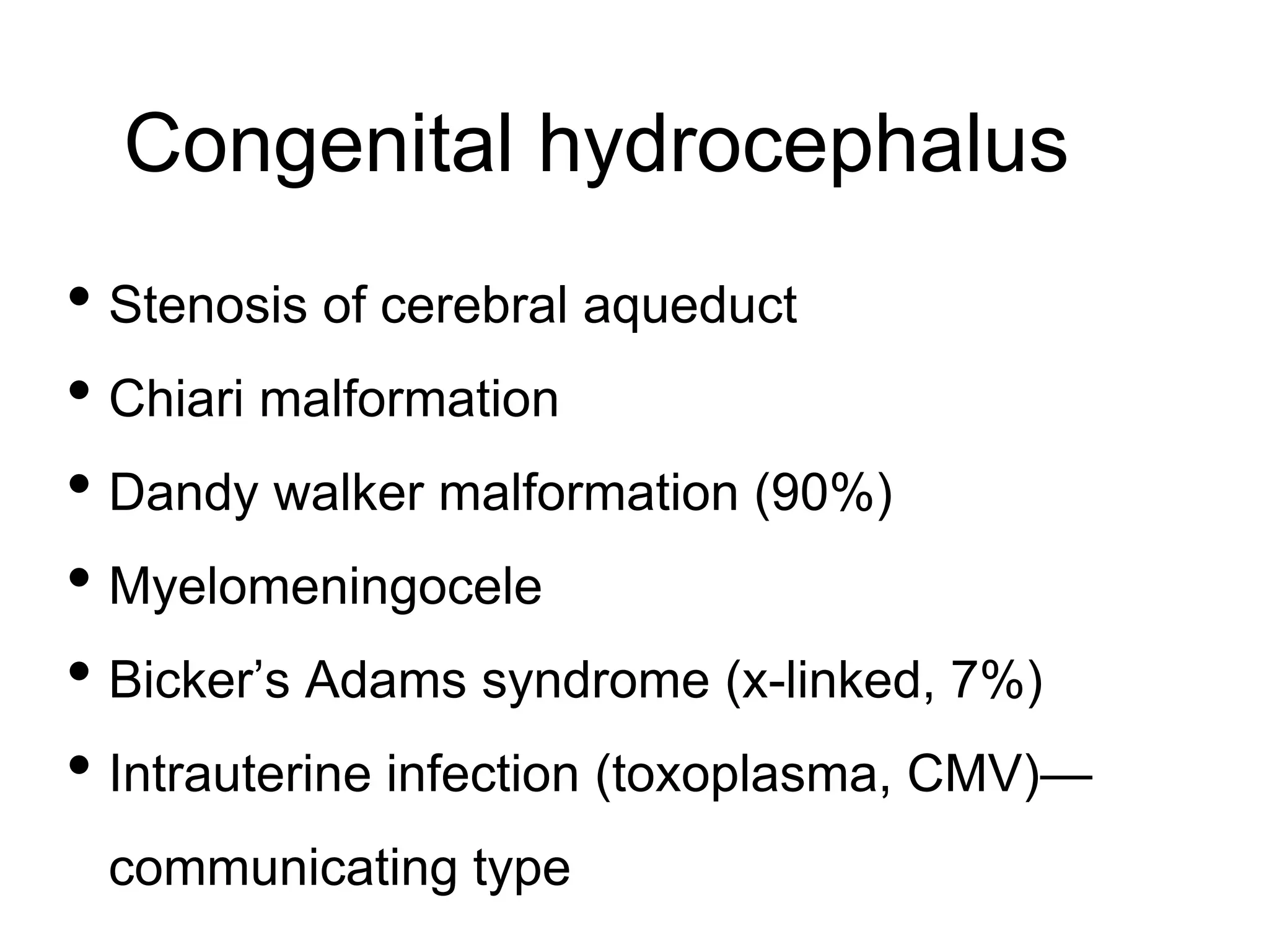
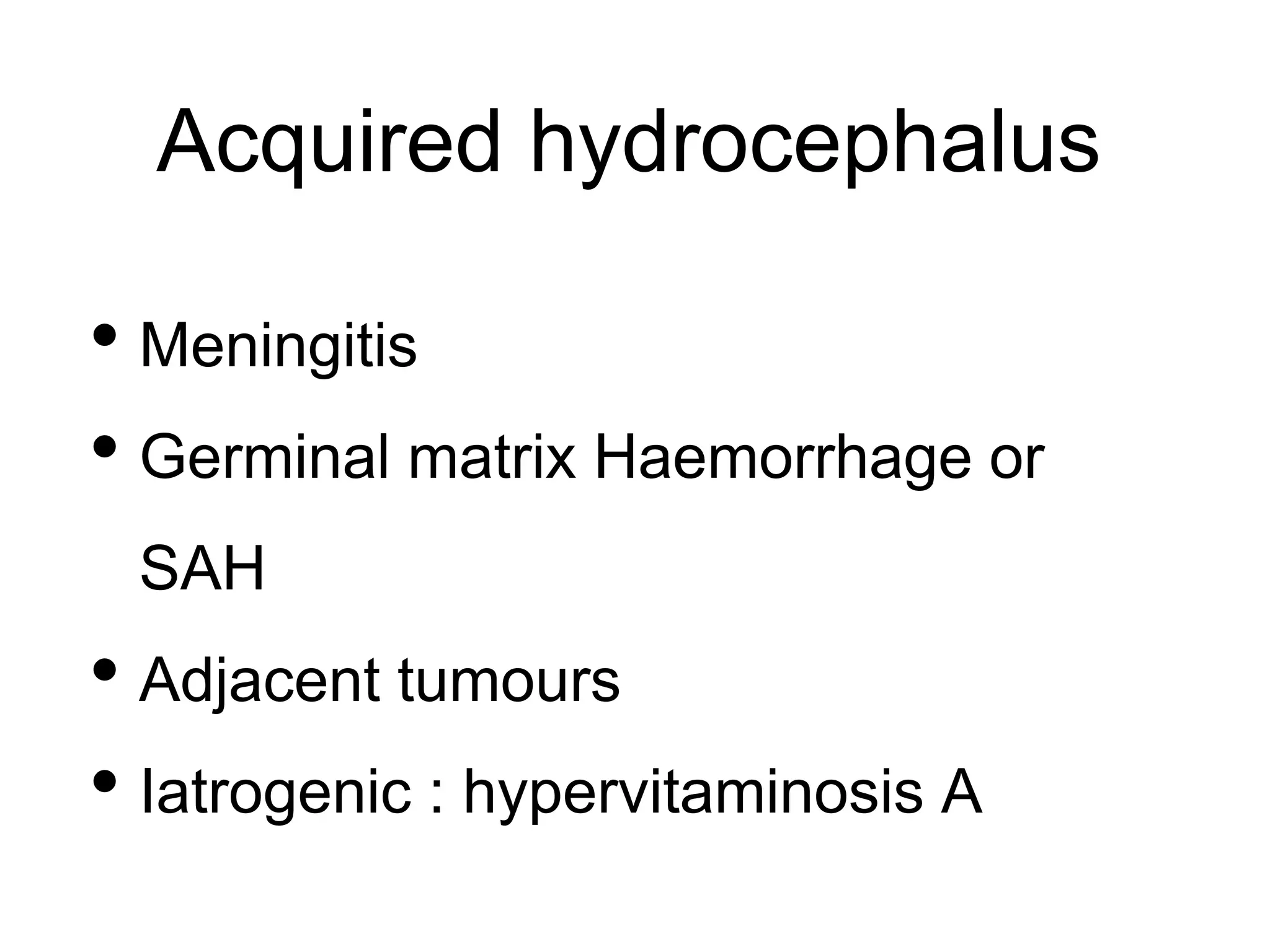
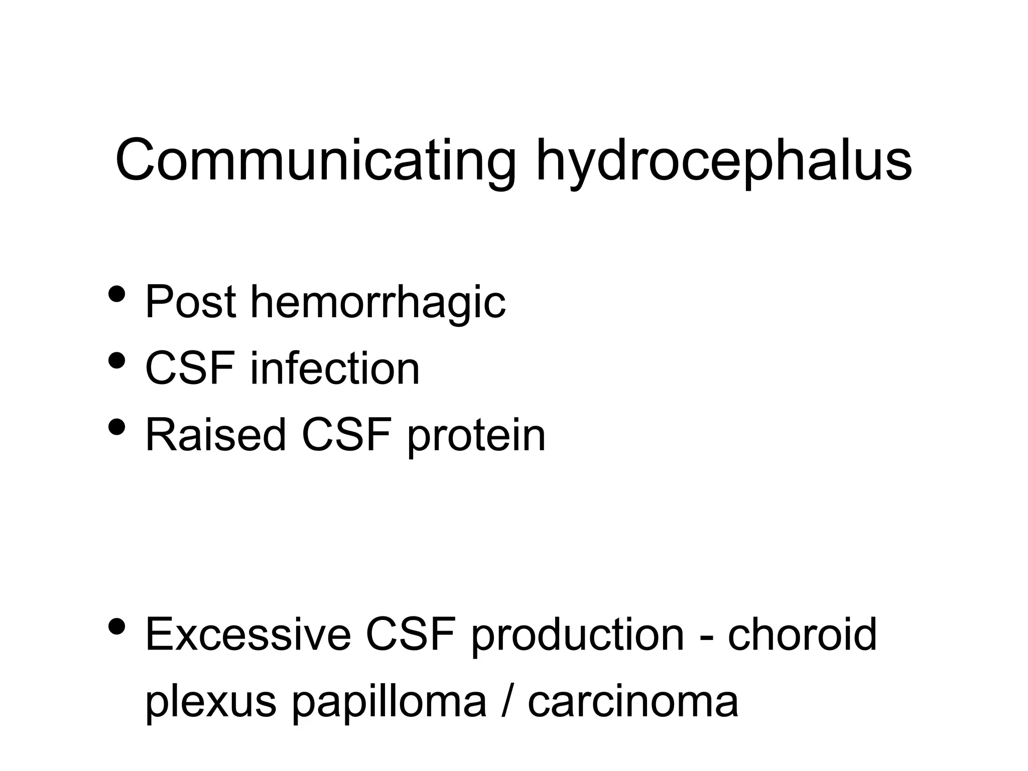
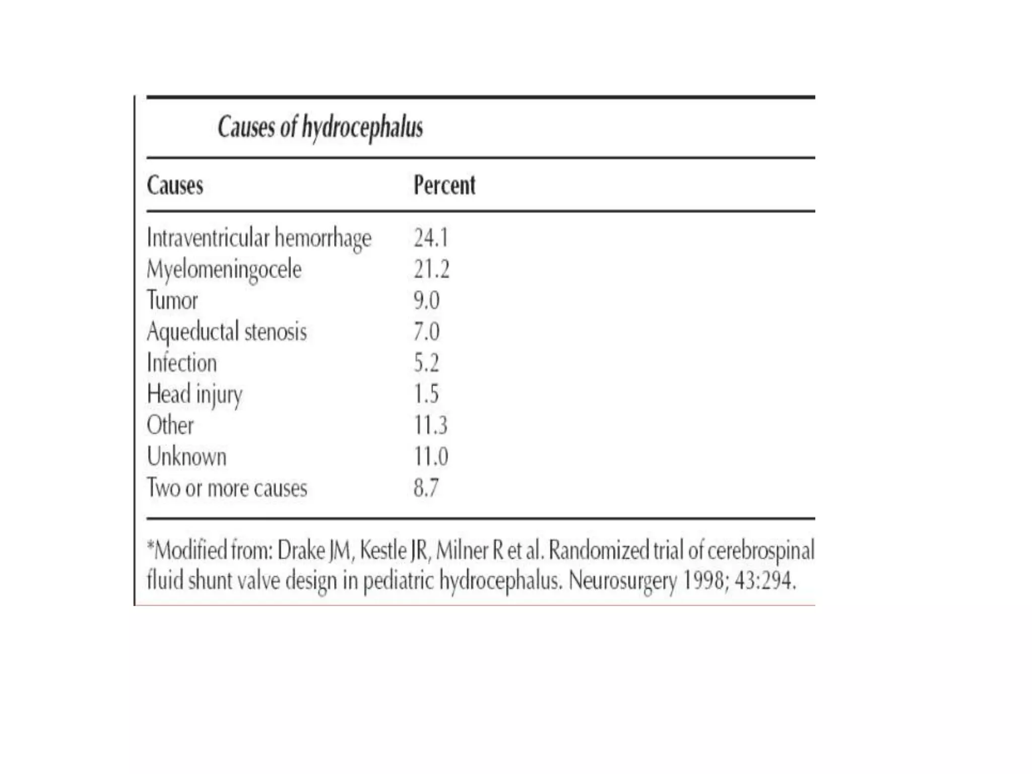
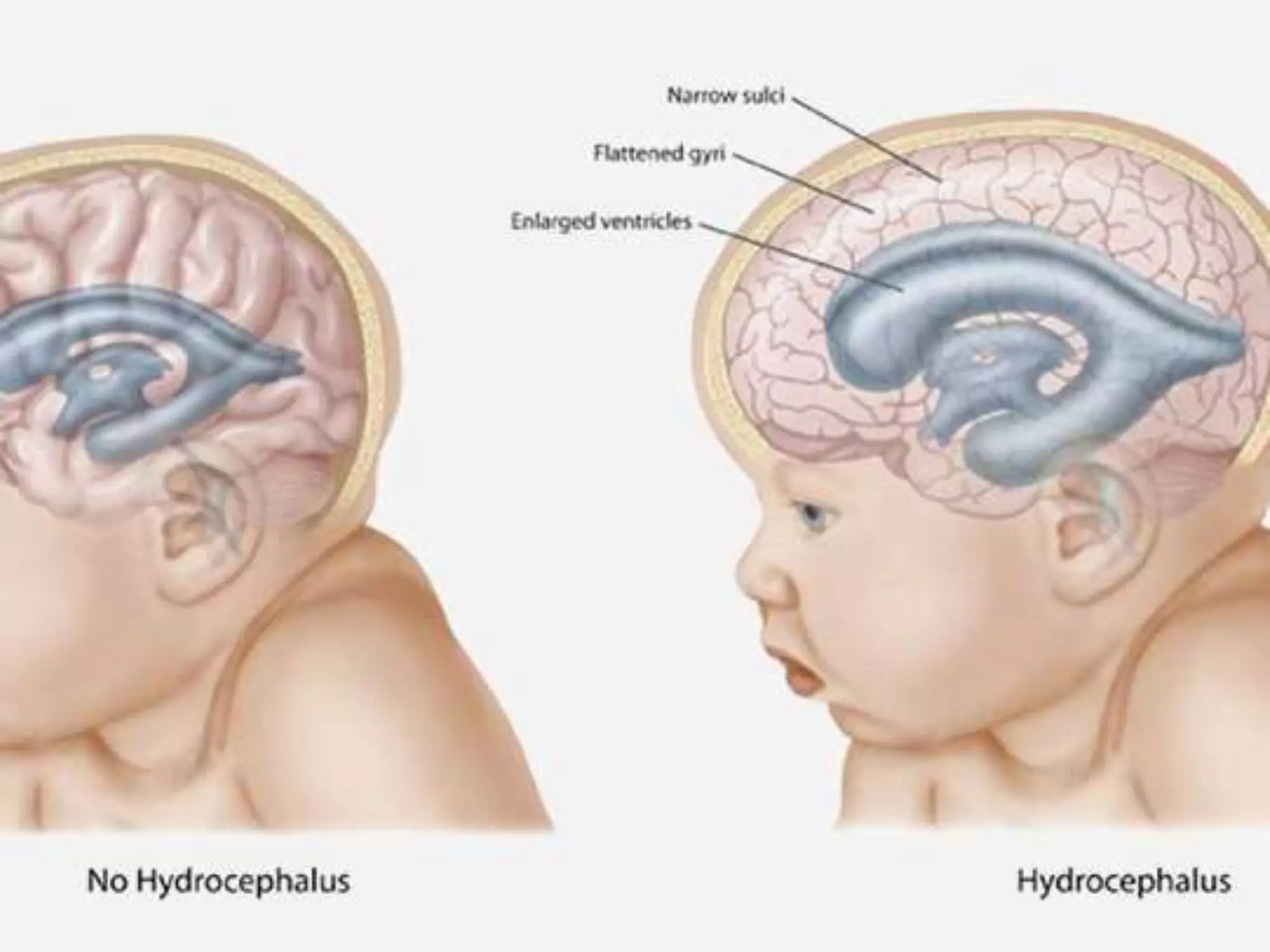
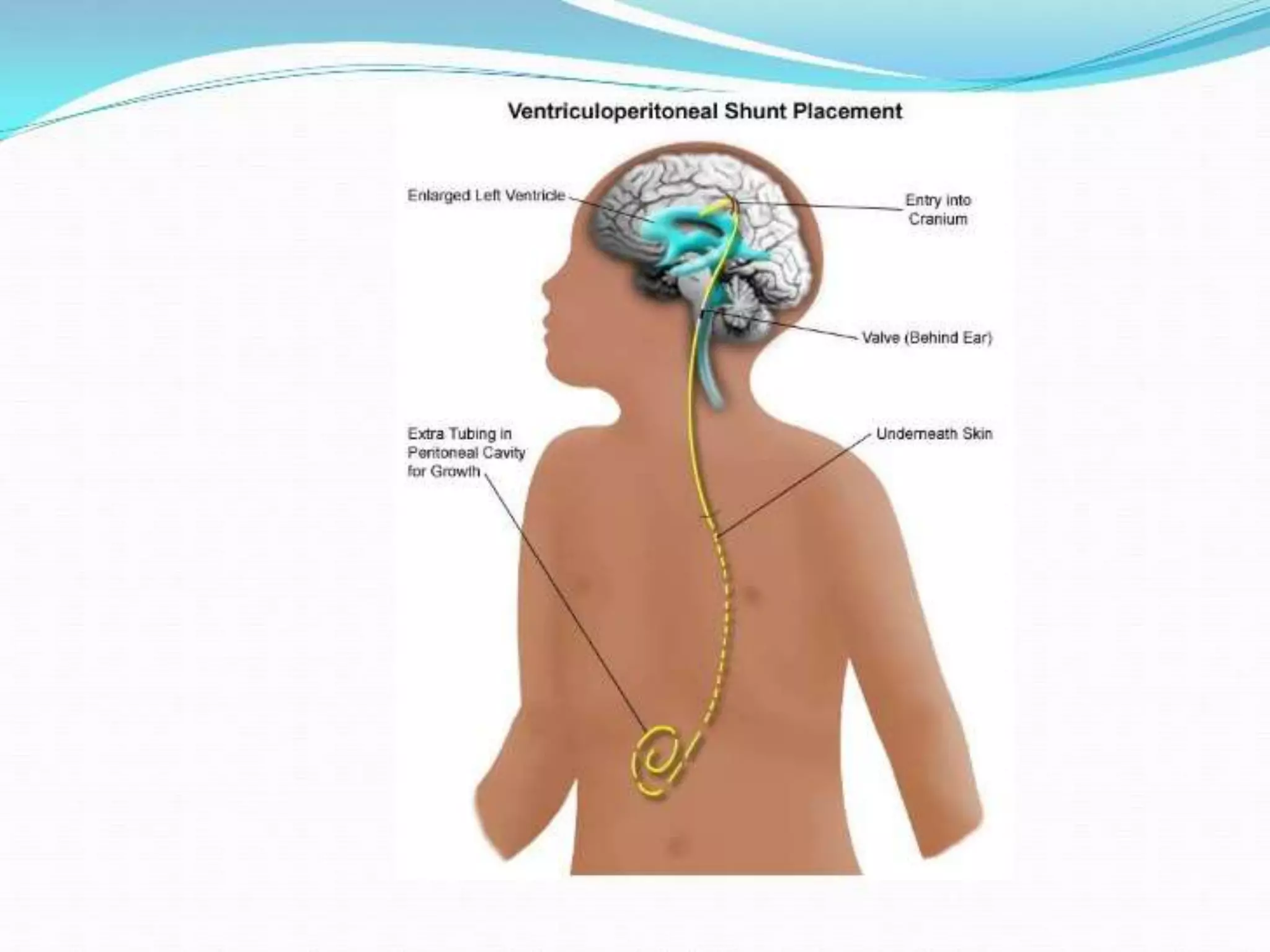
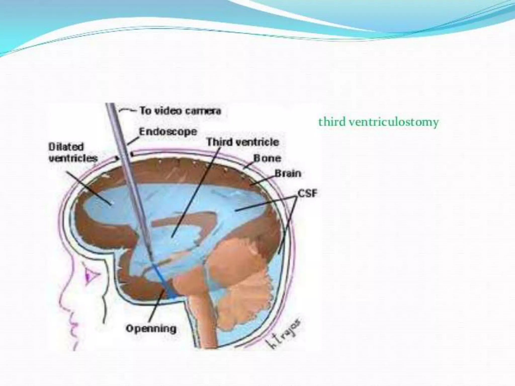
Seminar on
hydrocephalus
Presenter : Dr Biswajit Deka
PGT 2nd year
Deptt of surgery , SMCH
Definition
Hydrocephalus refers to an increase in
CSF volume with ventricular enlargement
Fluid accumulation
• Intracerebral (ventricular)
• Extracerebral (subarachnoid spaces &
cisterns)
Incidence : 3-4/1000 births
Physiology
•Total CSF volume : 150 ml
•Site : walls of ventricle & choroid
plexus
•Rate : 20 ml / hr (o.33ml/kg/hr)
•Circulation time : 8 hrs
Route of CSF flow
Lateral ventricle foramen of
Monro
Third ventricle cerebral
aqueduct
Fourth ventricle foramen of
Megendie & luschka
sub arachnoid space
Types
• Congenital & acquired
• Obstructive & communicating
• Acute (d/t tumour) and chronic (NPH)
Congenital hydrocephalus
• Stenosis of cerebral aqueduct
• Chiari malformation
• Dandy walker malformation (90%)
• Myelomeningocele
• Bicker’s Adams syndrome (x-linked, 7%)
• Intrauterine infection (toxoplasma, CMV)—
communicating type
Acquired hydrocephalus
• Meningitis
• Germinal matrix Haemorrhage or
SAH
• Adjacent tumours
• Iatrogenic : hypervitaminosis A
Obstructive hydrocephalus
• Lesions within the ventricle
• Lesions in the ventricular wall
• Lesions distant from ventricular wall
with a mass effect
Communicating hydrocephalus
• Post hemorrhagic
• CSF infection
• Raised CSF protein
• Excessive CSF production - choroid
plexus papilloma / carcinoma
Early
• Irritability
• Poor feeding
• Difficulty in focussing eye
• Excessive sleepiness
• Rapid increase in head circumference
• Seizures
Clinical presentation
Late
• High pressure headache - coughing /
bending forward
• Nausea & vomiting
• Blurred & double vision
• Eye movement abnormality
• Pupil abnormality
• Fundoscopy - papilloedema (absent in
acute phase)
Infant
• Tense & bulging fontanelle
• Increase in head circumference
• Bulging scalp veins
• Impaired conscious levels
• Sunsetting sign
• Parinaud's syndrome ( dorsal midbrain
syndrome / vertical gaze palsy / sunset
sign )is an inability to move the eyes up
and down.
• Caused by compression of the vertical
gaze center at the rostral interstitial
nucleus of medial longitudinal fasciculus
(riMLF).
Risk
• Cerebral herniation
• CVS instability
• Neurological deficit
• Vision - irreversible damage
• Death
Investigation
• CT brain - 1st line
• Degree of mass effect
• Potency of basal cisterns
• Spaces surrounding brainstem
• Cannot delineate exact site &
nature of obstruction.
• Assessment of shunt malfunction
• Lumber punture -( herniation )
• MRI - anatomy relating to potential
treatment ;eg third ventriculostomy,
aqueductoplasty & newly diagnosed case
• Cranial USG - new born
• ICP monitoring - transducer in brain
substance
Intracranial pressure
waveform
•P1 (percussion
wave) -arterial
pulsation
•P2 - brain
compliance
•P3 (dicrotic wave)-
venous pulsation
For communicating hydrocephalus
• LP : diagnostic & therapautic
• Deriving an opening pressure
• Assessment of CSF contents
• Drainage :10-30 ml CSF
• Aim : to make opening pressure half
Hydrocephalus ex vacuo
• A/k/a compensatory hydrocephalus
• Ventricle enlarge compulsory to overall
shrinking of the brain tissue .
• D/D : NPH
• Seen in -
• Advance age with brain atrophy
• After diffuse HI or stroke
• Neurodegenerative condition
Porencephaly
• Porencephalic cyst
• Loss of volume focal brain substance
— collection of CSF in the cavity
• Localised lesion
Arrested hydrocephalus
• Ventricles are enlarged
• No significant symptoms
• Do not require surgical Tx
• May develop symptoms during prolong
period or after acute ppt. factors (trauma
, infection) that alters CSF dynamics.
• Cause of dementia
• reversible
• Triad : gait disturbance , incontinence ,
cognitive decline
• De novo or previous brain insult (SAH,
HI, meningitis , tumour )
Normal pressure hydrocephalus
• Imaging : ventriculomegaly
• CSF pressure at LP : normal
• But , intermittent elevation in pressure
may be involved in etiology
• Lumbar infusion testing : insertion
of a fine drain at LP, f/b
measurement of CSF pressure
changes a/w a fluid challenge
administered through this.
• This allows evaluation of the likely
benefit from definitive treatment by
shunt insertion.
Idiopathic intracranial hypertension
• Pseudo tumour cerebri
• Benign intracranial hypertension
• Features of raised ICP with underlying
tumour
• Can cause blindness
• Young , over weight , female
• Headache & visual disturbance
• Examination :
• papilloedema
• Cranial nerve palsies
• Imaging : unremarkable
• LP : raised opening pressure > 25mm Hg
• Etiology : not well understood
• Diagnosis : is of exclusion
Treatment of IIH
• Weight loss
• Stop : OCPs
• Acetazolamide : to reduce CSF
production
• Shunting : to prevent visual loss
• Optic nerve sheath fenestration
Treatment of Hydrocephalus
Acute obstructive hydrocephalus - emergency
Hydrocephalic attack : coma & death
Aim : to treat underlying pathology
Temporary ventricular drainage
External ventricular drain
• EVD : effective temporary measure
• Inserted to the right of midline, anterior to coronal
suture, so that the catheter tip rests adjacent to the
fomamen of Monro in the lateral ventricle.
• Catheter connected to drain set such that CSF
drains when the ventricular pressure exceeds a
threshold, typically set at 10-20 mm Hg.
• Intrathecal antibiotics may be delivered
• Lumbar drains :
alternative means of temporary CSF
diversion , often used to manage CSF
leaks resulting from communicating
hydrocephalus
VP shunt
• Comprises insertion of a ventricular catheter into the
frontal or occipital horn of the lateral ventricle , while
a distal catheter is tunnelled subcutaneously to the
abdomen.
• A shunt valve, with an opening pressure that may be
high, medium or low , is inserted at the junction of
these catheters.
• Selection of shunt valve : patient specific
• High pressure valve may fail to allow adequate CSF
drainage ,whereas low pressure valve may
overdrain.
• Anti-siphon system incorporated : to prevent
excessive drainage in standing position.
• Programmable valve : offer variable opening
pressure , adjusted magnetically using a device
applied externally over the valve .
• Valve system will also incorporate a CSF reservoir ,
which allows for percutaneous sampling.
Shunt complication
• Overdrainage : low pressure headache
• Subdural hygroma / hematoma (d/t collapse of
ventricle that cause accumulation of blood / fluid in
subdural space)
• Slit ventricle syndrome : children treated with shunts,
whose ventricle & sub arachnoid spaces are
underdeveloped , resulting in poor brain compliance .
Here , normal fluctuation of ICP are exaggerated .
Any shunt blockage may not be evident on scan , as
ventricle fail to enlarge
• Disconnection
• Infection (75% within 1 month) - Staph Epidermidis
• Blockage
• 15-20% require replacement within 3 years
• Failure of valve mechanism
• Hydrocele and hernia
• distal end of shunt in the sac.
Endoscopic third ventriculostomy
• Useful in obstructive hydrocephalus d/t aqueduct
stenosis
• A neuroendoscope is inserted into the frontal horn of
the lateral ventricle & then into the third ventricle via
foramen of Monro.
• The floor of the ventricle is then opened between the
mammillary bodies & pituitary recess, which allows
free drainage between third ventricle & adjacent
subarachnoid cistern
• Position: supine with head flexed so that
the burr hole site is at the highest point.
• Generally, the width of the third ventricle
and foramen of Monro should be
approximately 7 mm or greater.
• Stereotactic guidance can be used as a
surgical adjunct to access ventricle.
• Burr hole made at or just anterior to the coronal
suture and about 2.5–3 cm lateral to the midline.
• Right side burr hole performed.
• Brain cannula is used to puncture ventricle and then
introduce endoscope with sheath.
• Peel-away sheath with a diameter, just slightly larger
than the endoscope, can be used. .
• If any significant hemorrhage occurs, the peel-away
sheath facilitates copious irrigation until the
hemorrhage is cleared.
• Lactate solution used for irrigation using gravity as
opposed to any pressure technique to avoid any
barotraumas to the brain or ventricles
• Foramen of Monro identified by confluence of thalamo-striate
vein, septal vein and choroid plexuses.
• Perforation made after negotiating endoscope through the
foramen of Monro.
• Fenestration: between mammillary bodies and infundibular
recess, at the most transparent site.
• Basilar artery identified to avoid an injury and the fenestration
made anterior to the artery complex.
• Microvascular Doppler probe: to locate basilar artery
• Position of dorsum sellae identified by gentle probing by the
blunt instrument, such as bipolar forceps if Doppler is not
available.
• Fenestration should be made just posterior to dorsum sellae.
• Water jet dissection technique can be used to prevent an injury
to vessel if the floor is thick and opaque
• The third ventricular floor is penetrated bluntly to
avoid vascular injury.
• Although thermal or electric energy are avoided ,
laser assisted third ventriculostomy was found to
be safe and effective.
• An initial fenestration is dilated up to
approximately 5 mm or more by using French
Fogarty catheter.
• An ultrasonic contact probe (NECUP-2) can be
used to create minimal and controlled lesion in
third ventriculostomy
• Re-blockage is common
• Less chance of infection
• Complication : damage to basilar artery
or fornicial damage resulting in
permanent memory impairment
Other new modalities
• Endoscopic aqueductoplasty ( 3 Fr Fogarty
catheter)
• Endoscopic aqueductal stenting
• Endsopic septostomy
Seminar on Hydrocephalus

Seminar on Hydrocephalus

  • 1.
    Seminar on hydrocephalus Presenter :Dr Biswajit Deka PGT 2nd year Deptt of surgery , SMCH
  • 2.
    Definition Hydrocephalus refers toan increase in CSF volume with ventricular enlargement Fluid accumulation • Intracerebral (ventricular) • Extracerebral (subarachnoid spaces & cisterns) Incidence : 3-4/1000 births
  • 3.
    Physiology •Total CSF volume: 150 ml •Site : walls of ventricle & choroid plexus •Rate : 20 ml / hr (o.33ml/kg/hr) •Circulation time : 8 hrs
  • 4.
    Route of CSFflow Lateral ventricle foramen of Monro Third ventricle cerebral aqueduct Fourth ventricle foramen of Megendie & luschka sub arachnoid space
  • 6.
    Types • Congenital &acquired • Obstructive & communicating • Acute (d/t tumour) and chronic (NPH)
  • 7.
    Congenital hydrocephalus • Stenosisof cerebral aqueduct • Chiari malformation • Dandy walker malformation (90%) • Myelomeningocele • Bicker’s Adams syndrome (x-linked, 7%) • Intrauterine infection (toxoplasma, CMV)— communicating type
  • 8.
    Acquired hydrocephalus • Meningitis •Germinal matrix Haemorrhage or SAH • Adjacent tumours • Iatrogenic : hypervitaminosis A
  • 9.
    Obstructive hydrocephalus • Lesionswithin the ventricle • Lesions in the ventricular wall • Lesions distant from ventricular wall with a mass effect
  • 10.
    Communicating hydrocephalus • Posthemorrhagic • CSF infection • Raised CSF protein • Excessive CSF production - choroid plexus papilloma / carcinoma
  • 14.
    Early • Irritability • Poorfeeding • Difficulty in focussing eye • Excessive sleepiness • Rapid increase in head circumference • Seizures Clinical presentation
  • 15.
    Late • High pressureheadache - coughing / bending forward • Nausea & vomiting • Blurred & double vision • Eye movement abnormality • Pupil abnormality • Fundoscopy - papilloedema (absent in acute phase)
  • 16.
    Infant • Tense &bulging fontanelle • Increase in head circumference • Bulging scalp veins • Impaired conscious levels • Sunsetting sign
  • 20.
    • Parinaud's syndrome( dorsal midbrain syndrome / vertical gaze palsy / sunset sign )is an inability to move the eyes up and down. • Caused by compression of the vertical gaze center at the rostral interstitial nucleus of medial longitudinal fasciculus (riMLF).
  • 22.
    Risk • Cerebral herniation •CVS instability • Neurological deficit • Vision - irreversible damage • Death
  • 23.
    Investigation • CT brain- 1st line • Degree of mass effect • Potency of basal cisterns • Spaces surrounding brainstem • Cannot delineate exact site & nature of obstruction. • Assessment of shunt malfunction
  • 24.
    • Lumber punture-( herniation ) • MRI - anatomy relating to potential treatment ;eg third ventriculostomy, aqueductoplasty & newly diagnosed case • Cranial USG - new born • ICP monitoring - transducer in brain substance
  • 26.
    Intracranial pressure waveform •P1 (percussion wave)-arterial pulsation •P2 - brain compliance •P3 (dicrotic wave)- venous pulsation
  • 27.
    For communicating hydrocephalus •LP : diagnostic & therapautic • Deriving an opening pressure • Assessment of CSF contents • Drainage :10-30 ml CSF • Aim : to make opening pressure half
  • 28.
    Hydrocephalus ex vacuo •A/k/a compensatory hydrocephalus • Ventricle enlarge compulsory to overall shrinking of the brain tissue . • D/D : NPH • Seen in - • Advance age with brain atrophy • After diffuse HI or stroke • Neurodegenerative condition
  • 29.
    Porencephaly • Porencephalic cyst •Loss of volume focal brain substance — collection of CSF in the cavity • Localised lesion
  • 30.
    Arrested hydrocephalus • Ventriclesare enlarged • No significant symptoms • Do not require surgical Tx • May develop symptoms during prolong period or after acute ppt. factors (trauma , infection) that alters CSF dynamics.
  • 31.
    • Cause ofdementia • reversible • Triad : gait disturbance , incontinence , cognitive decline • De novo or previous brain insult (SAH, HI, meningitis , tumour ) Normal pressure hydrocephalus
  • 32.
    • Imaging :ventriculomegaly • CSF pressure at LP : normal • But , intermittent elevation in pressure may be involved in etiology
  • 33.
    • Lumbar infusiontesting : insertion of a fine drain at LP, f/b measurement of CSF pressure changes a/w a fluid challenge administered through this. • This allows evaluation of the likely benefit from definitive treatment by shunt insertion.
  • 34.
    Idiopathic intracranial hypertension •Pseudo tumour cerebri • Benign intracranial hypertension • Features of raised ICP with underlying tumour • Can cause blindness • Young , over weight , female • Headache & visual disturbance
  • 35.
    • Examination : •papilloedema • Cranial nerve palsies • Imaging : unremarkable • LP : raised opening pressure > 25mm Hg • Etiology : not well understood • Diagnosis : is of exclusion
  • 36.
    Treatment of IIH •Weight loss • Stop : OCPs • Acetazolamide : to reduce CSF production • Shunting : to prevent visual loss • Optic nerve sheath fenestration
  • 37.
    Treatment of Hydrocephalus Acuteobstructive hydrocephalus - emergency Hydrocephalic attack : coma & death Aim : to treat underlying pathology Temporary ventricular drainage
  • 38.
    External ventricular drain •EVD : effective temporary measure • Inserted to the right of midline, anterior to coronal suture, so that the catheter tip rests adjacent to the fomamen of Monro in the lateral ventricle. • Catheter connected to drain set such that CSF drains when the ventricular pressure exceeds a threshold, typically set at 10-20 mm Hg. • Intrathecal antibiotics may be delivered
  • 39.
    • Lumbar drains: alternative means of temporary CSF diversion , often used to manage CSF leaks resulting from communicating hydrocephalus
  • 40.
    VP shunt • Comprisesinsertion of a ventricular catheter into the frontal or occipital horn of the lateral ventricle , while a distal catheter is tunnelled subcutaneously to the abdomen. • A shunt valve, with an opening pressure that may be high, medium or low , is inserted at the junction of these catheters. • Selection of shunt valve : patient specific
  • 41.
    • High pressurevalve may fail to allow adequate CSF drainage ,whereas low pressure valve may overdrain. • Anti-siphon system incorporated : to prevent excessive drainage in standing position. • Programmable valve : offer variable opening pressure , adjusted magnetically using a device applied externally over the valve . • Valve system will also incorporate a CSF reservoir , which allows for percutaneous sampling.
  • 48.
    Shunt complication • Overdrainage: low pressure headache • Subdural hygroma / hematoma (d/t collapse of ventricle that cause accumulation of blood / fluid in subdural space) • Slit ventricle syndrome : children treated with shunts, whose ventricle & sub arachnoid spaces are underdeveloped , resulting in poor brain compliance . Here , normal fluctuation of ICP are exaggerated .
  • 49.
    Any shunt blockagemay not be evident on scan , as ventricle fail to enlarge • Disconnection • Infection (75% within 1 month) - Staph Epidermidis • Blockage • 15-20% require replacement within 3 years • Failure of valve mechanism • Hydrocele and hernia • distal end of shunt in the sac.
  • 50.
    Endoscopic third ventriculostomy •Useful in obstructive hydrocephalus d/t aqueduct stenosis • A neuroendoscope is inserted into the frontal horn of the lateral ventricle & then into the third ventricle via foramen of Monro. • The floor of the ventricle is then opened between the mammillary bodies & pituitary recess, which allows free drainage between third ventricle & adjacent subarachnoid cistern
  • 51.
    • Position: supinewith head flexed so that the burr hole site is at the highest point. • Generally, the width of the third ventricle and foramen of Monro should be approximately 7 mm or greater. • Stereotactic guidance can be used as a surgical adjunct to access ventricle.
  • 52.
    • Burr holemade at or just anterior to the coronal suture and about 2.5–3 cm lateral to the midline. • Right side burr hole performed. • Brain cannula is used to puncture ventricle and then introduce endoscope with sheath. • Peel-away sheath with a diameter, just slightly larger than the endoscope, can be used. . • If any significant hemorrhage occurs, the peel-away sheath facilitates copious irrigation until the hemorrhage is cleared. • Lactate solution used for irrigation using gravity as opposed to any pressure technique to avoid any barotraumas to the brain or ventricles
  • 53.
    • Foramen ofMonro identified by confluence of thalamo-striate vein, septal vein and choroid plexuses. • Perforation made after negotiating endoscope through the foramen of Monro. • Fenestration: between mammillary bodies and infundibular recess, at the most transparent site. • Basilar artery identified to avoid an injury and the fenestration made anterior to the artery complex. • Microvascular Doppler probe: to locate basilar artery • Position of dorsum sellae identified by gentle probing by the blunt instrument, such as bipolar forceps if Doppler is not available. • Fenestration should be made just posterior to dorsum sellae. • Water jet dissection technique can be used to prevent an injury to vessel if the floor is thick and opaque
  • 54.
    • The thirdventricular floor is penetrated bluntly to avoid vascular injury. • Although thermal or electric energy are avoided , laser assisted third ventriculostomy was found to be safe and effective. • An initial fenestration is dilated up to approximately 5 mm or more by using French Fogarty catheter. • An ultrasonic contact probe (NECUP-2) can be used to create minimal and controlled lesion in third ventriculostomy
  • 57.
    • Re-blockage iscommon • Less chance of infection • Complication : damage to basilar artery or fornicial damage resulting in permanent memory impairment
  • 58.
    Other new modalities •Endoscopic aqueductoplasty ( 3 Fr Fogarty catheter) • Endoscopic aqueductal stenting • Endsopic septostomy