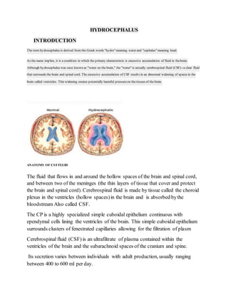
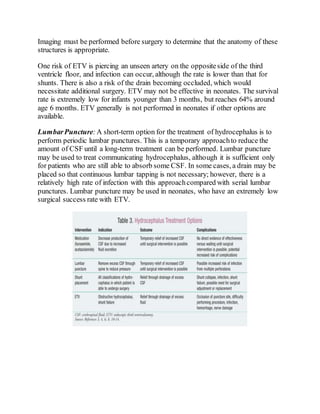
HYDROCEPHALUS
INTRODUCTION
The term hydrocephalus is derived from the Greek words "hydro" meaning water and "cephalus" meaning head.
As the name implies, it is a condition in which the primary characteristic is excessive accumulation of fluid in thebrain.
Although hydrocephalus was once known as "water on the brain," the "water" is actually cerebrospinal fluid (CSF)--a clear fluid
that surrounds the brain and spinal cord. The excessive accumulation of CSF results in an abnormal widening of spaces in the
brain called ventricles. This widening creates potentially harmful pressureon thetissues of the brain.
ANATOMY OF CSFFLUID
The fluid that flows in and around the hollow spaces of the brain and spinal cord,
and between two of the meninges (the thin layers of tissue that cover and protect
the brain and spinal cord). Cerebrospinal fluid is made by tissue called the choroid
plexus in the ventricles (hollow spaces)in the brain and is absorbed bythe
bloodstream Also called CSF.
The CP is a highly specialized simple cuboidal epithelium continuous with
ependymal cells lining the ventricles of the brain. This simple cuboidal epithelium
surrounds clusters of fenestrated capillaries allowing for the filtration of plasm
Cerebrospinal fluid (CSF) is an ultrafiltrate of plasma contained within the
ventricles of the brain and the subarachnoid spaces of the cranium and spine.
Its secretion varies between individuals with adult production, usually ranging
between 400 to 600 ml per day.
Adult CSF volume is estimated to be 150 ml with a distribution of 125 ml within
the subarachnoid spaces and 25 ml within the ventricles. The constant secretion of
CSF contributes to complete CSF renewal four to five times per 24-hour period in
the average young adult
FUNCTION OF CSF
 CSF acts as a shockabsorber, cushioning the brain against the skull.
 CSF allows the brain and spinal cord to become buoyant, reducing the
effective weight of the brain from its normal 1,500 grams to a much lesser
50 grams. The reduction in weight lessens the force applied to the brain
parenchyma and cerebral vessels during mechanical injury.
 Another function of CSF is to maintain homeostasis of the interstitial fluid
of the brain. A stable environment for brain parenchyma is imperative for
maintaining normal neuronal function.
A CSF analysis is a group of tests that look at your cerebrospinal fluid to help
diagnose diseases and conditions that affect the brain and spinal cord. Other
names: Spinal Fluid Analysis, CSF Analysis
TYPES
 Acquired Hydrocephalus: This is the type of hydrocephalus that develops
at birth or in adulthood and is typically caused by injury or disease.
 CongenitalHydrocephalus: It is present at birth and may be caused by
events that occurduring fetal development or as a result of genetic
abnormalities.
 Communicating Hydrocephalus: This type of hydrocephalus occurs when
there is no obstruction to the flow of CSF within the ventricular system. The
condition arises either due to inadequate absorptionor due to an abnormal
increase in the quantity of CSF produced.
 Non-communication (Obstructive) Hydrocephalus: It occurs when the
flow of CSF is blocked along one of more of the passages connecting the
ventricles, causing enlargement of the pathways upstream of the block and
leading to an increase in pressure within the skull.
 Normal Pressure Hydrocephalus: It is a form of communicating
hydrocephalus that can occurat any age, but is most common in the elderly.
It is characterized by dilated ventricles with normal pressure within the
spinal column.
 Hydrocephalus Ex-vacuo: It primarily affects adults and occurs when a
degenerative disease, like Alzheimer’s disease, stroke or trauma, causes
damage to the brain that may cause the brain tissue to shrink.
SYMPTOMS
The symptoms of hydrocephalus tend to vary greatly from person to personand
across different age groups. Infants and young children are more susceptible to
symptoms from increased intracranial pressure like vomiting and adults can
experience loss of function like walking or thinking.
Infants
 Unusually large head size
 Rapidly increasing head circumference
 Bulging and tense fontanelle or soft spot
 Prominent scalp veins
 Downward deviation of eyes or sunset sign
 Vomiting
 Sleepiness
 Irritability
 Seizures
Children and Adolescents
 Nausea and vomiting
 Swelling of the optic disc or papilledema
 Blurred or double vision
 Balance and gait abnormalities
 Slowing or loss of developmental progress
 Changes in personality
 Inability to concentrate
 Seizures
 Poorappetite
 Urinary incontinence
Adults
 Headache
 Nausea and vomiting
 Difficulty walking or gait disturbances
 Loss of balance or coordination
 Lethargy
 Bladder incontinence
 Impaired vision
 Impaired cognitive skills
 Memory loss
 Mild dementia
Normal pressure hydrocephalus (NPH)
This form of the condition usually begins slowly and is more common in adults
over the age of 60. One of the earliest signs is falling suddenly without losing
consciousness.Other common symptoms of normal pressure hydrocephalus
(NPH) include:
 changes in the way you walk
 impaired mental functions, such as memory problems
 trouble controlling urine
 trouble controlling stools
 headaches
DIAGNOSIS
 HC
 Physical examination
 Computed tomography scan (CT or CAT scan)
 Magnetic resonance imaging (MRI)
 Lumbar puncture (spinal tap)
 Intracranial pressure monitoring
 Isotopecisternography
 CT/MRI features
i) Increased frontal horn radius- (Mickey mouse ventricle)
ii) Dilatation of the temporal horns (>2mm)
iii) Acute ventricular angles
iv) Periventricular interstitial edema from the transependymal flow : high
T2 signal on MRI or low- density change on CT Intra-ventricular flow
void from CSF movement
v) Inferior displacement of the- floor of the 3rd ventricle Outward bowing
/ ballooning of the lateral walls & recesses of the third ventricle
(infundibular, optic and pineal recesses) Ballooning of the suprapineal
recess
vi) On mid-sagittal plane : Upward displacement of corpus callosum
Thinned out corpus callosum Depression of the posterior fornix
Decreased mamillopontine distance ( normal >5.5mm)
 CSF flow study
CSF flow studies are performed using a variety of MRI techniques and are
able to qualitatively assess and quantify pulsatile CSF flow. The most
common technique used is time-resolved 2D phase contrast MRI with
velocity encoding.
when referring to CSF flow in the context of imaging we are referring to
pulsatile to-and-fro flow due to vascular pulsations rather than bulk transport
of CSF (the mechanism by which secreted CSF circulates through the CNS
via the lymphatic pathway with absorption at arachnoid granulations). The
latter is too slow to be easily assessed clinically.
MANAGEMENT
The main goal is to minimize or prevent brain damage by decreasing ICP and
improving CSF flow.
Although many causes of hydrocephalus exist, the number of treatments is limited.
All successful, long-term treatments are surgical. There is little use for medication
in hydrocephalus. In some acquired cases, as with tumors and infections, resolving
the underlying condition will resolve the hydrocephalus, but most patients still
require surgical intervention.
MEDICAL MANGMENT:-
Acetazolamide Or Furosemide may be acceptable for CSF fluid reduction, but
these medications currently are used only on a temporary basis. Both drugs act to
reduce the productionof CSF by the choroid plexus.As with lumbar puncture,
these agents are typically used in low-birthweight infants who will have a low
success rate with shunt placement or ETV.
SURGICAL MANAGMENT:-
There are generally two approaches to treating hydrocephalus.
The most common treatment is the placement of a shunt.In use since the 1950s,
this approachis considered the best treatment option in most cases.
The other procedure, endoscopic third ventriculostomy (ETV), involves the
surgical creation of an opening in the floor of the third ventricle to enable the
passage of CSF.
ShuntPlacement:the standard treatment for hydrocephalus is shunt placement.
Shunts are usually placed in the lateral ventricle and can have one of three different
drainage points. The most common drainage site is the peritoneum, which is
connected to the shunt with subcutaneous tubing. This is known as
a ventriculoperitoneal shunt.
Two other types of shunts, ventriculopleural and ventriculoatrial, terminate in the
pleural spaceand the internal jugular vein,
The last type, the lumboperitoneal shunt, is placed in the lumbar intradural
space. Shunt systems include a valve that controls the rate of drainage. The valve
may have to be accessed surgically, or it may be placed so that adjustments can be
made without further surgery.
With current standards and infection control, the postoperative mortality rate for
shunt placement is less than 5%. Although shunt placement is considered the best
treatment for hydrocephalus
DrawbacksShunt failure occurs at a rate of approximately 20% and can be caused
by overdrainage, obstruction, or shunt collapse,Infection, which eventually can
lead to sepsis, occurs at a rate of 5% to 15%.Antibiotic-infused shunt catheters
appear to greatly reduce the rate of infection.
ETV:As mentioned previously, ETV is an alternative treatment for hydrocephalus.
It is indicated in patients with an obstruction that prevents CSF from draining
between the third ventricle and the cortical subarachnoid spaces. In this procedure,
the floor of the third ventricle is punctured, allowing CSF to flow into the cortical
subarachnoid space. ETV is currently approved in the treatment of obstructive
hydrocephalus and in patients who have had multiple shunt failures and
replacements.ETV also has been shown to be effective for treating NPH. The
procedureis difficult and requires that the third ventricle floor and the surrounding
structures have dimensions appropriate for successfulcompletion of the procedure.
Imaging must be performed before surgery to determine that the anatomy of these
structures is appropriate.
One risk of ETV is piercing an unseen artery on the oppositeside of the third
ventricle floor, and infection can occur, although the rate is lower than that for
shunts. There is also a risk of the drain becoming occluded, which would
necessitate additional surgery. ETV may not be effective in neonates. The survival
rate is extremely low for infants younger than 3 months, but reaches 64% around
age 6 months. ETV generally is not performed in neonates if other options are
available.
LumbarPuncture:A short-term option for the treatment of hydrocephalus is to
perform periodic lumbar punctures. This is a temporary approachto reduce the
amount of CSF until a long-term treatment can be performed. Lumbar puncture
may be used to treat communicating hydrocephalus, although it is sufficient only
for patients who are still able to absorb some CSF. In some cases, a drain may be
placed so that continuous lumbar tapping is not necessary; however, there is a
relatively high rate of infection with this approachcompared with serial lumbar
punctures. Lumbar puncture may be used in neonates, who have an extremely low
surgical success rate with ETV.
COMPLICATIONS
 Shunt collapse

Infection and occlusions, can cause a rise in csf pressure that may lead to
symptom recurrence and the need for additional surgeries.
 Hydrocephalus patients have reduced motor function,
 A lower-than-average adult iq, and decreased visual function
 They also are at risk for developing epilepsy.
 The extent of the complications observed is dependent upon the type of
hydrocephalus, but patients with epileptic seizures (approximately 30%)
have the worst clinical outcomes and, compared with patients who did not
develop seizures, are more likely to have an iq lower than 90.about 60% of
children with hydrocephalus are able to attend school(although many have
difficulties), and approximately 40% of children will lead relatively normal
lives.

Hydrocephalus

  • 1.
    HYDROCEPHALUS INTRODUCTION The term hydrocephalusis derived from the Greek words "hydro" meaning water and "cephalus" meaning head. As the name implies, it is a condition in which the primary characteristic is excessive accumulation of fluid in thebrain. Although hydrocephalus was once known as "water on the brain," the "water" is actually cerebrospinal fluid (CSF)--a clear fluid that surrounds the brain and spinal cord. The excessive accumulation of CSF results in an abnormal widening of spaces in the brain called ventricles. This widening creates potentially harmful pressureon thetissues of the brain. ANATOMY OF CSFFLUID The fluid that flows in and around the hollow spaces of the brain and spinal cord, and between two of the meninges (the thin layers of tissue that cover and protect the brain and spinal cord). Cerebrospinal fluid is made by tissue called the choroid plexus in the ventricles (hollow spaces)in the brain and is absorbed bythe bloodstream Also called CSF. The CP is a highly specialized simple cuboidal epithelium continuous with ependymal cells lining the ventricles of the brain. This simple cuboidal epithelium surrounds clusters of fenestrated capillaries allowing for the filtration of plasm Cerebrospinal fluid (CSF) is an ultrafiltrate of plasma contained within the ventricles of the brain and the subarachnoid spaces of the cranium and spine. Its secretion varies between individuals with adult production, usually ranging between 400 to 600 ml per day.
  • 2.
    Adult CSF volumeis estimated to be 150 ml with a distribution of 125 ml within the subarachnoid spaces and 25 ml within the ventricles. The constant secretion of CSF contributes to complete CSF renewal four to five times per 24-hour period in the average young adult FUNCTION OF CSF  CSF acts as a shockabsorber, cushioning the brain against the skull.  CSF allows the brain and spinal cord to become buoyant, reducing the effective weight of the brain from its normal 1,500 grams to a much lesser 50 grams. The reduction in weight lessens the force applied to the brain parenchyma and cerebral vessels during mechanical injury.  Another function of CSF is to maintain homeostasis of the interstitial fluid of the brain. A stable environment for brain parenchyma is imperative for maintaining normal neuronal function. A CSF analysis is a group of tests that look at your cerebrospinal fluid to help diagnose diseases and conditions that affect the brain and spinal cord. Other names: Spinal Fluid Analysis, CSF Analysis TYPES  Acquired Hydrocephalus: This is the type of hydrocephalus that develops at birth or in adulthood and is typically caused by injury or disease.  CongenitalHydrocephalus: It is present at birth and may be caused by events that occurduring fetal development or as a result of genetic abnormalities.
  • 3.
     Communicating Hydrocephalus:This type of hydrocephalus occurs when there is no obstruction to the flow of CSF within the ventricular system. The condition arises either due to inadequate absorptionor due to an abnormal increase in the quantity of CSF produced.  Non-communication (Obstructive) Hydrocephalus: It occurs when the flow of CSF is blocked along one of more of the passages connecting the ventricles, causing enlargement of the pathways upstream of the block and leading to an increase in pressure within the skull.  Normal Pressure Hydrocephalus: It is a form of communicating hydrocephalus that can occurat any age, but is most common in the elderly. It is characterized by dilated ventricles with normal pressure within the spinal column.  Hydrocephalus Ex-vacuo: It primarily affects adults and occurs when a degenerative disease, like Alzheimer’s disease, stroke or trauma, causes damage to the brain that may cause the brain tissue to shrink. SYMPTOMS The symptoms of hydrocephalus tend to vary greatly from person to personand across different age groups. Infants and young children are more susceptible to symptoms from increased intracranial pressure like vomiting and adults can experience loss of function like walking or thinking. Infants  Unusually large head size  Rapidly increasing head circumference  Bulging and tense fontanelle or soft spot  Prominent scalp veins  Downward deviation of eyes or sunset sign  Vomiting  Sleepiness  Irritability  Seizures Children and Adolescents  Nausea and vomiting  Swelling of the optic disc or papilledema  Blurred or double vision  Balance and gait abnormalities
  • 4.
     Slowing orloss of developmental progress  Changes in personality  Inability to concentrate  Seizures  Poorappetite  Urinary incontinence Adults  Headache  Nausea and vomiting  Difficulty walking or gait disturbances  Loss of balance or coordination  Lethargy  Bladder incontinence  Impaired vision  Impaired cognitive skills  Memory loss  Mild dementia Normal pressure hydrocephalus (NPH) This form of the condition usually begins slowly and is more common in adults over the age of 60. One of the earliest signs is falling suddenly without losing consciousness.Other common symptoms of normal pressure hydrocephalus (NPH) include:  changes in the way you walk  impaired mental functions, such as memory problems  trouble controlling urine  trouble controlling stools  headaches DIAGNOSIS  HC  Physical examination
  • 5.
     Computed tomographyscan (CT or CAT scan)  Magnetic resonance imaging (MRI)  Lumbar puncture (spinal tap)  Intracranial pressure monitoring  Isotopecisternography  CT/MRI features i) Increased frontal horn radius- (Mickey mouse ventricle) ii) Dilatation of the temporal horns (>2mm) iii) Acute ventricular angles iv) Periventricular interstitial edema from the transependymal flow : high T2 signal on MRI or low- density change on CT Intra-ventricular flow void from CSF movement v) Inferior displacement of the- floor of the 3rd ventricle Outward bowing / ballooning of the lateral walls & recesses of the third ventricle (infundibular, optic and pineal recesses) Ballooning of the suprapineal recess vi) On mid-sagittal plane : Upward displacement of corpus callosum Thinned out corpus callosum Depression of the posterior fornix Decreased mamillopontine distance ( normal >5.5mm)  CSF flow study CSF flow studies are performed using a variety of MRI techniques and are able to qualitatively assess and quantify pulsatile CSF flow. The most common technique used is time-resolved 2D phase contrast MRI with velocity encoding. when referring to CSF flow in the context of imaging we are referring to pulsatile to-and-fro flow due to vascular pulsations rather than bulk transport of CSF (the mechanism by which secreted CSF circulates through the CNS via the lymphatic pathway with absorption at arachnoid granulations). The latter is too slow to be easily assessed clinically. MANAGEMENT The main goal is to minimize or prevent brain damage by decreasing ICP and improving CSF flow.
  • 6.
    Although many causesof hydrocephalus exist, the number of treatments is limited. All successful, long-term treatments are surgical. There is little use for medication in hydrocephalus. In some acquired cases, as with tumors and infections, resolving the underlying condition will resolve the hydrocephalus, but most patients still require surgical intervention. MEDICAL MANGMENT:- Acetazolamide Or Furosemide may be acceptable for CSF fluid reduction, but these medications currently are used only on a temporary basis. Both drugs act to reduce the productionof CSF by the choroid plexus.As with lumbar puncture, these agents are typically used in low-birthweight infants who will have a low success rate with shunt placement or ETV. SURGICAL MANAGMENT:- There are generally two approaches to treating hydrocephalus. The most common treatment is the placement of a shunt.In use since the 1950s, this approachis considered the best treatment option in most cases. The other procedure, endoscopic third ventriculostomy (ETV), involves the surgical creation of an opening in the floor of the third ventricle to enable the passage of CSF. ShuntPlacement:the standard treatment for hydrocephalus is shunt placement. Shunts are usually placed in the lateral ventricle and can have one of three different drainage points. The most common drainage site is the peritoneum, which is
  • 7.
    connected to theshunt with subcutaneous tubing. This is known as a ventriculoperitoneal shunt. Two other types of shunts, ventriculopleural and ventriculoatrial, terminate in the pleural spaceand the internal jugular vein, The last type, the lumboperitoneal shunt, is placed in the lumbar intradural space. Shunt systems include a valve that controls the rate of drainage. The valve may have to be accessed surgically, or it may be placed so that adjustments can be made without further surgery. With current standards and infection control, the postoperative mortality rate for shunt placement is less than 5%. Although shunt placement is considered the best treatment for hydrocephalus DrawbacksShunt failure occurs at a rate of approximately 20% and can be caused by overdrainage, obstruction, or shunt collapse,Infection, which eventually can lead to sepsis, occurs at a rate of 5% to 15%.Antibiotic-infused shunt catheters appear to greatly reduce the rate of infection. ETV:As mentioned previously, ETV is an alternative treatment for hydrocephalus. It is indicated in patients with an obstruction that prevents CSF from draining between the third ventricle and the cortical subarachnoid spaces. In this procedure, the floor of the third ventricle is punctured, allowing CSF to flow into the cortical subarachnoid space. ETV is currently approved in the treatment of obstructive hydrocephalus and in patients who have had multiple shunt failures and replacements.ETV also has been shown to be effective for treating NPH. The procedureis difficult and requires that the third ventricle floor and the surrounding structures have dimensions appropriate for successfulcompletion of the procedure.
  • 8.
    Imaging must beperformed before surgery to determine that the anatomy of these structures is appropriate. One risk of ETV is piercing an unseen artery on the oppositeside of the third ventricle floor, and infection can occur, although the rate is lower than that for shunts. There is also a risk of the drain becoming occluded, which would necessitate additional surgery. ETV may not be effective in neonates. The survival rate is extremely low for infants younger than 3 months, but reaches 64% around age 6 months. ETV generally is not performed in neonates if other options are available. LumbarPuncture:A short-term option for the treatment of hydrocephalus is to perform periodic lumbar punctures. This is a temporary approachto reduce the amount of CSF until a long-term treatment can be performed. Lumbar puncture may be used to treat communicating hydrocephalus, although it is sufficient only for patients who are still able to absorb some CSF. In some cases, a drain may be placed so that continuous lumbar tapping is not necessary; however, there is a relatively high rate of infection with this approachcompared with serial lumbar punctures. Lumbar puncture may be used in neonates, who have an extremely low surgical success rate with ETV.
  • 9.
    COMPLICATIONS  Shunt collapse  Infectionand occlusions, can cause a rise in csf pressure that may lead to symptom recurrence and the need for additional surgeries.  Hydrocephalus patients have reduced motor function,  A lower-than-average adult iq, and decreased visual function  They also are at risk for developing epilepsy.  The extent of the complications observed is dependent upon the type of hydrocephalus, but patients with epileptic seizures (approximately 30%) have the worst clinical outcomes and, compared with patients who did not develop seizures, are more likely to have an iq lower than 90.about 60% of children with hydrocephalus are able to attend school(although many have difficulties), and approximately 40% of children will lead relatively normal lives.