Ejection fraction is one of the important measure of the health of heart. EF can be calculated from the 2D images of Echocardiogram using Image processing techniques.
Understanding Ventricular Pressure-Volume Catheter Calibrations and Experimen...InsideScientific
Cardiovascular researchers interested in acquiring baseline and load-independent ventricular pressure-volume data face a multitude of questions regarding technology, calibration and experimental design. Focusing on results and keeping the end in mind is often where the best answers lie.
In this webinar sponsored by ADInstruments, Dr. Dimitrios "Jim" Georgakopoulos -- an expert in the theory, history and application of ventricular pressure-volume science -- discusses conductance-based pressure-volume technology, the theory and importance behind resistivity and parallel volume calibration factors, how experimental conditions can (and should) influence your approach and the significance of proper calibration in acquiring quality results.
The fourth session in our "PV Loops to Measure Cardiac Function" Webinar Series touched on what is essential for the researcher to be aware of in order to collect valid Pressure-Volume Loop data that can be used with confidence in the ensuing analysis stage of their research project.
Dr. Filip Konecny and Peter Plouf present and offer discussion on best practices for obtaining quality and consistent Pressure-Volume loop data. The presentation is a distillation of more than 10 years of working with the PV Loop research community to develop better results, and insights from Dr. Konecny’s body of knowledge from collecting and publishing PV Loop study data across a wide spectrum of species and research models. This presentation touches on what is essential for researchers to be aware of in order to collect valid PV Loop data that can be used with confidence in the ensuing analysis stage of their research project.
Key Topics:
- surgical considerations for improved data stability and consistency between animals
- procedure checklists -- essential steps before, during, and post surgery
- how to properly check data integrity at the bench-top
- understanding conductance and admittance methodologies for deriving volume
Best Practices to Achieve Quality Pressure-Volume Loop Data in Large Animal M...InsideScientific
During this webinar sponsored by Transonic, Dr. Tim Hacker and Dr. Filip Konecny present common hemodynamic set ups for large animal models. Using case studies from dogs and swine models, they show surgical best-practices, tips for catheter navigation and how to correctly position a PV-catheter in the left or right ventricle. In addition, they explain how researchers can verify accurate and reliable PV loop data at the bench-side.
Large animal hemodynamic research models are on the rise. They are increasingly used in various preclinical studies including pharmaco-safety and drug discovery assessment, ventricular assist-device testing and models of pulmonary artery hyperthrophy and right ventricular overload. Important to these applications and all cardiovascular studies is the collection of both central and peripheral hemodynamics, with a focus on instantaneous pressure and volume measurements from the beating heart (PV Loops). Only with PV loops can scientists obtain the most comprehensive evaluation of cardiac function. It is therefore critical for cardiovascular scientists to understand how PV Loop data should be collected along with these peripheral hemodynamic measurements. This webinar aims to discuss these essential elements and how they should be applied.
Key Topics:
Basic and advanced set ups for large animal
hemodynamic studies
Essential hemodynamic equipment/technology
Anaesthetic and drug considerations for large animal PV studies
Surgical approaches -- which one is best for you?
How to successfully navigate the PV catheter and validate
correct position in the ventricle
Right ventricle PV loops -- important surgical, data collection
and analysis considerations
Unique attributes of 'admittance' derived volume
Spirometry and peak flow metry in bronchial asthmaDr Pankaj Yadav
SPIROMETRY AND PEAK FLOMETRY in BRONCHIAL ASTHMA
SPIROMETRY IS A PULMONARY FUNCTION TEST THAT MEASURES THE VOLUME OF AIR AN INDIVIDUAL INHALES OR EXHALES AS A FUNCTION OF TIME.
SPIROMETRY MEASURES HOW MUCH AND HOW QUICKLY AIR CAN BE EXPELLED FOLLOWING A DEEP BREATH.
FLOW , OR THE RATE AT WHICH VOLUME IS CHANGING AS A FUNCTION OF TIME, CAN ALSO BE MEASURED WITH SPIROMETRY.
Human Circulatory System/ Blood Vascular System Sonam
Human circulatory system, The study of blood vascular system or circulatory system is called angiology. The vascular system is made up of the....
ANATOMY OF HEART
Heart is a coned shaped hollow structure equal to the size of puzzle fit.
It is a mesodermally derived organ, is situated in the thoracic cavity, in between the two lungs (mediastinum), slightly tilled to the left......
The aorta arch is the section of the aorta between the ascending and descending aorta that distribute blood to the head and upper extremities via the brachiocephalic trunk, the left common carotid artery, and the left subclavian artery.
The aortic arch has 3 major branches.
The brachiocephalic trunk is........
overview of heart and its disease
The heart is a powerful muscle that pumps
blood throughout the body by means of a
coordinated contraction.
The contraction is generated by an
electrical activation, which is spread by a
wave of bioelectricity that propagates in a
coordinated manner throughout the
heart.
It is located in thoracic cavity,
posterior to the sternum ,superior
to the diaphragm between the
lungs.
A human heart beats an average of
100,000 times per day. During that
time, it pumps more than 4,300
gallons of blood throughout
the entire body.
EPICARDIUM:
It is the outer layer of the wall of the heart which is composed of
connective tissue covered by epithelium. It is also known as
visceral pericardium.
2) MYOCARDIUM:
It is the muscular middle layer of the wall of the heart. It is
composed of spontaneously contracting cardiac muscle fibers
which allow the heart to contract. It stimulates heart contraction
to pump blood from the ventricles and relaxes the heart to allow
the arteries to receive blood.
ENDOCARDIUM:
It is the inner layer of the heart which consist of epithelial
tissue and connective tissue.
Denoising of ECG -- A discrete time approach using DWTIJERA Editor
This paper is about denoising of ECG signal using DWT transform. In this paper, ECG signals are denoised using DWT transform.Ecg signals are taken and noise at different frequencies are generated which are superimposed on this original ecg signal.High frequency noise is of 4000 hertz and power line interference is of 50 hertz.Decomposition of noisy signal is achieved through wavelet packet .wavelet packets are reconstructed and appropriate wavelet packets are combined to obtain a signal, very similar to original ecg signal.This technique results in the minimization of mean square error in the filtered signals.
Denoising of ECG -- A discrete time approach using DWT.IJERA Editor
This paper is about denoising of ECG signal using DWT transform. In this paper, ECG signals are denoised using DWT transform.Ecg signals are taken and noise at different frequencies are generated which are superimposed on this original ecg signal.High frequency noise is of 4000 hertz and power line interference is of 50 hertz.Decomposition of noisy signal is achieved through wavelet packet .wavelet packets are reconstructed and appropriate wavelet packets are combined to obtain a signal, very similar to original ecg signal.This technique results in the minimization of mean square error in the filtered signals.
A moderately frequent illness called congestive heart failure occurs when the heart is unable to pump enough blood to meet the body's demands. It frequently happens as a result of a chronic illness or aging. The body makes an effort to make up for this by boosting blood salt levels and fluid retention.
Swelling, weight gain, and shortness of breath may result from this. Diabetes and high blood pressure are other conditions linked to congestive heart failure. Congestive heart failure, however, is most frequently brought on by coronary artery disease (CAD). This occurs when the arteries that carry blood to the heart start to constrict and narrow.
When calling a doctor is important to question Dr. Sumit shejol Cardiologist from Hrudaysparsh Clinic Suggests that if you recognize the majority of the symptoms of heart failure. Certain signs and symptoms, such as chest pain, acute breathlessness, an irregular heartbeat, extreme weakness, or fainting, demand rapid medical attention. Do not delay in seeking assistance, do not self-diagnose, and do not self-medicate if you feel any of that. Some of these symptoms may also be a sign of heart failure or another serious lung, heart, or cardiovascular disease. Your condition is stabilized as emergency room doctors try to identify the source of your symptoms. Call your doctor right away if you've already been given a heart failure diagnosis and you realize that your symptoms have gotten worse or a new symptom has appeared.
Congestive heart failure is a fatal condition with a high mortality rate. Congestive heart failure has a wide range of risk factors. Smoking, high blood pressure, diabetes, high cholesterol, being obese, and having experienced a heart attack in the past are some of them. It can also be brought on by a hereditary condition like cardiomyopathy. The condition can cause the heart muscle to expand and become excessively thick, which can result in heart failure. Congestive heart failure can be exacerbated by lifestyle choices including smoking, excessive alcohol intake, or tobacco use.
1. 1
Automated Ejection Fraction Calculations
from B-Mode (2-D) Echocardiography
Suhas Deshpande, Member, IEEE, IEEE-EMBS, CSULB, suhassd@ieee.org
Heart is basically the pump for the human circulatory
Abstract—Left Ventricular Ejection Fraction can be estimate system. It provides the blood and thus oxygen and vital
to acceptable accuracy using 2-D echocardiography images. 2-D nutrients from lungs to various parts of the body. The heart
Echocardiography due to its portability and versatility is used in physiologically consists of four parts- left and right ventricles
emergency operations and ICUs. The estimation of LVEF can and left and right Auricles. Right Auricle collects the
help the physians to get apriori knowledge of the start of
deoxygenated blood from all the parts of the body. Right
conditions such as Coronary heart disease or Cardiomyopathy.
The ejection fraction can be evaluated by using the simson’s
ventricle pumps the deoxygenated blood to lungs. The
approximation for calculating the volume of the 2-D image. The Oxygenated blood reaches the left auricle and is pumped out
two methods tried here are Ellipsoidal and Stacked disk method. to other parts of the body through left ventricle via the aorta.
Thus the left ventricle performance describes the amount of
Index Terms—Ejection Fraction, Echocardiography, Systole, oxygen reaching the body.
Diastole
A. Systole
Systole is a phase of the cardiac cycle in which the heart
I. INTRODUCTION muscle contracts in a coordinated manner in response to a
complex endogenous autonomic physiologic electrical
H eart diseases are by far the most common cause of deaths
in US. In majority of the cases the death can be
prevented if there is a priori knowledge of some of the cardiac
stimulus. Systole results in driving blood flow out of the heart
and into the body and lungs. All four chambers of a human
heart undergo systole and diastole in a timed fashion so that
functions. Left ventricular Ejection Fraction is one of the
blood is propelled forward and backward through the
most important cardiac function which gives a good idea
cardiovascular system.
about the cardiac problem in advance. The LVEF can be
estimated by using a 2D echocardiography, which is portable
and versatile, is widely used in emergency, operating, and B. Diastole
intensive care department. Diastole is the period of time when the heart fills with
blood after systole. Ventricular diastole is the period during
which the ventricles are relaxing, while atrial diastole is the
II. FUNCTIONING OF HEART period during which the atria are relaxing. During ventricular
diastole, the pressure in the ventricles drops from the peak in
systole to below the pressure in the left atrium, causing the
mitral valve to open. This forces the blood from the atrium to
flow into the ventricle.
III. ECHOCARDIOGRAM
Echocardiography is an ultrasound test done for cardiac
assessment. The ultrasound waves are transmitted by a
transmitter. The ultrasound waves are reflected off the cardiac
walls. The computer maps the ECHOs on a line & then takes
another sample on a different line thus building up a 2D
picture approx 50 times a second. A pseudo image can be
prepared on the basis of the received reflected ultrasound
wave. This image, echocardiogram or ECHO can be used in
diagnostics of the cardiac system. Since the system is simple
in design and easy to carry it is very popular with the cardiac
diagnosis.
Fig 1. Heart Anatomy and Blood Flow through Heart
The frequencies commonly used 2-10MHz are much
higher than the audible range of 2-18KHz. Higher
2. 2
frequencies allow better resolution. Lateral resolution of a 5 B. Parasternal Long Axis
MHz is 2 mm compared to 3mm of 3MHz whilst axial
resolution varies from 0.5 mm to 1 mm) but tissue penetration
is poorer.
The Echocardiogram can be optained by imaging the heart
by positioning the sensor at different position. Transthoracic
view and Parasternal view are commonly used. Trans
oesophagal view can be obtained by placing the sensor on a
catheter inside the oesophagus.
Fig 4. Parasternal Long Axis (Image:National University of
Singapore)
The transducer is placed in just to the left of the mid to
upper sternal border. The right ventricular outflow region, the
ventricular septum and the left atrium and ventricular are
well visualized. This is one of the best views to obtain an M
Mode and hence information on cardiac function.
Fig 2. Acoustic Windows (Image University of Minnesota) C. Parasternal Short Axis
The 2D pictures are taken from the various echo windows
and give "standard views" to build up a complete picture of
the cardiac anatomy. Not all chambers are visible in every
view.
A. Apical 4 Chamber
Fig 5. Parasternal short Axis (Image:National University of
Singapore)
From the parasternal long axis view the transduce is rotated
90˚ to point towards the left shoulder. The aorta and coronary
artery origins are seen well in cross section.
Fig 3. Apical 4 Chamber view (Image:National University of D. Subcostal View
Singapore)
The transducer is held at the apex of the heart and angled This is a good view to see lovely images - especially in
towards the right shoulder. The 4 chambers are readily seen babies as no ribs or lung tissue obscures the view. The atrial
and both the mitral and tricuspid valves. If the transducer is septum is particularly well seen. Unfortunately the
angled anteriorly then the aorta is also visualized. This view transducer is the furthest from the heart and in older children
is shows the ventricular septum well to look for septal defects. and adults the distance may be too great to allow detailed
imaging.
3. 3
mitral valve closure; (c) as the instant of maximum cardiac
dimension. End-systole is given by the instant preceding
mitral valve opening or by the instant of minimum cardiac
dimension.
IV. EJECTION FRACTION
One of the key indicators of cardiac health is measurement
of left ventricular (LV) volume and ejection fraction (EF).
The ejection fraction determines the amount of blood pumped
out of the ventricles with each cycle of the ECG.
The most commonly applied models for the computation of
the LV internal volume are derived from that proposed by
Simpson. According to the criterion usually called
Fig 6. Subcoastal view (Image:National University of “Simpson‟s rule,” the LV is approximated by a stack of
Singapore) circular (or elliptical) disks whose centers are all in the major
axis. The most important measurement of ventricular
function is the LVEF (Left Ventricle Ejection Function),
E. Arch View which is given by the normalized (nondimensional) difference
between End-Diastolic Volume (EDV) and the End-Systolic
Volume (ESV), both generally computed according to
Simpson‟s rule. The simple formula defining this parameter
is
LVEF =
A. Stacked disk method
In stacked disk method the left ventricle is divided into „n‟
Fig 7. Arch view (Image:National University of Singapore) disks. The number of disks „n‟ can be varied to get the
required accuracy level. Each disk diameter is matched with
This is obtained by sliding the transducer towards the upper the respective contour of the left ventricle. Each disk is
sternal edge and suprasternal notch. It allows the ascending considered to be an ideal circular shape. The LV volume is
aorta, arch and neck vessels to be imaged. estimated using the Simpson‟s equation
F. Importance
Echocardiography is the preferred method for the
documentation of cardiac function at rest. In particular, 2D
Transthoracic Echocardiography (TTE), because of its
portability and versatility, is widely used in emergency,
operating, and intensive care departments. The TTE
technique provides numerous highly significant quantitative
parameters. In fact, it has been demonstrated that cardiac risk
increases significantly when the values of certain parameters
are abnormal. A number of these parameters are connected to
properties of the LV. Thus LV dimensions, volumes, and wall Figure 8. Stacked disc method for Ejection Fraction
thickness are widely used in clinical practice and research. estimation
With regard to LV volume quantification, the most important
windows are the apical (4-chamber view and the 2-chamber
view) and the left parasternal (short-axis view at the papillary Stacked-disk estimation of LV volume
muscle level). The various quantitative parameters are = ((first disk area + last disk area)/2
generally measured at end-diastole and at end-systole. End- + ∑in-1 disk area) x d
diastole can be defined in three ways: (a) as the onset of the d = disk spacing; n = number of disks; disk are = *r2
QRS Complex in the ECG signal; (b) as the instant after
5. 5
Imaging: From Nano to Macro - Proceedings, vol. 2006, pp. 97-100, 2006.
[6] http://www.med.nus.edu.sg/paed/resources/cardiac_thumbnail/
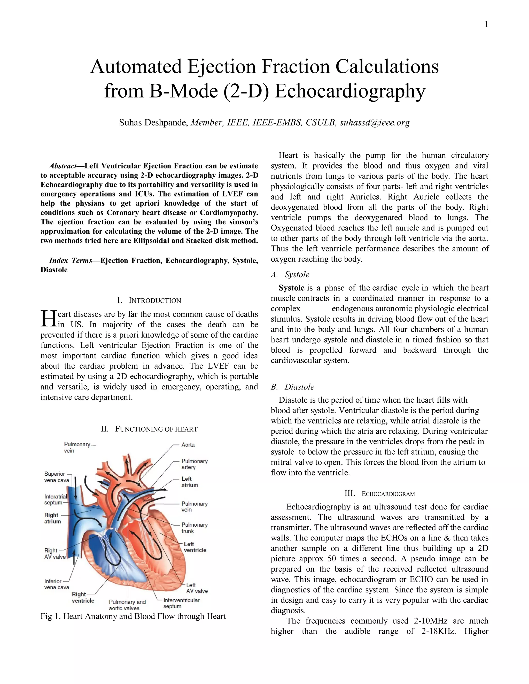
Table 1. Ejection Fraction by Slotted disk method, Ellipsoid investigations/echo.htm
method and provided by Phillips [7] http://www.vhlab.umn.edu/atlas/echotutorial/echotutorial1.shtml
Method Slotted Disk Ellipsoid Original [8] http://rwjms1.umdnj.edu/shindler/imageproc.html#edgefin3.m
EF(Phillips)
Ejection 55.1% 61.45% 54%
fraction
The End Systolic Volume and End Diastolic value was
calculated by calculating the pixels in the image. The pixels
along one axis give the dimension of the left ventricle along
that axis. For e.g to calculate the disk diameter the pixels
along the x axis were calculated. The number of disks was
chosen to be 20.
VII. CONCLUSION
The ejection fraction calculated using the two methods is
around 54% which is the EF measured by the Phillips
Ultrasound (Data imprinted on the image). There are still
some discrepancies in the code and the image. The apical 2
chamber view would have been a better choice for the
estimation of the volume of LV volume. This is because the
ROI is the left ventricle. The repeatability is poor as the final
result depends much on the cropping of the image to get the
ROI as the left ventricle. There are some problems regarding
the cropping the image to the ROI. If the cropping border
touches the left ventricular edge, the estimation goes wrong.
This is because the ROI is not evaluated precisely due to
opening in the region. There are several discontinuities (black
subregions) present in the filtered image. These subregions
need to be removed to get an accurate EF measurement. A
better filtering approach may remove the discontinuities.
ACKNOWLEDGMENT
I am thankful to Dr. Lobodzinsky for conducting the course
of Digital Image Processing which helped me to base my
study in the project. Further I would like to thank Dr. David
Hull and Tibor Duliskovich from Phillips Radiology
department to provide assistance in providing the images for
the project.
REFERENCES
[1] Arthur J. Vander, “Cardiovascular Physiology,” Human Physiology: Th e
Mechanisms of Body Function, 9th edition: MGH, pp 375-399, 2004
[2] Bonita Anderson, “Two dimensional echocardiographic measurements
and calculations,” Echocardiography: The Normal Examination and
Echocardiographic Measurements: Wiley, pp 96- 103, 2002
[3] T. L. Szabo, "Improving ejection fraction estimation for 2d ultrasound
using a computer-generated cardiac model," Proceedings - IEEE Ultrasonics
Symposium, pp. 1757-1760, 2008.
[4] U. Barcaro, "Automatic computation of left ventricle ejection fraction
from dynamic ultrasound images," Pattern Recognition and Image Analysis, vol.
18, pp. 351-358, 2008.
[5] M. Jolly, "Assisted ejection fraction in B-mode and contrast
echocardiography," 2006 3rd IEEE International Symposium on Biomedical