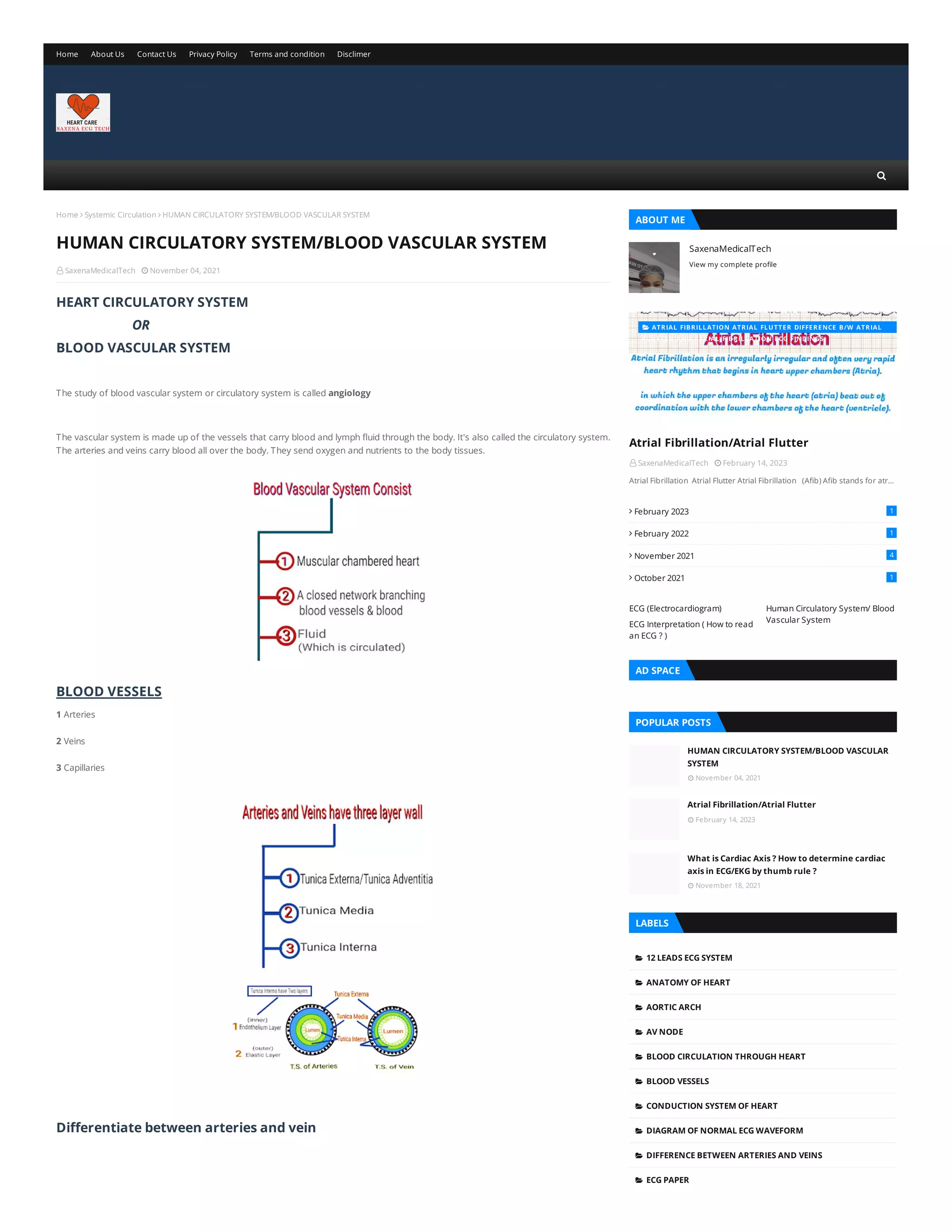
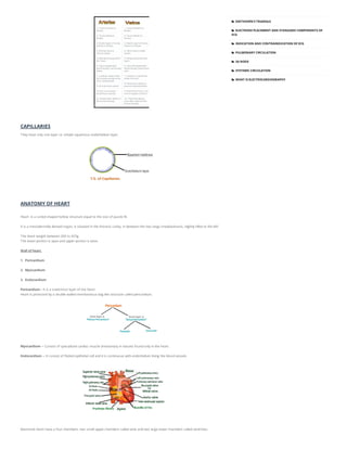
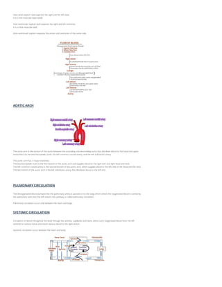
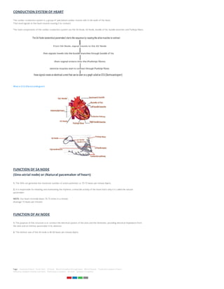
 SaxenaMedicalTech  November 04, 2021
Home  Systemic Circulation  HUMAN CIRCULATORY SYSTEM/BLOOD VASCULAR SYSTEM
HEART CIRCULATORY SYSTEM
OR
BLOOD VASCULAR SYSTEM
The study of blood vascular system or circulatory system is called angiology
The vascular system is made up of the vessels that carry blood and lymph fluid through the body. It's also called the circulatory system.
The arteries and veins carry blood all over the body. They send oxygen and nutrients to the body tissues.
BLOOD VESSELS
1 Arteries
2 Veins
3 Capillaries
Differentiate between arteries and vein
HUMAN CIRCULATORY SYSTEM/BLOOD VASCULAR SYSTEM SaxenaMedicalTech
View my complete profile
ABOUT ME
 SaxenaMedicalTech  February 14, 2023
Atrial Fibrillation/Atrial Flutter
Atrial Fibrillation Atrial Flutter Atrial Fibrillation (Afib) Afib stands for atr…




February 2023
February 2022
November 2021
October 2021
1
1
4
1
ECG (Electrocardiogram) Human Circulatory System/ Blood
Vascular System
ECG Interpretation ( How to read
an ECG ? )
AD SPACE
 November 04, 2021
 February 14, 2023
 November 18, 2021
HUMAN CIRCULATORY SYSTEM/BLOOD VASCULAR
SYSTEM
Atrial Fibrillation/Atrial Flutter
What is Cardiac Axis ? How to determine cardiac
axis in ECG/EKG by thumb rule ?
POPULAR POSTS
LABELS
12 LEADS ECG SYSTEM

ANATOMY OF HEART

AORTIC ARCH

AV NODE

BLOOD CIRCULATION THROUGH HEART

BLOOD VESSELS

CONDUCTION SYSTEM OF HEART

DIAGRAM OF NORMAL ECG WAVEFORM

DIFFERENCE BETWEEN ARTERIES AND VEINS

ECG PAPER

Home About Us Contact Us Privacy Policy Terms and condition Disclimer
 ATRIAL FIBRILLATION ATRIAL FLUTTER DIFFERENCE B/W ATRIAL
FLUTTER AND ATRIAL FIBRILLATION ECG FINDINGS

CAPILLARIES
They have only one layer i.e. simple squamous endothelium layer.
ANATOMY OF HEART
Heart is a coned shaped hollow structure equal to the size of puzzle fit.
It is a mesodermally derived organ, is situated in the thoracic cavity, in between the two lungs (mediastinum), slightly tilled to the left.
The heart weight between 200 to 425g.
The lower portion is apex and upper portion is base.
Wall of heart
1. Pericardium
2. Myocardium
3. Endocardium
Pericardium-- It is a outermost layer of the heart.
Heart is protected by a double walled membranous bag like structure called pericardium.
Myocardium -- Consist of specialized cardiac muscle (involuntary in nature) found only in the heart.
Endocardium -- It consist of flatted epithelial cell and it is continuous with endothelium lining the blood vessels.
Mammals heart have a four chambers, two small upper chambers called atria and two large lower chambers called ventricles.
EINTHOVEN'S TRIANGLE

ELECTRODE PLACEMENT AND STANDARD COMPONENTS OF
ECG

INDICATION AND CONTRAINDICATION OF ECG

PULMONARY CIRCULATION

SA NODE

SYSTEMIC CIRCULATION

WHAT IS ELECTROCARDIOGRAPHY

Inter-atrial septum wall separate the right and the left atria.
It is a thin muscular layer (wall).
Inter-ventricular septum wall separate the right and left ventricles.
It is a thick muscular wall.
Atrio-ventricualr septum separate the atrium and ventricles of the same side.
AORTIC ARCH
The aorta arch is the section of the aorta between the ascending and descending aorta that distribute blood to the head and upper
extremities via the brachiocephalic trunk, the left common carotid artery, and the left subclavian artery.
The aortic arch has 3 major branches.
The brachiocephalic trunk is the first branch of the aortic arch and supplies blood to the right arm and right head and neck.
The left common carotid artery is the second branch of the aortic arch, which supplies blood to the left side of the head and the neck.
The last branch of the aortic arch is the left subclavian artery that distribute blood to the left arm.
PULMONARY CIRCULATION
The deoxygenated blood pumped into the pulmonary artery is passed on to the lungs (from where the oxygenated blood is carried by
the pulmonary veins into the left atrium) this pathway is called pulmonary circulation.
Pulmonary circulation occur only between the heart and lungs.
SYSTEMIC CIRCULATION
Circulation of blood throughout the body through the arteries, capillaries and veins, which carry oxygenated blood from the left
ventricle to various tissue and return venous blood to the right atrium.
Systemic circulation occur between the heart and body.
CONDUCTION SYSTEM OF HEART
The cardiac conduction system is a group of specialized cardiac muscle cells in the walls of the heart.
That send signals to the heart muscle causing it to contract.
The main components of the cardiac conduction system are the SA Node, AV Node, bundle of his, bundle branches and Purkinje fibers.
What is ECG (Electrocardiogram)
FUNCTION OF SA NODE
(Sino-atrial node) or (Natural pacemaker of heart)
1. The SAN can generate the maximum number of action potential. i.e. 70-75 beats per minute (bpm).
2. It is responsible for initiating and maintaining the rhythmic contractile activity of the heart that's why it is called the natural
pacemaker.
NOTE- Our heart normally beats 70-75 times in a minute.
(Average 72 beats per minute)
FUNCTION OF AV NODE
1. The purpose of this structure is to connect the electrical system of the atria and the Ventricles, providing electrical impedance from
the atria and an intrinsic pacemaker in its absence.
2. The intrinsic rate of the AV node is 40-60 beats per minute (bpm).
Tags: Anatomy of heart Aortic Arch AV node Blood circulation through heart Blood Vessels Conduction system of heart
Difference between Arteries and Veins Pulmonary Circulation SA node Systemic Circulation
0 Comments
To leave a comment, click the button below to sign in with Google.
SIGN IN WITH GOOGLE
 Facebook  Twitter    
POST A COMMENT
 November 04, 2021
 November 14, 2021
 October 31, 2021
HUMAN CIRCULATORY SYSTEM/BLOOD VASCULAR
SYSTEM
ECG Interpretation/ How to read an ECG or EKG/
Characteristics of the normal ECG
ELECTROCARDIOGRAPHY BASIC
TAGS
INCUBATION PERIOD FOR COVID-19

LIFE SPAN OF CORONAVIRUS

PRECAUTION & PREVENTION OF CORONAVIRUS

SIGN SYMPTOMS & COMPLICATIONS OF CORONAVIRUS

TYPES OF COVID-19 VARIANTS

WHAT IS CORONAVIRUS?

POPULAR POSTS LABELS
CARDIAC AXIS THUMB RULE

CHARACTERISTICS OF NORMAL ECG (P WAVE PR INTERVAL ST
SEGMENT QRS COMPLEX T WAVE U WAVE)

ECG INTERPRETATION METHOD

HOW TO DETERMINE CARDIAC AXIS IN ECG OR EKG BY
THUMB RULE

NORMAL SINUS RHYTHM

THE EFFECT OF REVERSING THE ELECTRODE

WHAT IS CARDIAC AXIS

WHAT IS ELECTROCARDIOGRAPHY

WHAT IS LEFT AXIS DEVIATION

WHAT IS NORMAL AXIS

WHAT IS RIGHT AXIS DEVIATION

WHY WORRY ABOUT THE CARDIAC AXIS

Created By | Distributed By Gooyaabi Templates Home About Us Contact Us
Blogging

Human Circulatory System/ Blood Vascular System

  • 1.
     SaxenaMedicalTech November 04, 2021 Home  Systemic Circulation  HUMAN CIRCULATORY SYSTEM/BLOOD VASCULAR SYSTEM HEART CIRCULATORY SYSTEM OR BLOOD VASCULAR SYSTEM The study of blood vascular system or circulatory system is called angiology The vascular system is made up of the vessels that carry blood and lymph fluid through the body. It's also called the circulatory system. The arteries and veins carry blood all over the body. They send oxygen and nutrients to the body tissues. BLOOD VESSELS 1 Arteries 2 Veins 3 Capillaries Differentiate between arteries and vein HUMAN CIRCULATORY SYSTEM/BLOOD VASCULAR SYSTEM SaxenaMedicalTech View my complete profile ABOUT ME  SaxenaMedicalTech  February 14, 2023 Atrial Fibrillation/Atrial Flutter Atrial Fibrillation Atrial Flutter Atrial Fibrillation (Afib) Afib stands for atr…     February 2023 February 2022 November 2021 October 2021 1 1 4 1 ECG (Electrocardiogram) Human Circulatory System/ Blood Vascular System ECG Interpretation ( How to read an ECG ? ) AD SPACE  November 04, 2021  February 14, 2023  November 18, 2021 HUMAN CIRCULATORY SYSTEM/BLOOD VASCULAR SYSTEM Atrial Fibrillation/Atrial Flutter What is Cardiac Axis ? How to determine cardiac axis in ECG/EKG by thumb rule ? POPULAR POSTS LABELS 12 LEADS ECG SYSTEM  ANATOMY OF HEART  AORTIC ARCH  AV NODE  BLOOD CIRCULATION THROUGH HEART  BLOOD VESSELS  CONDUCTION SYSTEM OF HEART  DIAGRAM OF NORMAL ECG WAVEFORM  DIFFERENCE BETWEEN ARTERIES AND VEINS  ECG PAPER  Home About Us Contact Us Privacy Policy Terms and condition Disclimer  ATRIAL FIBRILLATION ATRIAL FLUTTER DIFFERENCE B/W ATRIAL FLUTTER AND ATRIAL FIBRILLATION ECG FINDINGS 
  • 2.
    CAPILLARIES They have onlyone layer i.e. simple squamous endothelium layer. ANATOMY OF HEART Heart is a coned shaped hollow structure equal to the size of puzzle fit. It is a mesodermally derived organ, is situated in the thoracic cavity, in between the two lungs (mediastinum), slightly tilled to the left. The heart weight between 200 to 425g. The lower portion is apex and upper portion is base. Wall of heart 1. Pericardium 2. Myocardium 3. Endocardium Pericardium-- It is a outermost layer of the heart. Heart is protected by a double walled membranous bag like structure called pericardium. Myocardium -- Consist of specialized cardiac muscle (involuntary in nature) found only in the heart. Endocardium -- It consist of flatted epithelial cell and it is continuous with endothelium lining the blood vessels. Mammals heart have a four chambers, two small upper chambers called atria and two large lower chambers called ventricles. EINTHOVEN'S TRIANGLE  ELECTRODE PLACEMENT AND STANDARD COMPONENTS OF ECG  INDICATION AND CONTRAINDICATION OF ECG  PULMONARY CIRCULATION  SA NODE  SYSTEMIC CIRCULATION  WHAT IS ELECTROCARDIOGRAPHY 
  • 3.
    Inter-atrial septum wallseparate the right and the left atria. It is a thin muscular layer (wall). Inter-ventricular septum wall separate the right and left ventricles. It is a thick muscular wall. Atrio-ventricualr septum separate the atrium and ventricles of the same side. AORTIC ARCH The aorta arch is the section of the aorta between the ascending and descending aorta that distribute blood to the head and upper extremities via the brachiocephalic trunk, the left common carotid artery, and the left subclavian artery. The aortic arch has 3 major branches. The brachiocephalic trunk is the first branch of the aortic arch and supplies blood to the right arm and right head and neck. The left common carotid artery is the second branch of the aortic arch, which supplies blood to the left side of the head and the neck. The last branch of the aortic arch is the left subclavian artery that distribute blood to the left arm. PULMONARY CIRCULATION The deoxygenated blood pumped into the pulmonary artery is passed on to the lungs (from where the oxygenated blood is carried by the pulmonary veins into the left atrium) this pathway is called pulmonary circulation. Pulmonary circulation occur only between the heart and lungs. SYSTEMIC CIRCULATION Circulation of blood throughout the body through the arteries, capillaries and veins, which carry oxygenated blood from the left ventricle to various tissue and return venous blood to the right atrium. Systemic circulation occur between the heart and body.
  • 4.
    CONDUCTION SYSTEM OFHEART The cardiac conduction system is a group of specialized cardiac muscle cells in the walls of the heart. That send signals to the heart muscle causing it to contract. The main components of the cardiac conduction system are the SA Node, AV Node, bundle of his, bundle branches and Purkinje fibers. What is ECG (Electrocardiogram) FUNCTION OF SA NODE (Sino-atrial node) or (Natural pacemaker of heart) 1. The SAN can generate the maximum number of action potential. i.e. 70-75 beats per minute (bpm). 2. It is responsible for initiating and maintaining the rhythmic contractile activity of the heart that's why it is called the natural pacemaker. NOTE- Our heart normally beats 70-75 times in a minute. (Average 72 beats per minute) FUNCTION OF AV NODE 1. The purpose of this structure is to connect the electrical system of the atria and the Ventricles, providing electrical impedance from the atria and an intrinsic pacemaker in its absence. 2. The intrinsic rate of the AV node is 40-60 beats per minute (bpm). Tags: Anatomy of heart Aortic Arch AV node Blood circulation through heart Blood Vessels Conduction system of heart Difference between Arteries and Veins Pulmonary Circulation SA node Systemic Circulation
  • 5.
    0 Comments To leavea comment, click the button below to sign in with Google. SIGN IN WITH GOOGLE  Facebook  Twitter     POST A COMMENT  November 04, 2021  November 14, 2021  October 31, 2021 HUMAN CIRCULATORY SYSTEM/BLOOD VASCULAR SYSTEM ECG Interpretation/ How to read an ECG or EKG/ Characteristics of the normal ECG ELECTROCARDIOGRAPHY BASIC TAGS INCUBATION PERIOD FOR COVID-19  LIFE SPAN OF CORONAVIRUS  PRECAUTION & PREVENTION OF CORONAVIRUS  SIGN SYMPTOMS & COMPLICATIONS OF CORONAVIRUS  TYPES OF COVID-19 VARIANTS  WHAT IS CORONAVIRUS?  POPULAR POSTS LABELS CARDIAC AXIS THUMB RULE  CHARACTERISTICS OF NORMAL ECG (P WAVE PR INTERVAL ST SEGMENT QRS COMPLEX T WAVE U WAVE)  ECG INTERPRETATION METHOD  HOW TO DETERMINE CARDIAC AXIS IN ECG OR EKG BY THUMB RULE  NORMAL SINUS RHYTHM  THE EFFECT OF REVERSING THE ELECTRODE  WHAT IS CARDIAC AXIS  WHAT IS ELECTROCARDIOGRAPHY  WHAT IS LEFT AXIS DEVIATION  WHAT IS NORMAL AXIS  WHAT IS RIGHT AXIS DEVIATION  WHY WORRY ABOUT THE CARDIAC AXIS  Created By | Distributed By Gooyaabi Templates Home About Us Contact Us Blogging