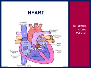
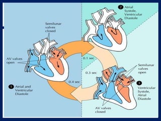
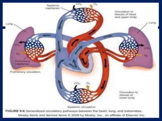
2. The heart is a roughly cone shaped hollow
muscular organ.
Length: 10 cm
Weight: 225 g in women
310 g in men
Position/location: the heart lies in the thoracic
cavity in the mediastinum( the space between
two lungs).
It lies obliquely little more to the left that right
and presents a base above and apex below.
INTRODUCTION
3. Inferiorly: the apex rests on the central tendon of
the diaphragm.
Superiorly: the great blood vessels e.g. aorta,
superior venacava, pulmonary artery and
pulmonary veins
Posteriorly: the oesophagus, trachea, left and
right bronchus, descending aorta, inferior
venacava and thoracic vertebra
Laterally: the lungs- the left lungs overlaps the
left side of the heart
Anteriorly: the sternum , ribs and intercostal
muscles.
ORGANS ASSOCIATED
WITH THE HEART
4. The heart wall:
The heart wall is composed of three layers of the
tissue.
1. Pericardium
2. Myocardium
3. endocardium
STRUCTURE OF THE HEART
5.
6. The pericardium is the outermost layer of the
heart and is made up with two sacs.
Outer sac consist of : fibrous tissue
Inner sac consists of : continues double layer of
serous membrane
Outer layer is known as parietal pericardium
Inner layer is known as visceral pericardium.
the space between two sacs is known as
pericardial space, which is filled with serous
fluid, which is secreted from serous membrane of
inner sac.
1. PERICARDIUM
7. The myocardium is composed of specialised
cardiac muscles founded only in heart.
It is not under voluntary control but striated like
skeletal muscle. Each fibres cells has nucleus
and one or more braches.
The sheet arrangements of the myocardium
enables the atria and ventricular to contract in a
co ordinated and efficient manner.
Myocardium is network of specialised conducting
fibres responsible for transmitting the heart
electrical signals.
Myocardium is thickest at the apex and thins
out towards the base.
2. MYOCARDIUM
8. This lines the chambers and valves of the hearts.
It is a thin smooth membrane to ensure smooth
flow of blood through the heart. It consists of
flattened epithelial cells and it is continuous
with the endothelium lining the blood vessels.
3. ENDOCARDIUM:
9. The heart is divide into right and left sided by
the septum, a partition consisting of myocardium
converted by endocardium.
Each side is divide by an antrioventricular valve
into upper atrium and the ventricle below.
Atrioventricular valve: these are formed by
double layer of endocardium.
Strengthen by a little fibrous tissue.
INTERIOR OF THE HEART
10.
11. AV VLAVES: right side AV valve has three flaps
is known as tricuspid valve, which separates
right atrium and right ventricle.
Left sided AV valve has two flaps is called as
mitral valve or bicuspid valve, which separates
left atrium and left ventricle.
12. There are four chambers of the heart.
1. right atrium – 2- 3 mm size
2.rigth ventricle-4-5 mm
3. left atrium – 2-3 mm
Left ventricle- 10-15 mm
CHAMBERS OF THE HEART
13. The two largest veins of the body, the superior
vencava and inferior vencava empty their
contents into the right atrium.
This blood passes via right atrioventriculer vlave
into right ventricle and from there blood is
pumped out into the pulmonary artery( the only
artery in the body which caries deoxygenated
blood).
BLOOD FLOW THROUGH THE
HEART
14. At the opening of pulmonary artery pulmonary
valve is present which prevents backflow of the
blood into right ventricle , it is also known as
semi-luner valve.
After leaving the heart the pulmonary artery
divides into left and right pulmonary arteries,
which carry the blood to the lungs where
exchange of gases takes place, carbon dioxide
excreted and oxygen is absorbed.
15. After purification of blood in the lungs two
pulmonary veins carry oxygenated blood back to
the left atrium.
Blood than passes through left atrioventriculer
valve ( mitral valve, bicuspid valve) into the left
ventricle and from there blood Is pumped out into
the aorta( the first artery of general circulation).
The opening of aorta is guarded by the aortic
valve.( at the opening of aorta there are valve
present which is called as semilunar or aortic
valve.
16. However , it should be noted that both atria
contract at the same time and this followed by
the simultaneous contraction of both ventricles.
17.
18. arterial supply: the heart I supplied with arterial
blood by the right and left coronary arteries.
The coronary arteries branch from the ascending
aorta.
The coronary arteries receives about 5% of the
blood pumped from the heart.
Large amount of blood supply especially to the left
ventricle.
There are two coronary arteries :
1. Right coronary artery
2. Left coronary artery
BLOOD CIRCULATION TO THE
HEART/ CORONARY CIRCULATION
19.
20. 1. right coronary artery:
It supplies small branches to the right side heart.
Right arteries having two branches:
Posterior inter ventricular branch
Marginal branches
The posterior inter ventricular branches supplies
blood to the wall of two ventricles.
The marginal branches supplies blood to the
right ventricles.
21. 2. left coronary artery:
The left coronary artery passes inferior to the
left circle & divides into the anterior inter
ventricular branch & the circumflex branches.
The anterior inter ventriculer branches will
supplies oxygenated blood to the walls of both
ventricles.
The circumflex branch will supplies oxygenated
blood to the walls of the left ventricle & left
atrium.
22.
23. Venous drainage: most of venous blood is
collected into number of cardiac veins that join to
form the coronary sinus, which opens into the
right atrium.
24. Autorythmicity: THE heart posses the property
of generating its own electrical impulse & beats
independently of nervous or hormonal control.
It is sullied with both sympathetic & Para
sympathetic automic nerve fibres.
Components: SA NODE: sino-atrial node
AV NODE: atrio-ventricular node
AV bundle( bundle of his): atrio ventricular
bandle
CONDUCTIVE SYSTEM OF HEART
25.
26. SA NODE: SINO ATRIAL NODE
This is small mass of specialise cells lies in the opening of
the superior venacava.
The Sino atrial cells generates these regular impulses
because they are electrically unstable.
This is also known as parameters.
AV NODE: atrioventriculer node
This is small mass of neuromuscular tissue is situated in
the wall of the atrial septum near the arioventricular
valve.
Normally AV node merely transmit the electrical signals
from the atria into ventricle.
27. 3. atriovenriculaer bundle( bundle of his)
This mass of specialised fibres originated from the
AV node.
The AV node crosses the fibrous ring that separates
atria and ventricles , then at the upper end of
ventricular septum, it divides into right and left
bundle branches.
Within the ventricular myocardium the branches
break up into the fibres called parkinje fibres, which
supplies electrical impulses into the apex of the
heart.
28. Heart is influenced by the autonomic( sympathetic &
para sympathetic nerves originating in the
cardiovascular centre in the medulla oblongata.
The vegus nerve: supply mainly the SA node & AV
nodes & atrial muscles.
NERVE SUPPLY TO THE HEART
30. At rest the healthy adult heart is likely to beat at a
rate of 60-70 bpm.
Each heart beat cycle is known as cardiac cycle .
During this time heart will contract & relax.
The period of contraction is called as systole &
relaxation is called as diastole.
31. Each cardiac cycle lasts about 0.8. sec consists of :
1.Atrial systole: contraction of atria 0.1 sec
2. Ventricular systole : contraction of ventricles 0.3
secs
3. Complete cardiac diastole:
Relaxation of atria & ventricles 0.4 secs
STAGES OF CARDIAC CYCLE
32.
33. The superior venacava & inferior vencava transport deoxygenated
blood into the right atrium at the same time four pulmonary
veins bring oxygenated blood into the left atrium.
Through AV valves
Blood flows passively into left ventricle(SA node spreads a wave
of impulse into myocardium of left atrium & starts)
Atrial contraction
AV node spreads , impulses quickly into ventricular muscle via
the AV bundle & purkinje fibres
34. A wave of contraction sweeps upward from apex of the heart
across the both ventricles
Ventricular contraction
Blood pumps into pulmonary artery & aorta
After contraction of ventricles the atrium & ventricles relax
Myocardium recover in preparation for the next heartbeat &
atria refill for next cycle
35. Heart sound is a involuntary action.
There are four heart sounds. Each corresponding to a
particular event in the cardiac cycle.
LUB: first sound , fairly loud occurs due to closure of
atrioventriculer vlave.
DUP: softer , sound due to closure of aortic and
pulmonary valves.
HEART SOUND
36. It is the report of electrical activity of the heart.
The pattern of electrical activity may be displayed on
an oscilloscope screen or traced on paper.
The normal ECG showing five waves by convention ,
have been named PQ,R,S, &T.
P wave: it arise when impulse from the SA NODE
sweeps over the atria( atrial depolarisation)
The QRS wave: it represent the very rapid spread of
the impulse from the AV node through the a bundle &
the purkinjie fibres & the electrical activities of the
ventricular muscle.
ECG: ELETROCARDIOGRAM
38. T WAVE: it represents the relaxation of the ventricular
muscle.( ventricular repolarization)
The ECG described above originated from the SA NODE
and is known as sinus rhythm.
P: atrial contraction
QRS: ventricular contraction
T: ventricular relaxation
39. The cardiac output is the amount of blood ejected
from each ventricle every minute.
The amount of blood expelled by each contraction of
each ventricle is the stroke volume.
Cardiac output is expressed in litters per minutes.
Cardiac out put = stroke volume * heart beat
In healthy adult heart at rest the stroke volume is
approximately 70ml and per minute heart rate is 72
per minute
Cardiac out put will be 5 liter/ minute
CARDIAC OUTPUT
41. Blood pressure is the force pressure that the blood
exerts on the walls of the blood vessels.
Blood pressure varies according to the times of the
day, posture, gender, age of the individual.
Blood pressure= systolic pressure
diastolic pressure
Systolic pressure:
When the left ventricular contracts & pushes blood
into the aorta, the pressure produced within the
arterial system is called the systolic blood pressure.
In adult it will be 120 mm of Hg or 16 Kpa( kilo
paskal)
BLOOD PRESSURE
42. Diastolic pressure:
When complete cardiac diastole occurs & the heart
is resting following the ejection of the blood , the
pressure within the arteries is much lower & is called
diastole blood pressure. In adult it will be 80 mm of
Hg 0r 11 Kpa.
BP= 120 mm of HG/ 80 mm of Hg
Blood pressure = cardiac output * peripheral
resistance
43. blood pressure is controlled in two ways.
1. short term control:
Moment to moment basis which mainly involves
1. baroreceptor reflex
2. chemo receptor reflex
3. higher centres in the brain
2. Long term control:
it involves regulation of blood by kidney and renin
angiotensin – aldosterone system( RAAS)
CONTROL OF BLOOD PRESSURE
44. Cardio vascular centre (CVC): it is collection of
interconnected nervous in the brain & situated in the
medulla & pons.
BARORECEPTORS:
These are nerve ending sensitive to pressure
changes ( stretch) within the vessel, situated in the
arch of aorta and in the carotid sinuses.( these are
body’s principal moment to moment regulatory
mechanism for controlling blood pressure.
1. SHORT TERM BP REGULATION
45. Chemoreceptors: these are the nerve ending situated in the
carotid and aortic bodies.
1. BARORECEPTORS:
When BP increases(BP more than 120 mm of hg)
Stimulation of baroreceptors in the aorta
Sending the impulses in to the cardiovascular centre in the
brain
CVC stimulates the parasympathetic nervous system
Relaxation of muscle of blood vessels ( tunica media)
Vasodilation
Blood pressure will decrease
46. when BP decreases less than 120/80 mm of Hg
Stimulation of brain receptors in the aorta
Sending impulses into the CVC in the brain
CVC will pass message into sympathetic nervous system
Stimulation of sympathetic nerve system
Contraction of muscles of blood vessels ( tunica media)
Vasoconstriction
BP will increase
47.
48. When co2 level increases & O2 level decrease in the blood stream
Tissue hypoxia
Need to improve tissue perfusion
Blood pressure should increase in this situation,
chemoreceptor will recognize this changes in CO2 & O2 in the blood
stream.
Pass this contraction to CVC in the brain
CVC will stimulate sympathetic nerve system
Vasoconstriction
BP will increase
CHEMORECEPTORS:
49.
50. when emotional status such as fear, anxiety, pain , anger,
occurs.
Hypothalamus will get stimulated
Brain need more blood supply
It will stimulate sympathetic nerve system
Vasoconstriction
BP increases thus brain will get more blood supply,
HIGHER CENTRE IN THE BRAIN
51. RAAS: renin angiotensin – aldosterone system
It is the mechanism where involvement of kidney is possible.
When kidney is unable to get blood supply , only this mechanism
will be activated.
Low renal blood flow
Decreased blood volume
Decreased BP
Kidney will secret renin
Angiotensinogen _____ angiotensin I
ACE
LONG TERM MECHANISM
52. Angiotensin II
Adrenal cortex vasoconstriction
Secretion of aldosterone increased BP
Kidney tubules reabsorbs kidney will get adequate blood supply
Na & water
Increased blood volume
Increased BP
53. Although circulation of blood around the body is
continues. It is described into two parts.
A. pulmonary circulation
B. systemic circulation
CIRCULATION OF BLOOD
54.
55.
56. This consist of the circulation of blood from the right
ventricle of the heart to the lungs & back to the left
atrium.
The pulmonary arteries: there are two branches for the
pulmonary trunk,
1. right pulmonary artery: it passes to the root of the
right lungs & divides into the branches.
- larger branches carries blood into middle & lower lobes
- smaller branches carries into upper lobe
2. the left pulmonary artery; it runs to the left lungs
where it divides into two branches.
One passing into each lobe.
A. PULMONARY CIRCULATION
57. Within the lungs these arteries divides & subdivide into smaller
arteries,
Arterioles
Capillaries
the inter change of O2 & CO2 takes place between capillary
blood & air in the alveoli of the lungs.
After gas exchanges each capillaries will join together & forms
vessels.
Capillaries
Veinules
veins
58. there are two pulmonary veins.
Right & left pulmonary veins
This both will carry oxygenated blood into left atrium
of the heart.
59. It is the circulation of blood all around the body.
The blood pumped out from the left ventricle is
carried by the branches of the aorta around the body
& is returned to the right atrium of the heart by
superior & anterior vencava is known as systemic
circulation.
2. SYSTEMIC / GENERAL CIRCULATION
60. Blood vessels are acting as pumps to supply blood
through out the body.
Blood vessels vary in structure size, function & these
are several types
Arteries
Arterioles
Capillaries
Venules
veins
BLOOD VESSELS
61.
62.
63. These are the blood vessels that transport blood
away from the heart.
They vary in considerably in size & their wall consist
of their layers of tissue.
Tunica adventitia: outer layer : fibrous tissue
Tunica media : middle layer: smooth muscle &
elastic tissue
Tunica intima: inner layer: squamous epithelial tissue
ARTERIES & ARTERIOLES
64.
65.
66. Anastomosis are arteries that form a link between
main arteries supplying an area.
End arteries are the arteries with no anastomoses or
those beyond the most distal anastomosis.
ANASTOMOSIS & END ARTERIES
67.
68. The smallest arterioles break into a number of
minotic vessels called capillaries.
The walls consist of a single layers of endothelial
cells sitting on a very thin basement membrane.
Through which water & other small molecules can
pass.
Entry to capillaries bed is grounded by rings of
smooth muscle( pre capillary sphincters) that directs
the blood vessels.
In certain places, including liver & bone marrow, the
capillaries are significantly wider & thicker than
normal. These are known as SINUSOIDS.
CAPILLARIES & SINUSOIDS
69.
70.
71. Veins are blood vessles that returns blood at low
pressure to the heart. The walls of the veins are the
thinner than those of arteries but have the same
three layers of tissue.
Veins are having valves, which prevent backflow of
blood. These are formed by folds of tunica intima &
strengthen by connective tissue.
Smallest veins are called as venules.
VEINS & VENULES
72. The outer layer of tissue of thick walled blood
vessels receive & the their blood walled blood
supply via network of blood vessels called the vasa
vasorum.
Thin walled vessels & the endothelium of other
receives O2 & nutrients by diffusion from the blood
passing trough them.
BLOOD SUPPLY
73. Vasomotor centre in medulla oblongata
Autonomic nerve system
Sympathetic stimulation para sympathetic stimulation
Contraction of smooth relaxation of smooth muscle
tunica media muscle of tunica media
Vasoconstriction vasodilatation
Lumen of blood vessel lumen of blood vessel increase
decreased
NERVE SUPPLY
74. The aorta is biggest artery in the human body &
begins at the upper part of the left ventricle &
passing up wards for short way , it arches back
wards and to the left.
It then descends behind the heart through the
thoracic cavity a little to the left of the thoracic
vertebrae.
At the level of 12th thoracic vertebra it passes behind
the diaphragm then downwards in the abdominal
cavity to the level of 4th lumber vertebra, where it
divides into the right and left common iliac arteries.
AORTA
75. The aorta will be described here according to its
location.
1. thoracic aorta:
This is the part of aorta lies above the diaphragm
and is described in three parts.
- ascending aorta
- arch of aorta
- descending aorta in the thorax
76. A. ascending aorta:
It is about 5 cm long , lies behind the sternum.
Two blood vessels stating from ascending aorta that are, right
and left coronary artery.
77.
78. B. Arch of aorta:
The arch of aorta is continuation of ascending aorta.
It begins behind the manubrium of the sternum and runs up
wards, back wards and to the left in front of trachea.
And it is continuous with the descending aorta.
- brachiocephalic artery
-Left common carotid artery
-Left subclvian artery
79. The venous blood from head & neck is returned by deep &
superficial veins.
The superficial veins will drain blood from superficial area of
the brain & deep vein drain from deep area of deep of brain.
Veins of head and neck:
1. right and left middle temporal vein
2. right and left supra orbital veins
3. right and left maxillary vein
4. right and left facial vein
5. right and left lingual vein
6. right and left occipital veins
VENUS RETURNS FROM THE HEAD &
NECK
80.
81.
82. The greater part of the brain is supplied by
arrangement of arteries called the circle of
willis. It consist of following arteries:
Two internal carotid arteries
Two vertebral arteries
CIRCULUS ARTERIOSOS (CIRCLE OF
WILLI)
87. The veins of upper limb is divided into two groups:
1. Deep veins:
Metacarpal veins
Deep palmar venous arch
Brachial veins
Axillary veins
2. Superficial veins
Cephalic vein
Basilica veins
Median veins
Medial cubital veins
Both join together & forms subclavian veins.
VENUS SUPPLY:
88. This is the part of aorta which continuous with
arch of aorta & begins at the 4 the thoracic
vertebra & extends downwards on the anterior
surface of the thoracic vertebra.
The descending aorta in the thorax gives of many
paired branches those are,
Bronchial arteries: supply to brachia & lungs
Oesophagus arteries: oesophagus
Intercostal artery: ribs, intercostal muscles
DESCENDING AORTA:
89.
90. There are two veins to drain venous blood in
thoracic cavity.
Azygos vein & hemizygos vein
The brachial vein, oesophageal vein & intercostal
vein join together & forms these two main veins.
Azygos vein joins with superior venacva &
hemizyagose vein join with left brachiocephalic
vein.
VENOUS RETURNS:
91.
92.
93.
94. The abdominal aorta is the continuation of thoracic
aorta& entering behind diaphragm at the 12th
thoracic vertebrae , at the level of 4ht lumber
vertebrae. it will divide into two common iliac artery.
Many branches arises from the abdominal aorta.
Paired branches:
Inferior phrenic artery : diaphragm
Renal artery: kidney
Superior renal artery: adrenal gland
Testicular artery: testes
Ovarian artery: ovary
ABDOMINAL AORTA:
95. Unpaired branches:
1. The coeliac artery: is short, thick, 1.25cm long & it rises
immediate below the diaphragm 7, divides into 3 branches.
Left gastric artery: stomach
Splenic artery: pancreas & spleen
Hepatic artery: liver, gall bladder, duodenum
2. Superior mesenteric artery:
It is a branch from aorta between the coeliac artery & renal
artery & supply blood supply to small intestine & proximal
part of large intestine.
3. Inferior mesenteric artery:
It arises from aorta about 4cm above its division into common
iliac artery. It supplies distal half of large intestine & part of
rectum.
96.
97. Paired testicular, ovarian, renal & adrenal veins
join into directly inferior venacava.
Blood from remaining organs in the abdominal
cavity passes through the liver via PORTAL
CIRCULATION & enter into interior venacva.
VENUS RETURN:
98. In the portal circulation, venous blood passes
from the capillary beds of the abdominal part of
the digestive system, spleen, pancreas to the
liver.
In this way blood with a high concentration of
nutrients, absorbed from the stomach and
intestines goes to the liver first.
In liver certain modification take place including
the regulation of blood nutrients levels.
It passes in second capillary bed , the hepatic
sinusoids & from portal vein.
PORTAL CIRCULATION:
99.
100. PORTAL VEIN:
This is formed by the union of the following veins, each of
which drains blood from the area supplied by
corresponding artery.
Splenic vein: drains blood spleen , pancreas, part of
stomach
Inferior mesenteric vein: rectum, pelvic, descending colon
of large intestine.
Superior mesenteric vein: small intestine, proximal part
of large intestine
Gastric vein: stomach. Distal end of oesophagus
Cystic vein: gall bladder
101. HEPATIC VEIN:
These are very short veins that leave the
posterior surface of the liver & almost
immediately enter the inferior vencava.
102. Abdominal aorta will divide into right & left
common iliac artery. These are divided into
further two branches.
Internal iliac artery
External iliac artery
Internal iliac artery:
It will supply organs in the pelvic cavity. In
female: uterine artery
CIRCULATION OF PELVIS & LOWER LIMB:
103. External iliac artery:
It will obliquely downwards & passes into where it
divides femoral artery & branches into popliteal
artery which supply blood to the knee area and
popliteal fossa. It further divides into :
The femoral artery: begins at midpoint at the
inguinal ligament & extends downwards in front of
the thigh. Then it eventually passes through
popliteal space where it becomes the popliteal artery.
Popliteal artery: it passes through the popliteal fossa
behind the knee, where pulse can be felt. At lower
border of the popliteal fossa it divides into the
anterior and posterior tibial arteries.
104. Anterior tibial artery: which supplies blood
supply to the tibia, ankle joint. It will divide over
the dorsum of foot & forms dorsalis pedis artery.
Posterior tibial artery: it runs medially back of
legs & gives a branch peroneal artery which
supplies lateral aspect of the leg & forms a new
branch- planter artery, which supplies blood to
the sole of the foot.
106. 1. Deep veins:
Digital vein
Planter venous arch
Post tibial vein
Anterior tibial vein
Popliteal vein
Femoral vein
External iliac vein
Internal iliac vein
Common iliac vein
Both right & left common iliac veins joins into inferior
venacava at the sacro iliac joint.
VENUS RETURN:
107.
108. 2. Superficial vein: there are two main superficial
veins are draining blood from the lower limbs are,
Small saphenous vein: the small saphenous vein
begins behind the ankle joint& joins into
popliteal vein.
Great saphenous vein: it is the longest vein of
the body, it begins at the medial half of the
dorsum of the foot & runs upwards crossing over
thigh & joins into political vein.