SlideShare a Scribd company logo
1 of 53
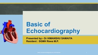
Basic of
Echocardiography
Presented by:- Dr HIMANSHU SAMAIYA
Resident : SGMH Rewa M.P.
basic of echocardiography by dr.himanshu 1
INTRODUCTION
It is a type of ultrasound test that uses high pitched
sound waves to produce an image of the heart.
 The sound waves are sent through a device called a
transducer and are reflected off the various structures
of the heart.
 These echoes are converted into pictures of the heart
that can be seen on a video monitor.
basic of echocardiography by dr.himanshu 2
COMPONENTS
 Pulse generator - applies high amplitude voltage to energize the
crystals
 Transducer - converts electrical energy to mechanical (ultrasound)
energy and vice versa
 Receiver - detects and amplifies weak signals
 Display - displays ultrasound signals in a variety of modes
 Memory - stores video display
basic of echocardiography by dr.himanshu 3
INDICATION
 Cardiac chamber size and contents
 Ejection fraction
 Pericardial sac i.e. pericardial effusion, constrictive pericarditis
 Ascending aorta assessment of known or suspected adult congenital heart diease.
 Evaluation of suspected complication of myocardial ischemia/infarction.
 Evaluation of valvular or structural heart disease.
 Infective endocarditis
 Suspected tumor or thrombus
 Cardiomyopathy : dilated, restrictive, and hypertrophic
 Pulmonary hypertension
basic of echocardiography by dr.himanshu 4
PROCEDURE
 A standard echocardiogram is also known as a
transthoracic echocardiogram (TTE), or cardiac
ultrasound.
 The subject is asked to lie in the semi recumbent position
on his or her left side with the head elevated.
 Ultrasound is transmitted from a transducer having a frequency
of 2.5 to 3.5 MHz for echo in adults.
 To study deep seated structures because of better penetration.
 A transducer frequency of 5.0 MHz is suitable for pediatric echo,
since the heart is more superficial in children
basic of echocardiography by dr.himanshu 5
WINDOWS OF ECHO
 Evaluation of the heart with
echocardiography requires
"acoustic windows" of the heart.
 Bone reflects the ultrasound waves
and so all structures directly behind
bone are not visible with ultrasound.
basic of echocardiography by dr.himanshu 6
Parasternal Long-Axis View
(PLAX)
Transducer position: left sternal
edge; 2nd – 4th intercostal space
Marker dot direction: points
towards right shoulder
Most echo studies begin with this
view
It sets the stage for subsequent
echo views
Many structures seen from this
view
basic of echocardiography by dr.himanshu 7
Measurements in PLAX view can be used to quantify the
heart:
 Left ventricular size and wall thickness
 Left atrial linear dimension (as opposed to area)
 Left ventricular outflow tract diameter (used to
calculate aortic valve area by the continuity equation)
 Aortic annulus, sinus of Valsalva, and aortic root
sizes
basic of echocardiography by dr.himanshu 8
Parasternal Short Axis View
(PSAX)
 Transducer position: left sternal edge;
2nd – 4th intercostal space.
 Marker dot direction: points towards left
shoulder(90˚ clockwise from PLAX view)
 By tilting transducer on an axis between
the left hip and right shoulder, short axis
views are obtained at different levels,
from the aorta to the LV apex.
 The aortic valve, right ventricular inflow
& outflow tracts visible with the tricuspid
valve
basic of echocardiography by dr.himanshu 9
Papillary Muscle (PM)level
•PSAX at the level of the
papillary muscles showing how
the respective LV segments are
identified, usually for the
purposes of describing
abnormal LV wall motion
• LV wall thickness can also be
assessed
basic of echocardiography by dr.himanshu 10
Apical 4-Chamber View (AP4CH)
• Transducer position: apex of
heart
• Marker dot direction: points
towards left shoulder
• The AP5CH view is obtained from
this view by slight anterior
angulation of the transducer
towards the chest wall.
• The LVOT can then be visualised
basic of echocardiography by dr.himanshu 11
Apical 2-Chamber View (AP2CH)
• Transducer position: apex of the
heart
• Marker dot direction: points
towards left side of neck (45˚
anticlockwise from AP4CH view)
• Good for assessment of LV
anterior wall and LV inferior wall
basic of echocardiography by dr.himanshu 12
Sub–Costal 4 Chamber View(SC4CH)
• Transducer position: under the
xiphisternum
• Marker dot position: points
towards left shoulder
• The subject lies supine with head
slightly low (no pillow). With feet
on the bed, the knees are slightly
elevated
• Better images are obtained with
the abdomen relaxed and during
inspiration
• Interatrial septum, pericardial
effusion, desc abdominal aorta
basic of echocardiography by dr.himanshu 13
Suprasternal View
• Transducer position: suprasternal
notch
• Marker dot direction: points towards
left jaw
• The subject lies supine with the neck
hyperextended. The head is rotated
slightly towards the left
• The position of arms or legs and the
phase of respiration have no bearing
on this echo window
• Arch of aorta,ascending
aorta,pulmonary artery
basic of echocardiography by dr.himanshu 14
TYPES OF ECHOCARDIOGRAPHY
Transthoracic echocardiography
Transesophageal echocardiography
Stress echocardiography
Three-dimensional echocardiography
basic of echocardiography by dr.himanshu 15
TRANSESOPHAGEAL ECHOCARDIOGRAPHY
• The oesophagus in its mid-course is located posterior to the heart and
anterior to the descending aorta.
• This provides an opportunity to interrogate the heart and related
mediastinal structures with a high frequency transducer positioned in the
esophagus for better image resolution.
• The transducer to be inserted down the throat into the esophagus (the
swallowing tube that connects the mouth to the stomach).
basic of echocardiography by dr.himanshu 16
basic of echocardiography by dr.himanshu 17
Advantages of TEE
 Useful alternative to transthoracic echo in case of obesity, chest wall
deformity, emphysema or pulmonary fibrosis.
 Useful complement to transthoracic echo because of better image
quality and resolution due to two reasons:
 – absence of acoustic barrier between the ultrasound beam and the
rib cage, chest wall and lung tissue.
 – greater proximity to the heart and therefore the ability to use higher
frequency probe with vastly improved image quality and precise
spatial resolution. basic of echocardiography by dr.himanshu 18
 Useful supplement to transthoracic echo, which
cannot examine the posterior aspect of the heart.
 Structures such as left atrial appendage, descending
aorta and pulmonary veins can only be visualized by
TEE.
 very high sensitivity for locating a blood clot inside
the left atrium.
basic of echocardiography by dr.himanshu 19
Disadvantages
 It takes longer to perform a TEE than a TTE.
 It may be uncomfortable for the patient.
 It requires short-term sedation, oxygen
administration and ECG monitoring since, there are
chances of hypoxia, arrhythmia and angina.
 TEE images require a comprehensive understanding
of the spatial relationship between cardiac structures.
basic of echocardiography by dr.himanshu 20
Complications with TEE
Major :-
 Esophageal rupture or perforation
 • Laryngospasm or bronchopasm
 • Sustained ventricular tachycardia
Minor:-
 Retching and vomiting
 • Sore-throat and hoarseness
 • Blood-tinged sputum
 • Tachycardia or bradycardia
 • Hypoxia and ischemia
 • Transient BP rise or fall basic of echocardiography by dr.himanshu 21
Contraindications to TEE
 Unrepaired tracheoesophageal fistula
 History prior esophageal surgery
 Esophageal obstruction or stricture
 Perforated hollow viscus
 Gastric or esophageal bleeding
 Poor airway control
 Severe respiratory depression
 Oropharyngeal pathology
 Uncooperative, unsedated patient
 Severe coagulopathy
 Cervical spine injury
basic of echocardiography by dr.himanshu 22
STRESS ECHOCARDIOGRAM
 A stress test accompanied by echocardiography.
 During a stress.echo, patient exercise on a treadmill or statio
nary bike with blood pressure and heart rhythm monitoring.
 The echocardiography is performed both before and after the
exercise to compare structural differences.
 To assess for any abnormalities in wall motion of the heart.
 This is used to detect obstructive coronary artery disease
basic of echocardiography by dr.himanshu 23
Technique:
 NPO for four hours before the test.
 Do not drink or eat caffeine products (cola, chocolate, coffee, tea)
for 24 hours before the test.
 Do not take any over-the-counter medications that contain caffeine
for 24 hours before the test.
 Do not take the following heart medications for 24 hour before the
test unless doctor tells.
 *Beta-blockers * Isosorbide dinitrate
 *Isosorbide mononitrate *Nitroglycerin
basic of echocardiography by dr.himanshu 24
basic of echocardiography by dr.himanshu 25
DOBUTAMINE STRESS ECHOCARDIOGRAM
 A form of stress echocardiogram.
 The test is used to evaluate heart and valve function when
unable to exercise on a treadmill or stationary bike.
 Most dobutamine stress protocols start at an infusion rate
of 5 ug/kg/min and increase to a peak dose of 40
or 50 ug / kg / min.
 To further increase heart rate, a bolus injection of 0.25—
1 .0 mg atropine is added
basic of echocardiography by dr.himanshu 26
INTRAVASCULAR ULTRASOUND
 A form of echocardiography performed during cardiac
catheterization.
 During this procedure, the transducer is threaded into
the heart blood vessels via a femoral catheter.
 Used to provide detailed information about the
atherosclerosis (blockage) inside the blood vessels.
basic of echocardiography by dr.himanshu 27
THE MODALITIES OF ECHO
1. Conventional echo
 Two-Dimensional echo (2-D echo)
 Motion- mode echo (M-mode echo)
2 Doppler Echo
 Continuous wave (CW) Doppler
 Pulsed wave (PW) Doppler
 Colour flow(CF) Doppler
basic of echocardiography by dr.himanshu 28
MOTION-MODE (M MODE) ECHO
 In the M-mode tracing, ultrasound is transmitted and
received along only one scan line.
 This line is obtained by applying the cursor to the 2-D
image and aligning it perpendicular to the structure
being studied.
 M-mode is displayed as a continuous tracing with two
axes.
 The vertical axis represents distance between the
moving structure and the transducer. The horizontal
axis represents time.
basic of echocardiography by dr.himanshu 29
 Since only one scan line is imaged, M-mode echo provides greater
sensitivity than 2-D echo for studying the motion of moving cardiac
structures.
 Motion and thickness of ventricular walls, changing size of cardiac
chambers and opening and closure of valves is better displayed on
M mode.
 Simultaneous ECG recording facilitates accurate timing of cardiac
events.
 Similarly, the flow pattern on color flow mapping can be timed in
relation to the cardiac cycle.
basic of echocardiography by dr.himanshu 30
Motion-mode echo (M-mode Echo) levels:
A. Mitral valve (MV) level
B. Aortic valve (AV) level
basic of echocardiography by dr.himanshu 31
DOPPLER ECHOCARDIOGRAPHY
 Doppler echocardiography is a method for detecting the direction and
velocity of moving blood within the heart.
 PULSED WAVE (PW):
 useful for low velocity flow e.g. MV flow.
 PW Doppler transmits ultrasound in pulses and waits to receive the
returning ultrasound after each pulse.
 PW Doppler provides a better spectral tracing than CW Doppler, which is
used for calculations.
 PW Doppler modality is used to localize velocity signals and
 Abnormal flow patterns picked up by CW Doppler and color flow mapping,
respectively.
basic of echocardiography by dr.himanshu 32
Continuous Wave (CW)
 Useful for high velocity flow e.g aortic stenosis
 CW Doppler transmits and receives ultrasound continuously
 This Doppler modality is used for rapid scanning of the heart in
search of high velocity signals and abnormal flow patterns.
 CW Doppler is used for grading the severity of valvular stenosis and
assessing the degree of valvular regurgitation.
 An intracardiac left-to-right shunt such as a ventricular septal defect
can be quantified.
 By using CW Doppler signal of the tricuspid valve, pulmonary artery
pressure can be calculated.
basic of echocardiography by dr.himanshu 33
Color Flow (CF)
 It is also known as real-time Doppler imaging.
 Color Doppler provides a visual display of blood flow within the
heart, in the form of a color flow map.
 Different colors are used to designate the direction of blood
flow.
 Red is flow toward, and Blue is flow away from the transducer
with turbulent flow shown as a mosaic pattern. (BART)
basic of echocardiography by dr.himanshu 34
Color flow map of a normal mitral valve from A4CH view
showing a red-colored jet
basic of echocardiography by dr.himanshu 35
Color flow map of ventricular outflow tract from A5CH view
showing a blue jet
basic of echocardiography by dr.himanshu 36
APPLICATIONS OF COLOR DOPPLER
Stenotic Lesions:
 Color Doppler can identify, localize and quantitate stenotic lesions of
the cardiac valves.
 It visually displays the stenotic area and the resultant jet as distinct
from normal flow.
Regurgitant Lesions:
 Color Doppler can diagnose and estimate the severity of regurgitant
lesions of the valves
Intercardiac shunts
basic of echocardiography by dr.himanshu 37
THREE DIMENSION ECHO
 Future direction in echo
 Obviates the need for cognitive 3 D construction of 2
D image plane
 Useful in:-
1. ventricular volume assessment
2. Study of asymmetrical stenotic valve
3. Complex structural relationships in congenital
heart disease .
basic of echocardiography by dr.himanshu 38
MYOCARDIAL CONTRAST ECHO
 Application of ultrasonic
contrast agent, to accurately
delineate areas of reduced
myocardial blood flow or
perfusion defect related to
coronary occlusion
 Contrast exist as microbubbles
basic of echocardiography by dr.himanshu 39
Echo findings in Heart
diseases
basic of echocardiography by dr.himanshu 40
DIALATED CARDIOMYOPATHY
 The ventricles are dilated more
than the normal.
basic of echocardiography by dr.himanshu 41
HYPERTROPHIC CARDIOMYOPATHY
• The intra ventricular
septum appears thickened
• IVS:LVPW ratio >1.5
basic of echocardiography by dr.himanshu 42
RESTRICTIVE CARDIOMYOPATHY
• Thick and bright septum
• Reduced size of ventricles
• Dilated RA and LA
basic of echocardiography by dr.himanshu 43
LEFT VENTRICULAR HYPERTROPHY
basic of echocardiography by dr.himanshu 44
CARDIAC TAMPONADE
basic of echocardiography by dr.himanshu 45
INFECTIVE ENDOCARDITIS
basic of echocardiography by dr.himanshu 46
MITRAL STENOSIS
• Thickening of valve leaflets
• Restricted opening of valve
• Dilatation of left atrium
• Diastolic doming of anterior
leaflets
basic of echocardiography by dr.himanshu 47
MITRAL REGURGITATION
• Regurgitant jet in LA on
A4CH view.
• Extent of MR jet fills the LA
Cavity indicates severity of
MR.
basic of echocardiography by dr.himanshu 48
basic of echocardiography by dr.himanshu 49
VSD
ASD
basic of echocardiography by dr.himanshu 50
LEFT ATRIAL CLOT LEFT VENTRICULAR CLOT
basic of echocardiography by dr.himanshu 51
CORONARY ARTERY DISEASE
basic of echocardiography by dr.himanshu 52
basic of echocardiography by dr.himanshu 53

More Related Content

What's hot

LEFT VENTRICULOGRAPHY.pptx
LEFT VENTRICULOGRAPHY.pptxLEFT VENTRICULOGRAPHY.pptx
LEFT VENTRICULOGRAPHY.pptxnomusasibanda
 
Transesophageal echocardiography
Transesophageal echocardiographyTransesophageal echocardiography
Transesophageal echocardiographyNeeraj Varyani
 
Tissue doppler imaging
Tissue doppler imagingTissue doppler imaging
Tissue doppler imagingFuad Farooq
 
Diagnostic catheters for coronary angiography
Diagnostic catheters for coronary angiography Diagnostic catheters for coronary angiography
Diagnostic catheters for coronary angiography Aswin Rm
 
Peripheral angiography
Peripheral angiographyPeripheral angiography
Peripheral angiographyInosRagan
 
Echocardiographic measurements
Echocardiographic measurementsEchocardiographic measurements
Echocardiographic measurementsabdidandena
 
Tissue doppler Echocardiography (TDE)
Tissue doppler Echocardiography (TDE)Tissue doppler Echocardiography (TDE)
Tissue doppler Echocardiography (TDE)sruthiMeenaxshiSR
 
Basics of echo & principles of doppler echocardiography
Basics of echo & principles of doppler echocardiographyBasics of echo & principles of doppler echocardiography
Basics of echo & principles of doppler echocardiographyabrahahailu
 
Shunt Detection And Quantification
Shunt Detection And QuantificationShunt Detection And Quantification
Shunt Detection And QuantificationDang Thanh Tuan
 
Echo views
Echo viewsEcho views
Echo viewsnmonty02
 
Hemodynamic assessment in cardiology
Hemodynamic assessment in cardiologyHemodynamic assessment in cardiology
Hemodynamic assessment in cardiologySujit Sahu
 
Echocardiography in ER 2020
Echocardiography in ER 2020Echocardiography in ER 2020
Echocardiography in ER 2020islamali260580
 

What's hot (20)

LEFT VENTRICULOGRAPHY.pptx
LEFT VENTRICULOGRAPHY.pptxLEFT VENTRICULOGRAPHY.pptx
LEFT VENTRICULOGRAPHY.pptx
 
Transesophageal echocardiography
Transesophageal echocardiographyTransesophageal echocardiography
Transesophageal echocardiography
 
Tissue doppler imaging
Tissue doppler imagingTissue doppler imaging
Tissue doppler imaging
 
Diagnostic catheters for coronary angiography
Diagnostic catheters for coronary angiography Diagnostic catheters for coronary angiography
Diagnostic catheters for coronary angiography
 
Peripheral angiography
Peripheral angiographyPeripheral angiography
Peripheral angiography
 
Echocardiographic measurements
Echocardiographic measurementsEchocardiographic measurements
Echocardiographic measurements
 
Echo in pericardial diseases
Echo in pericardial diseasesEcho in pericardial diseases
Echo in pericardial diseases
 
Tissue doppler Echocardiography (TDE)
Tissue doppler Echocardiography (TDE)Tissue doppler Echocardiography (TDE)
Tissue doppler Echocardiography (TDE)
 
Basic echocardiography
Basic echocardiographyBasic echocardiography
Basic echocardiography
 
Basics of echo & principles of doppler echocardiography
Basics of echo & principles of doppler echocardiographyBasics of echo & principles of doppler echocardiography
Basics of echo & principles of doppler echocardiography
 
Cath hemodynamics vir
Cath hemodynamics virCath hemodynamics vir
Cath hemodynamics vir
 
Contrast echocardiography
Contrast echocardiography Contrast echocardiography
Contrast echocardiography
 
Basics of echocardiograghy
Basics of echocardiograghyBasics of echocardiograghy
Basics of echocardiograghy
 
3D Echocardiography
3D Echocardiography3D Echocardiography
3D Echocardiography
 
Shunt Detection And Quantification
Shunt Detection And QuantificationShunt Detection And Quantification
Shunt Detection And Quantification
 
Transesophageal echocardiography
Transesophageal echocardiographyTransesophageal echocardiography
Transesophageal echocardiography
 
Echo views
Echo viewsEcho views
Echo views
 
Echocardiogram Basics
Echocardiogram BasicsEchocardiogram Basics
Echocardiogram Basics
 
Hemodynamic assessment in cardiology
Hemodynamic assessment in cardiologyHemodynamic assessment in cardiology
Hemodynamic assessment in cardiology
 
Echocardiography in ER 2020
Echocardiography in ER 2020Echocardiography in ER 2020
Echocardiography in ER 2020
 

Similar to Echocardiography basics

invasive non invasive procedures.pdf for bsc nursing students
invasive non invasive procedures.pdf for bsc nursing studentsinvasive non invasive procedures.pdf for bsc nursing students
invasive non invasive procedures.pdf for bsc nursing studentsshanmukhadevi
 
non invasive and invasive cardiac monitoring.pptx
non invasive and invasive cardiac monitoring.pptxnon invasive and invasive cardiac monitoring.pptx
non invasive and invasive cardiac monitoring.pptxshafina27
 
ECHO in Cardiac Failure
ECHO in Cardiac Failure ECHO in Cardiac Failure
ECHO in Cardiac Failure MANULALVS
 
Introduction to echocardiography
Introduction to echocardiography Introduction to echocardiography
Introduction to echocardiography Bibini Bab
 
Cardiovascular assessment and diagnostic investigations ppt slideshare
Cardiovascular assessment and diagnostic investigations ppt slideshareCardiovascular assessment and diagnostic investigations ppt slideshare
Cardiovascular assessment and diagnostic investigations ppt slidesharesonam
 
26 USCardio (1).ppt
26 USCardio (1).ppt26 USCardio (1).ppt
26 USCardio (1).pptsharifi3
 
Transthoracic Echocardiogram, 3D, Doppler, TEE.pptx
Transthoracic Echocardiogram, 3D, Doppler, TEE.pptxTransthoracic Echocardiogram, 3D, Doppler, TEE.pptx
Transthoracic Echocardiogram, 3D, Doppler, TEE.pptxSalmanMajid2
 
Echocardiogram or Echocardiography or Heart Ultrasound
Echocardiogram or Echocardiography or Heart UltrasoundEchocardiogram or Echocardiography or Heart Ultrasound
Echocardiogram or Echocardiography or Heart UltrasoundNishuVerma20
 
emergency echo in critically ill patients.ppt
emergency echo in critically ill patients.pptemergency echo in critically ill patients.ppt
emergency echo in critically ill patients.pptShivani Rao
 
Cardiac diagnostics and laboratory tests
Cardiac diagnostics and laboratory tests Cardiac diagnostics and laboratory tests
Cardiac diagnostics and laboratory tests Dr. Rima Das
 
Diagnostic measures in cardiology
Diagnostic measures in cardiologyDiagnostic measures in cardiology
Diagnostic measures in cardiologySUDESHNA BANERJEE
 
Presentation2.pptx technique chest 2
Presentation2.pptx technique chest 2Presentation2.pptx technique chest 2
Presentation2.pptx technique chest 2Yashawant Yadav
 
Basics of echocardiography
Basics of echocardiographyBasics of echocardiography
Basics of echocardiographyaasrithakotha2
 
basicechocardiography-170913182344 (2).pdf
basicechocardiography-170913182344 (2).pdfbasicechocardiography-170913182344 (2).pdf
basicechocardiography-170913182344 (2).pdflittlealphonsa
 
Pedia Cardiac Imaging by Caffey.pptx
Pedia Cardiac Imaging by Caffey.pptxPedia Cardiac Imaging by Caffey.pptx
Pedia Cardiac Imaging by Caffey.pptxFreceGaming1
 

Similar to Echocardiography basics (20)

invasive non invasive procedures.pdf for bsc nursing students
invasive non invasive procedures.pdf for bsc nursing studentsinvasive non invasive procedures.pdf for bsc nursing students
invasive non invasive procedures.pdf for bsc nursing students
 
non invasive and invasive cardiac monitoring.pptx
non invasive and invasive cardiac monitoring.pptxnon invasive and invasive cardiac monitoring.pptx
non invasive and invasive cardiac monitoring.pptx
 
ECHO in Cardiac Failure
ECHO in Cardiac Failure ECHO in Cardiac Failure
ECHO in Cardiac Failure
 
Introduction to echocardiography
Introduction to echocardiography Introduction to echocardiography
Introduction to echocardiography
 
Cardiovascular assessment and diagnostic investigations ppt slideshare
Cardiovascular assessment and diagnostic investigations ppt slideshareCardiovascular assessment and diagnostic investigations ppt slideshare
Cardiovascular assessment and diagnostic investigations ppt slideshare
 
26 USCardio (1).ppt
26 USCardio (1).ppt26 USCardio (1).ppt
26 USCardio (1).ppt
 
Cardiac catheterization
Cardiac catheterizationCardiac catheterization
Cardiac catheterization
 
Transthoracic Echocardiogram, 3D, Doppler, TEE.pptx
Transthoracic Echocardiogram, 3D, Doppler, TEE.pptxTransthoracic Echocardiogram, 3D, Doppler, TEE.pptx
Transthoracic Echocardiogram, 3D, Doppler, TEE.pptx
 
Echocardiogram or Echocardiography or Heart Ultrasound
Echocardiogram or Echocardiography or Heart UltrasoundEchocardiogram or Echocardiography or Heart Ultrasound
Echocardiogram or Echocardiography or Heart Ultrasound
 
emergency echo in critically ill patients.ppt
emergency echo in critically ill patients.pptemergency echo in critically ill patients.ppt
emergency echo in critically ill patients.ppt
 
Cardiac diagnostics and laboratory tests
Cardiac diagnostics and laboratory tests Cardiac diagnostics and laboratory tests
Cardiac diagnostics and laboratory tests
 
Diagnostic measures in cardiology
Diagnostic measures in cardiologyDiagnostic measures in cardiology
Diagnostic measures in cardiology
 
TRANSESOPHAGEAL ECHOCARDIOGRAPHY.pptx
TRANSESOPHAGEAL ECHOCARDIOGRAPHY.pptxTRANSESOPHAGEAL ECHOCARDIOGRAPHY.pptx
TRANSESOPHAGEAL ECHOCARDIOGRAPHY.pptx
 
Echocardiogram
EchocardiogramEchocardiogram
Echocardiogram
 
Cardio2
Cardio2Cardio2
Cardio2
 
Presentation2.pptx technique chest 2
Presentation2.pptx technique chest 2Presentation2.pptx technique chest 2
Presentation2.pptx technique chest 2
 
2D ECHO Basics
2D ECHO Basics2D ECHO Basics
2D ECHO Basics
 
Basics of echocardiography
Basics of echocardiographyBasics of echocardiography
Basics of echocardiography
 
basicechocardiography-170913182344 (2).pdf
basicechocardiography-170913182344 (2).pdfbasicechocardiography-170913182344 (2).pdf
basicechocardiography-170913182344 (2).pdf
 
Pedia Cardiac Imaging by Caffey.pptx
Pedia Cardiac Imaging by Caffey.pptxPedia Cardiac Imaging by Caffey.pptx
Pedia Cardiac Imaging by Caffey.pptx
 

Recently uploaded

How to Make a Pirate ship Primary Education.pptx
How to Make a Pirate ship Primary Education.pptxHow to Make a Pirate ship Primary Education.pptx
How to Make a Pirate ship Primary Education.pptxmanuelaromero2013
 
MENTAL STATUS EXAMINATION format.docx
MENTAL     STATUS EXAMINATION format.docxMENTAL     STATUS EXAMINATION format.docx
MENTAL STATUS EXAMINATION format.docxPoojaSen20
 
Sanyam Choudhary Chemistry practical.pdf
Sanyam Choudhary Chemistry practical.pdfSanyam Choudhary Chemistry practical.pdf
Sanyam Choudhary Chemistry practical.pdfsanyamsingh5019
 
Call Girls in Dwarka Mor Delhi Contact Us 9654467111
Call Girls in Dwarka Mor Delhi Contact Us 9654467111Call Girls in Dwarka Mor Delhi Contact Us 9654467111
Call Girls in Dwarka Mor Delhi Contact Us 9654467111Sapana Sha
 
_Math 4-Q4 Week 5.pptx Steps in Collecting Data
_Math 4-Q4 Week 5.pptx Steps in Collecting Data_Math 4-Q4 Week 5.pptx Steps in Collecting Data
_Math 4-Q4 Week 5.pptx Steps in Collecting DataJhengPantaleon
 
Concept of Vouching. B.Com(Hons) /B.Compdf
Concept of Vouching. B.Com(Hons) /B.CompdfConcept of Vouching. B.Com(Hons) /B.Compdf
Concept of Vouching. B.Com(Hons) /B.CompdfUmakantAnnand
 
Software Engineering Methodologies (overview)
Software Engineering Methodologies (overview)Software Engineering Methodologies (overview)
Software Engineering Methodologies (overview)eniolaolutunde
 
Separation of Lanthanides/ Lanthanides and Actinides
Separation of Lanthanides/ Lanthanides and ActinidesSeparation of Lanthanides/ Lanthanides and Actinides
Separation of Lanthanides/ Lanthanides and ActinidesFatimaKhan178732
 
“Oh GOSH! Reflecting on Hackteria's Collaborative Practices in a Global Do-It...
“Oh GOSH! Reflecting on Hackteria's Collaborative Practices in a Global Do-It...“Oh GOSH! Reflecting on Hackteria's Collaborative Practices in a Global Do-It...
“Oh GOSH! Reflecting on Hackteria's Collaborative Practices in a Global Do-It...Marc Dusseiller Dusjagr
 
Employee wellbeing at the workplace.pptx
Employee wellbeing at the workplace.pptxEmployee wellbeing at the workplace.pptx
Employee wellbeing at the workplace.pptxNirmalaLoungPoorunde1
 
Introduction to ArtificiaI Intelligence in Higher Education
Introduction to ArtificiaI Intelligence in Higher EducationIntroduction to ArtificiaI Intelligence in Higher Education
Introduction to ArtificiaI Intelligence in Higher Educationpboyjonauth
 
Crayon Activity Handout For the Crayon A
Crayon Activity Handout For the Crayon ACrayon Activity Handout For the Crayon A
Crayon Activity Handout For the Crayon AUnboundStockton
 
18-04-UA_REPORT_MEDIALITERAСY_INDEX-DM_23-1-final-eng.pdf
18-04-UA_REPORT_MEDIALITERAСY_INDEX-DM_23-1-final-eng.pdf18-04-UA_REPORT_MEDIALITERAСY_INDEX-DM_23-1-final-eng.pdf
18-04-UA_REPORT_MEDIALITERAСY_INDEX-DM_23-1-final-eng.pdfssuser54595a
 
BASLIQ CURRENT LOOKBOOK LOOKBOOK(1) (1).pdf
BASLIQ CURRENT LOOKBOOK  LOOKBOOK(1) (1).pdfBASLIQ CURRENT LOOKBOOK  LOOKBOOK(1) (1).pdf
BASLIQ CURRENT LOOKBOOK LOOKBOOK(1) (1).pdfSoniaTolstoy
 
The basics of sentences session 2pptx copy.pptx
The basics of sentences session 2pptx copy.pptxThe basics of sentences session 2pptx copy.pptx
The basics of sentences session 2pptx copy.pptxheathfieldcps1
 
Kisan Call Centre - To harness potential of ICT in Agriculture by answer farm...
Kisan Call Centre - To harness potential of ICT in Agriculture by answer farm...Kisan Call Centre - To harness potential of ICT in Agriculture by answer farm...
Kisan Call Centre - To harness potential of ICT in Agriculture by answer farm...Krashi Coaching
 
Accessible design: Minimum effort, maximum impact
Accessible design: Minimum effort, maximum impactAccessible design: Minimum effort, maximum impact
Accessible design: Minimum effort, maximum impactdawncurless
 
Paris 2024 Olympic Geographies - an activity
Paris 2024 Olympic Geographies - an activityParis 2024 Olympic Geographies - an activity
Paris 2024 Olympic Geographies - an activityGeoBlogs
 
ECONOMIC CONTEXT - LONG FORM TV DRAMA - PPT
ECONOMIC CONTEXT - LONG FORM TV DRAMA - PPTECONOMIC CONTEXT - LONG FORM TV DRAMA - PPT
ECONOMIC CONTEXT - LONG FORM TV DRAMA - PPTiammrhaywood
 

Recently uploaded (20)

How to Make a Pirate ship Primary Education.pptx
How to Make a Pirate ship Primary Education.pptxHow to Make a Pirate ship Primary Education.pptx
How to Make a Pirate ship Primary Education.pptx
 
MENTAL STATUS EXAMINATION format.docx
MENTAL     STATUS EXAMINATION format.docxMENTAL     STATUS EXAMINATION format.docx
MENTAL STATUS EXAMINATION format.docx
 
Sanyam Choudhary Chemistry practical.pdf
Sanyam Choudhary Chemistry practical.pdfSanyam Choudhary Chemistry practical.pdf
Sanyam Choudhary Chemistry practical.pdf
 
Call Girls in Dwarka Mor Delhi Contact Us 9654467111
Call Girls in Dwarka Mor Delhi Contact Us 9654467111Call Girls in Dwarka Mor Delhi Contact Us 9654467111
Call Girls in Dwarka Mor Delhi Contact Us 9654467111
 
_Math 4-Q4 Week 5.pptx Steps in Collecting Data
_Math 4-Q4 Week 5.pptx Steps in Collecting Data_Math 4-Q4 Week 5.pptx Steps in Collecting Data
_Math 4-Q4 Week 5.pptx Steps in Collecting Data
 
Concept of Vouching. B.Com(Hons) /B.Compdf
Concept of Vouching. B.Com(Hons) /B.CompdfConcept of Vouching. B.Com(Hons) /B.Compdf
Concept of Vouching. B.Com(Hons) /B.Compdf
 
Software Engineering Methodologies (overview)
Software Engineering Methodologies (overview)Software Engineering Methodologies (overview)
Software Engineering Methodologies (overview)
 
Separation of Lanthanides/ Lanthanides and Actinides
Separation of Lanthanides/ Lanthanides and ActinidesSeparation of Lanthanides/ Lanthanides and Actinides
Separation of Lanthanides/ Lanthanides and Actinides
 
“Oh GOSH! Reflecting on Hackteria's Collaborative Practices in a Global Do-It...
“Oh GOSH! Reflecting on Hackteria's Collaborative Practices in a Global Do-It...“Oh GOSH! Reflecting on Hackteria's Collaborative Practices in a Global Do-It...
“Oh GOSH! Reflecting on Hackteria's Collaborative Practices in a Global Do-It...
 
Employee wellbeing at the workplace.pptx
Employee wellbeing at the workplace.pptxEmployee wellbeing at the workplace.pptx
Employee wellbeing at the workplace.pptx
 
Introduction to ArtificiaI Intelligence in Higher Education
Introduction to ArtificiaI Intelligence in Higher EducationIntroduction to ArtificiaI Intelligence in Higher Education
Introduction to ArtificiaI Intelligence in Higher Education
 
Crayon Activity Handout For the Crayon A
Crayon Activity Handout For the Crayon ACrayon Activity Handout For the Crayon A
Crayon Activity Handout For the Crayon A
 
18-04-UA_REPORT_MEDIALITERAСY_INDEX-DM_23-1-final-eng.pdf
18-04-UA_REPORT_MEDIALITERAСY_INDEX-DM_23-1-final-eng.pdf18-04-UA_REPORT_MEDIALITERAСY_INDEX-DM_23-1-final-eng.pdf
18-04-UA_REPORT_MEDIALITERAСY_INDEX-DM_23-1-final-eng.pdf
 
BASLIQ CURRENT LOOKBOOK LOOKBOOK(1) (1).pdf
BASLIQ CURRENT LOOKBOOK  LOOKBOOK(1) (1).pdfBASLIQ CURRENT LOOKBOOK  LOOKBOOK(1) (1).pdf
BASLIQ CURRENT LOOKBOOK LOOKBOOK(1) (1).pdf
 
The basics of sentences session 2pptx copy.pptx
The basics of sentences session 2pptx copy.pptxThe basics of sentences session 2pptx copy.pptx
The basics of sentences session 2pptx copy.pptx
 
Kisan Call Centre - To harness potential of ICT in Agriculture by answer farm...
Kisan Call Centre - To harness potential of ICT in Agriculture by answer farm...Kisan Call Centre - To harness potential of ICT in Agriculture by answer farm...
Kisan Call Centre - To harness potential of ICT in Agriculture by answer farm...
 
Model Call Girl in Tilak Nagar Delhi reach out to us at 🔝9953056974🔝
Model Call Girl in Tilak Nagar Delhi reach out to us at 🔝9953056974🔝Model Call Girl in Tilak Nagar Delhi reach out to us at 🔝9953056974🔝
Model Call Girl in Tilak Nagar Delhi reach out to us at 🔝9953056974🔝
 
Accessible design: Minimum effort, maximum impact
Accessible design: Minimum effort, maximum impactAccessible design: Minimum effort, maximum impact
Accessible design: Minimum effort, maximum impact
 
Paris 2024 Olympic Geographies - an activity
Paris 2024 Olympic Geographies - an activityParis 2024 Olympic Geographies - an activity
Paris 2024 Olympic Geographies - an activity
 
ECONOMIC CONTEXT - LONG FORM TV DRAMA - PPT
ECONOMIC CONTEXT - LONG FORM TV DRAMA - PPTECONOMIC CONTEXT - LONG FORM TV DRAMA - PPT
ECONOMIC CONTEXT - LONG FORM TV DRAMA - PPT
 

Echocardiography basics

  • 1. Basic of Echocardiography Presented by:- Dr HIMANSHU SAMAIYA Resident : SGMH Rewa M.P. basic of echocardiography by dr.himanshu 1
  • 2. INTRODUCTION It is a type of ultrasound test that uses high pitched sound waves to produce an image of the heart.  The sound waves are sent through a device called a transducer and are reflected off the various structures of the heart.  These echoes are converted into pictures of the heart that can be seen on a video monitor. basic of echocardiography by dr.himanshu 2
  • 3. COMPONENTS  Pulse generator - applies high amplitude voltage to energize the crystals  Transducer - converts electrical energy to mechanical (ultrasound) energy and vice versa  Receiver - detects and amplifies weak signals  Display - displays ultrasound signals in a variety of modes  Memory - stores video display basic of echocardiography by dr.himanshu 3
  • 4. INDICATION  Cardiac chamber size and contents  Ejection fraction  Pericardial sac i.e. pericardial effusion, constrictive pericarditis  Ascending aorta assessment of known or suspected adult congenital heart diease.  Evaluation of suspected complication of myocardial ischemia/infarction.  Evaluation of valvular or structural heart disease.  Infective endocarditis  Suspected tumor or thrombus  Cardiomyopathy : dilated, restrictive, and hypertrophic  Pulmonary hypertension basic of echocardiography by dr.himanshu 4
  • 5. PROCEDURE  A standard echocardiogram is also known as a transthoracic echocardiogram (TTE), or cardiac ultrasound.  The subject is asked to lie in the semi recumbent position on his or her left side with the head elevated.  Ultrasound is transmitted from a transducer having a frequency of 2.5 to 3.5 MHz for echo in adults.  To study deep seated structures because of better penetration.  A transducer frequency of 5.0 MHz is suitable for pediatric echo, since the heart is more superficial in children basic of echocardiography by dr.himanshu 5
  • 6. WINDOWS OF ECHO  Evaluation of the heart with echocardiography requires "acoustic windows" of the heart.  Bone reflects the ultrasound waves and so all structures directly behind bone are not visible with ultrasound. basic of echocardiography by dr.himanshu 6
  • 7. Parasternal Long-Axis View (PLAX) Transducer position: left sternal edge; 2nd – 4th intercostal space Marker dot direction: points towards right shoulder Most echo studies begin with this view It sets the stage for subsequent echo views Many structures seen from this view basic of echocardiography by dr.himanshu 7
  • 8. Measurements in PLAX view can be used to quantify the heart:  Left ventricular size and wall thickness  Left atrial linear dimension (as opposed to area)  Left ventricular outflow tract diameter (used to calculate aortic valve area by the continuity equation)  Aortic annulus, sinus of Valsalva, and aortic root sizes basic of echocardiography by dr.himanshu 8
  • 9. Parasternal Short Axis View (PSAX)  Transducer position: left sternal edge; 2nd – 4th intercostal space.  Marker dot direction: points towards left shoulder(90˚ clockwise from PLAX view)  By tilting transducer on an axis between the left hip and right shoulder, short axis views are obtained at different levels, from the aorta to the LV apex.  The aortic valve, right ventricular inflow & outflow tracts visible with the tricuspid valve basic of echocardiography by dr.himanshu 9
  • 10. Papillary Muscle (PM)level •PSAX at the level of the papillary muscles showing how the respective LV segments are identified, usually for the purposes of describing abnormal LV wall motion • LV wall thickness can also be assessed basic of echocardiography by dr.himanshu 10
  • 11. Apical 4-Chamber View (AP4CH) • Transducer position: apex of heart • Marker dot direction: points towards left shoulder • The AP5CH view is obtained from this view by slight anterior angulation of the transducer towards the chest wall. • The LVOT can then be visualised basic of echocardiography by dr.himanshu 11
  • 12. Apical 2-Chamber View (AP2CH) • Transducer position: apex of the heart • Marker dot direction: points towards left side of neck (45˚ anticlockwise from AP4CH view) • Good for assessment of LV anterior wall and LV inferior wall basic of echocardiography by dr.himanshu 12
  • 13. Sub–Costal 4 Chamber View(SC4CH) • Transducer position: under the xiphisternum • Marker dot position: points towards left shoulder • The subject lies supine with head slightly low (no pillow). With feet on the bed, the knees are slightly elevated • Better images are obtained with the abdomen relaxed and during inspiration • Interatrial septum, pericardial effusion, desc abdominal aorta basic of echocardiography by dr.himanshu 13
  • 14. Suprasternal View • Transducer position: suprasternal notch • Marker dot direction: points towards left jaw • The subject lies supine with the neck hyperextended. The head is rotated slightly towards the left • The position of arms or legs and the phase of respiration have no bearing on this echo window • Arch of aorta,ascending aorta,pulmonary artery basic of echocardiography by dr.himanshu 14
  • 15. TYPES OF ECHOCARDIOGRAPHY Transthoracic echocardiography Transesophageal echocardiography Stress echocardiography Three-dimensional echocardiography basic of echocardiography by dr.himanshu 15
  • 16. TRANSESOPHAGEAL ECHOCARDIOGRAPHY • The oesophagus in its mid-course is located posterior to the heart and anterior to the descending aorta. • This provides an opportunity to interrogate the heart and related mediastinal structures with a high frequency transducer positioned in the esophagus for better image resolution. • The transducer to be inserted down the throat into the esophagus (the swallowing tube that connects the mouth to the stomach). basic of echocardiography by dr.himanshu 16
  • 17. basic of echocardiography by dr.himanshu 17
  • 18. Advantages of TEE  Useful alternative to transthoracic echo in case of obesity, chest wall deformity, emphysema or pulmonary fibrosis.  Useful complement to transthoracic echo because of better image quality and resolution due to two reasons:  – absence of acoustic barrier between the ultrasound beam and the rib cage, chest wall and lung tissue.  – greater proximity to the heart and therefore the ability to use higher frequency probe with vastly improved image quality and precise spatial resolution. basic of echocardiography by dr.himanshu 18
  • 19.  Useful supplement to transthoracic echo, which cannot examine the posterior aspect of the heart.  Structures such as left atrial appendage, descending aorta and pulmonary veins can only be visualized by TEE.  very high sensitivity for locating a blood clot inside the left atrium. basic of echocardiography by dr.himanshu 19
  • 20. Disadvantages  It takes longer to perform a TEE than a TTE.  It may be uncomfortable for the patient.  It requires short-term sedation, oxygen administration and ECG monitoring since, there are chances of hypoxia, arrhythmia and angina.  TEE images require a comprehensive understanding of the spatial relationship between cardiac structures. basic of echocardiography by dr.himanshu 20
  • 21. Complications with TEE Major :-  Esophageal rupture or perforation  • Laryngospasm or bronchopasm  • Sustained ventricular tachycardia Minor:-  Retching and vomiting  • Sore-throat and hoarseness  • Blood-tinged sputum  • Tachycardia or bradycardia  • Hypoxia and ischemia  • Transient BP rise or fall basic of echocardiography by dr.himanshu 21
  • 22. Contraindications to TEE  Unrepaired tracheoesophageal fistula  History prior esophageal surgery  Esophageal obstruction or stricture  Perforated hollow viscus  Gastric or esophageal bleeding  Poor airway control  Severe respiratory depression  Oropharyngeal pathology  Uncooperative, unsedated patient  Severe coagulopathy  Cervical spine injury basic of echocardiography by dr.himanshu 22
  • 23. STRESS ECHOCARDIOGRAM  A stress test accompanied by echocardiography.  During a stress.echo, patient exercise on a treadmill or statio nary bike with blood pressure and heart rhythm monitoring.  The echocardiography is performed both before and after the exercise to compare structural differences.  To assess for any abnormalities in wall motion of the heart.  This is used to detect obstructive coronary artery disease basic of echocardiography by dr.himanshu 23
  • 24. Technique:  NPO for four hours before the test.  Do not drink or eat caffeine products (cola, chocolate, coffee, tea) for 24 hours before the test.  Do not take any over-the-counter medications that contain caffeine for 24 hours before the test.  Do not take the following heart medications for 24 hour before the test unless doctor tells.  *Beta-blockers * Isosorbide dinitrate  *Isosorbide mononitrate *Nitroglycerin basic of echocardiography by dr.himanshu 24
  • 25. basic of echocardiography by dr.himanshu 25
  • 26. DOBUTAMINE STRESS ECHOCARDIOGRAM  A form of stress echocardiogram.  The test is used to evaluate heart and valve function when unable to exercise on a treadmill or stationary bike.  Most dobutamine stress protocols start at an infusion rate of 5 ug/kg/min and increase to a peak dose of 40 or 50 ug / kg / min.  To further increase heart rate, a bolus injection of 0.25— 1 .0 mg atropine is added basic of echocardiography by dr.himanshu 26
  • 27. INTRAVASCULAR ULTRASOUND  A form of echocardiography performed during cardiac catheterization.  During this procedure, the transducer is threaded into the heart blood vessels via a femoral catheter.  Used to provide detailed information about the atherosclerosis (blockage) inside the blood vessels. basic of echocardiography by dr.himanshu 27
  • 28. THE MODALITIES OF ECHO 1. Conventional echo  Two-Dimensional echo (2-D echo)  Motion- mode echo (M-mode echo) 2 Doppler Echo  Continuous wave (CW) Doppler  Pulsed wave (PW) Doppler  Colour flow(CF) Doppler basic of echocardiography by dr.himanshu 28
  • 29. MOTION-MODE (M MODE) ECHO  In the M-mode tracing, ultrasound is transmitted and received along only one scan line.  This line is obtained by applying the cursor to the 2-D image and aligning it perpendicular to the structure being studied.  M-mode is displayed as a continuous tracing with two axes.  The vertical axis represents distance between the moving structure and the transducer. The horizontal axis represents time. basic of echocardiography by dr.himanshu 29
  • 30.  Since only one scan line is imaged, M-mode echo provides greater sensitivity than 2-D echo for studying the motion of moving cardiac structures.  Motion and thickness of ventricular walls, changing size of cardiac chambers and opening and closure of valves is better displayed on M mode.  Simultaneous ECG recording facilitates accurate timing of cardiac events.  Similarly, the flow pattern on color flow mapping can be timed in relation to the cardiac cycle. basic of echocardiography by dr.himanshu 30
  • 31. Motion-mode echo (M-mode Echo) levels: A. Mitral valve (MV) level B. Aortic valve (AV) level basic of echocardiography by dr.himanshu 31
  • 32. DOPPLER ECHOCARDIOGRAPHY  Doppler echocardiography is a method for detecting the direction and velocity of moving blood within the heart.  PULSED WAVE (PW):  useful for low velocity flow e.g. MV flow.  PW Doppler transmits ultrasound in pulses and waits to receive the returning ultrasound after each pulse.  PW Doppler provides a better spectral tracing than CW Doppler, which is used for calculations.  PW Doppler modality is used to localize velocity signals and  Abnormal flow patterns picked up by CW Doppler and color flow mapping, respectively. basic of echocardiography by dr.himanshu 32
  • 33. Continuous Wave (CW)  Useful for high velocity flow e.g aortic stenosis  CW Doppler transmits and receives ultrasound continuously  This Doppler modality is used for rapid scanning of the heart in search of high velocity signals and abnormal flow patterns.  CW Doppler is used for grading the severity of valvular stenosis and assessing the degree of valvular regurgitation.  An intracardiac left-to-right shunt such as a ventricular septal defect can be quantified.  By using CW Doppler signal of the tricuspid valve, pulmonary artery pressure can be calculated. basic of echocardiography by dr.himanshu 33
  • 34. Color Flow (CF)  It is also known as real-time Doppler imaging.  Color Doppler provides a visual display of blood flow within the heart, in the form of a color flow map.  Different colors are used to designate the direction of blood flow.  Red is flow toward, and Blue is flow away from the transducer with turbulent flow shown as a mosaic pattern. (BART) basic of echocardiography by dr.himanshu 34
  • 35. Color flow map of a normal mitral valve from A4CH view showing a red-colored jet basic of echocardiography by dr.himanshu 35
  • 36. Color flow map of ventricular outflow tract from A5CH view showing a blue jet basic of echocardiography by dr.himanshu 36
  • 37. APPLICATIONS OF COLOR DOPPLER Stenotic Lesions:  Color Doppler can identify, localize and quantitate stenotic lesions of the cardiac valves.  It visually displays the stenotic area and the resultant jet as distinct from normal flow. Regurgitant Lesions:  Color Doppler can diagnose and estimate the severity of regurgitant lesions of the valves Intercardiac shunts basic of echocardiography by dr.himanshu 37
  • 38. THREE DIMENSION ECHO  Future direction in echo  Obviates the need for cognitive 3 D construction of 2 D image plane  Useful in:- 1. ventricular volume assessment 2. Study of asymmetrical stenotic valve 3. Complex structural relationships in congenital heart disease . basic of echocardiography by dr.himanshu 38
  • 39. MYOCARDIAL CONTRAST ECHO  Application of ultrasonic contrast agent, to accurately delineate areas of reduced myocardial blood flow or perfusion defect related to coronary occlusion  Contrast exist as microbubbles basic of echocardiography by dr.himanshu 39
  • 40. Echo findings in Heart diseases basic of echocardiography by dr.himanshu 40
  • 41. DIALATED CARDIOMYOPATHY  The ventricles are dilated more than the normal. basic of echocardiography by dr.himanshu 41
  • 42. HYPERTROPHIC CARDIOMYOPATHY • The intra ventricular septum appears thickened • IVS:LVPW ratio >1.5 basic of echocardiography by dr.himanshu 42
  • 43. RESTRICTIVE CARDIOMYOPATHY • Thick and bright septum • Reduced size of ventricles • Dilated RA and LA basic of echocardiography by dr.himanshu 43
  • 44. LEFT VENTRICULAR HYPERTROPHY basic of echocardiography by dr.himanshu 44
  • 45. CARDIAC TAMPONADE basic of echocardiography by dr.himanshu 45
  • 46. INFECTIVE ENDOCARDITIS basic of echocardiography by dr.himanshu 46
  • 47. MITRAL STENOSIS • Thickening of valve leaflets • Restricted opening of valve • Dilatation of left atrium • Diastolic doming of anterior leaflets basic of echocardiography by dr.himanshu 47
  • 48. MITRAL REGURGITATION • Regurgitant jet in LA on A4CH view. • Extent of MR jet fills the LA Cavity indicates severity of MR. basic of echocardiography by dr.himanshu 48
  • 49. basic of echocardiography by dr.himanshu 49
  • 51. LEFT ATRIAL CLOT LEFT VENTRICULAR CLOT basic of echocardiography by dr.himanshu 51
  • 52. CORONARY ARTERY DISEASE basic of echocardiography by dr.himanshu 52
  • 53. basic of echocardiography by dr.himanshu 53