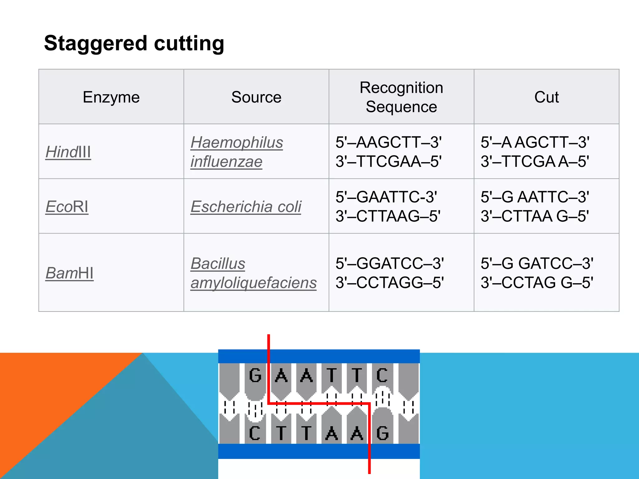
NUCLEASES &
RIBONUCLEASES
CONTENT
Contents
1 History
2 Numerical Classification System
3 Structure
4 Site recognition
 4.1Structure specific nuclease
 4.2Sequence specific nuclease
5 Endonucleases
 5.1Staggered cutting
6 Role in nature
 6.1DNA repair
7 Meganucleases
HISTORY
 In the late 1960s, scientists Stuart Linn and Werner
Arber isolated examples of the two types of enzymes responsible
for phage growth restriction in Escherichia coli (E. coli) bacteria.
One of these enzymes added a methyl group to the DNA,
generating methylated DNA, while the other cleaved
unmethylated DNA at a wide variety of locations along the length
of the molecule. The first type of enzyme was called a
"methylase" and the other a "restriction nuclease". These
enzymatic tools were important to scientists who were gathering
the tools needed to "cut and paste" DNA molecules.
 An important development came when H.O. Smith, K.W. Wilcox,
and T.J. Kelley, working at Johns Hopkins University in 1968,
isolated and characterized the first restriction nuclease whose
functioning depended on a specific DNA nucleotide sequence.
Working with Haemophilus influenzae bacteria, this group isolated
an enzyme, called HindII, that always cut DNA molecules at a
particular point within a specific sequence of six base pairs.
NUMERICAL CLASSIFICATION SYSTEM
 Most nucleases are classified by the Enzyme
Commission number of the "Nomenclature
Committee of the International Union of
Biochemistry and Molecular Biology"
as hydrolases .
 The nucleases belong just
like phosphodiesterase, lipase and phosphatase to
the esterases , a subgroup of the hydrolases.
STRUCTURE
Nuclease primary structure is large poorly
conserved and minimally conserved at
active sites, the surfaces of which primarily
comprise acidic and basic amino acid
residues. Nucleases can be classified into
folding families.
SITE RECOGNITION
A nuclease must associate with a nucleic acid
before it can cleave the molecule. That entails a
degree of recognition. Nucleases variously employ
both nonspecific and specific associations in their
modes of recognition and binding. Both modes play
important roles in living organisms, especially in
DNA repair
Nonspecific endonucleases involved in DNA repair can scan
DNA for target sequences or damage. Such a nuclease
diffuses along DNA until it encounters a target, upon which
the residues of its active site interact with the chemical groups
of the DNA.
A site-specific nuclease forms far stronger associations by
contrast. It draws DNA into the deep groove of its DNA-binding
domain. This results in significant deformation of the
DNA tertiary structure and is accomplished with a surfaces rich
in basic (positively charged) residues. It engages in extensive
electrostatic interaction with the DNA
SEQUENCE SPECIFIC NUCLEASE
There are more than 900 restriction enzymes, some
sequence specific and some not, have been isolated from
over 230 strains of bacteria since the initial discovery
of HindII. These restriction enzymes generally have names
that reflect their origin—The first letter of the name comes
from the genus and the second two letters come from the
species of the prokaryotic cell from which they were isolated.
For example, EcoRI comes from Escherichia coli RY13
bacteria, while HindII comes from Haemophilus
influenzae strain Rd. Numbers following the nuclease names
indicate the order in which the enzymes were isolated from
single strains of bacteria: EcoRI, EcoRII.
ENDONUCLEASES
A restriction endonuclease functions by "scanning" the
length of a DNA molecule. Once it encounters its particular
specific recognition sequence, it will bind to the DNA
molecule and makes one cut in each of the two sugar-
phosphate backbones. The positions of these two cuts,
both in relation to each other, and to the recognition
sequence itself, are determined by the identity of the
restriction endonuclease. Different endonucleases yield
different sets of cuts, but one endonuclease will always cut
a particular base sequence the same way, no matter what
DNA molecule it is acting on. Once the cuts have been
made, the DNA molecule will break into fragments.
Enzyme Source
Recognition
Sequence
Cut
HindIII
Haemophilus
influenzae
5'–AAGCTT–3'
3'–TTCGAA–5'
5'–A AGCTT–3'
3'–TTCGAA–5'
EcoRI Escherichia coli
5'–GAATTC-3'
3'–CTTAAG–5'
5'–G AATTC–3'
3'–CTTAA G–5'
BamHI
Bacillus
amyloliquefaciens
5'–GGATCC–3'
3'–CCTAGG–5'
5'–G GATCC–3'
3'–CCTAG G–5'
Staggered cutting
RIBONUCLEASE
Ribonuclease (commonly
abbreviated RNase) is a type
of nuclease that catalyzes the degradation
of RNA into smaller components.
Ribonucleases can be divided
into endoribonucleases and exoribonucleas
es, and comprise several sub-classes
 Phosphorolytic enzyme
 Hydrolytic enzyme
FUNCTION
 All organisms studied contain many RNases of
two different classes, showing that RNA
degradation is a very ancient and important
process.
 RNases play key roles in the maturation of all
RNA molecules, both messenger RNAs that carry
genetic material for making proteins, and non-
coding RNAs that function in varied cellular
processes.
 Similar to restriction enzymes, which cleave highly
specific sequences of double-stranded DNA, a
variety of endoribonucleases that recognize and
cleave specific sequences of single-stranded RNA
have been recently classified.
CLASSIFICATION
Major types of endoribonucleases
 Rnase A
 Rnase H
 Rnase III
Major types of exoribonucleases
 Exoribonuclease I
 Exoribonuclease II
 Oligoribonuclease
RNASE SPECIFICITY
The active site looks like a rift valley where all the
active site residues create the wall and bottom of
the valley. the rift is very thin and the small
substrate fits perfectly in the middle of the active
site, which allows for perfect interaction with the
residues. It actually has a little curvature to the site
which the substrate also has. Although, usually
most of exo- and endo ribonucleases are not
sequenced specific, recently CRISPR/Cas system
natively recognizing and cutting DNA was
engineered to cleave ssRNA in a sequence-specific
manner

Nucleases

  • 1.
  • 2.
    CONTENT Contents 1 History 2 NumericalClassification System 3 Structure 4 Site recognition  4.1Structure specific nuclease  4.2Sequence specific nuclease 5 Endonucleases  5.1Staggered cutting 6 Role in nature  6.1DNA repair 7 Meganucleases
  • 3.
    HISTORY  In thelate 1960s, scientists Stuart Linn and Werner Arber isolated examples of the two types of enzymes responsible for phage growth restriction in Escherichia coli (E. coli) bacteria. One of these enzymes added a methyl group to the DNA, generating methylated DNA, while the other cleaved unmethylated DNA at a wide variety of locations along the length of the molecule. The first type of enzyme was called a "methylase" and the other a "restriction nuclease". These enzymatic tools were important to scientists who were gathering the tools needed to "cut and paste" DNA molecules.  An important development came when H.O. Smith, K.W. Wilcox, and T.J. Kelley, working at Johns Hopkins University in 1968, isolated and characterized the first restriction nuclease whose functioning depended on a specific DNA nucleotide sequence. Working with Haemophilus influenzae bacteria, this group isolated an enzyme, called HindII, that always cut DNA molecules at a particular point within a specific sequence of six base pairs.
  • 4.
    NUMERICAL CLASSIFICATION SYSTEM Most nucleases are classified by the Enzyme Commission number of the "Nomenclature Committee of the International Union of Biochemistry and Molecular Biology" as hydrolases .  The nucleases belong just like phosphodiesterase, lipase and phosphatase to the esterases , a subgroup of the hydrolases.
  • 5.
    STRUCTURE Nuclease primary structureis large poorly conserved and minimally conserved at active sites, the surfaces of which primarily comprise acidic and basic amino acid residues. Nucleases can be classified into folding families.
  • 6.
    SITE RECOGNITION A nucleasemust associate with a nucleic acid before it can cleave the molecule. That entails a degree of recognition. Nucleases variously employ both nonspecific and specific associations in their modes of recognition and binding. Both modes play important roles in living organisms, especially in DNA repair
  • 7.
    Nonspecific endonucleases involvedin DNA repair can scan DNA for target sequences or damage. Such a nuclease diffuses along DNA until it encounters a target, upon which the residues of its active site interact with the chemical groups of the DNA. A site-specific nuclease forms far stronger associations by contrast. It draws DNA into the deep groove of its DNA-binding domain. This results in significant deformation of the DNA tertiary structure and is accomplished with a surfaces rich in basic (positively charged) residues. It engages in extensive electrostatic interaction with the DNA
  • 8.
    SEQUENCE SPECIFIC NUCLEASE Thereare more than 900 restriction enzymes, some sequence specific and some not, have been isolated from over 230 strains of bacteria since the initial discovery of HindII. These restriction enzymes generally have names that reflect their origin—The first letter of the name comes from the genus and the second two letters come from the species of the prokaryotic cell from which they were isolated. For example, EcoRI comes from Escherichia coli RY13 bacteria, while HindII comes from Haemophilus influenzae strain Rd. Numbers following the nuclease names indicate the order in which the enzymes were isolated from single strains of bacteria: EcoRI, EcoRII.
  • 9.
    ENDONUCLEASES A restriction endonucleasefunctions by "scanning" the length of a DNA molecule. Once it encounters its particular specific recognition sequence, it will bind to the DNA molecule and makes one cut in each of the two sugar- phosphate backbones. The positions of these two cuts, both in relation to each other, and to the recognition sequence itself, are determined by the identity of the restriction endonuclease. Different endonucleases yield different sets of cuts, but one endonuclease will always cut a particular base sequence the same way, no matter what DNA molecule it is acting on. Once the cuts have been made, the DNA molecule will break into fragments.
  • 10.
    Enzyme Source Recognition Sequence Cut HindIII Haemophilus influenzae 5'–AAGCTT–3' 3'–TTCGAA–5' 5'–A AGCTT–3' 3'–TTCGAA–5' EcoRIEscherichia coli 5'–GAATTC-3' 3'–CTTAAG–5' 5'–G AATTC–3' 3'–CTTAA G–5' BamHI Bacillus amyloliquefaciens 5'–GGATCC–3' 3'–CCTAGG–5' 5'–G GATCC–3' 3'–CCTAG G–5' Staggered cutting
  • 11.
    RIBONUCLEASE Ribonuclease (commonly abbreviated RNase)is a type of nuclease that catalyzes the degradation of RNA into smaller components. Ribonucleases can be divided into endoribonucleases and exoribonucleas es, and comprise several sub-classes  Phosphorolytic enzyme  Hydrolytic enzyme
  • 12.
    FUNCTION  All organismsstudied contain many RNases of two different classes, showing that RNA degradation is a very ancient and important process.  RNases play key roles in the maturation of all RNA molecules, both messenger RNAs that carry genetic material for making proteins, and non- coding RNAs that function in varied cellular processes.  Similar to restriction enzymes, which cleave highly specific sequences of double-stranded DNA, a variety of endoribonucleases that recognize and cleave specific sequences of single-stranded RNA have been recently classified.
  • 13.
    CLASSIFICATION Major types ofendoribonucleases  Rnase A  Rnase H  Rnase III Major types of exoribonucleases  Exoribonuclease I  Exoribonuclease II  Oligoribonuclease
  • 14.
    RNASE SPECIFICITY The activesite looks like a rift valley where all the active site residues create the wall and bottom of the valley. the rift is very thin and the small substrate fits perfectly in the middle of the active site, which allows for perfect interaction with the residues. It actually has a little curvature to the site which the substrate also has. Although, usually most of exo- and endo ribonucleases are not sequenced specific, recently CRISPR/Cas system natively recognizing and cutting DNA was engineered to cleave ssRNA in a sequence-specific manner