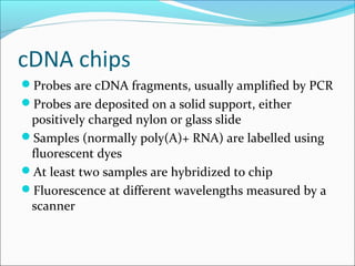
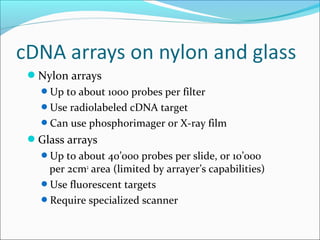
Technologies and utility
By: Harshit Jain
IBS Business School
Gurgaon.
Historical perspective
DNA hybridization (1960s)
Detection of hybrids
hydroxyapatite
radioactive labelling
enzyme-linked detection
fluorescent labelling
Fixing sample on solid support
Southern blots (1970s)
Northern blots
Dot blots
Basic principles
Main novelty is one of scale
hundreds or thousands of probes rather than tens
Probes are attached to solid supports
Robotics are used extensively
Informatics is a central component at all stages
Major technologies
cDNA probes (> 200 nt), usually produced by PCR,
attached to either nylon or glass supports
Oligonucleotides (25-80 nt) attached to glass support
Oligonucleotides (25-30 nt) synthesized in situ on
silica wafers (Affymetrix)
Probes attached to tagged beads
Principal uses of chips
Genome-scale gene expression analysis
Differentiation
Responses to environmental factors
Disease processes
Effects of drugs
Detection of sequence variation
Genetic typing
Detection of somatic mutations (e.g. in oncogenes)
Direct sequencing
cDNA chips
Probes are cDNA fragments, usually amplified by PCR
Probes are deposited on a solid support, either
positively charged nylon or glass slide
Samples (normally poly(A)+ RNA) are labelled using
fluorescent dyes
At least two samples are hybridized to chip
Fluorescence at different wavelengths measured by a
scanner
Probe selection
Non-redundant set of probes
Includes genes of interest to project
Corresponds to physically available clones
Chip layout
Grouping of probes by function
Correspondance between wells in microtitre
plates and spots on the chip
Nylon arrays
Up to about 1000 probes per filter
Use radiolabeled cDNA target
Can use phosphorimager or X-ray film
Glass arrays
Up to about 40’000 probes per slide, or 10’000
per 2cm2
area (limited by arrayer’s capabilities)
Use fluorescent targets
Require specialized scanner
Volume per spot: 250 nl
Spot size: 1100 µm
Spot density: 70/cm2
Volume per spot: 0.5 nl
Spot size: 115
µm
Spot density:
4800/cm2Labelled BSA
7x11 microarray consisting of
identical Cy5-BSA spots (pitch 500
µm)
Typical CV: ≤ 5%
0
0.5
1.0
1.5
rel.fluor.int.(a.u.)
Targets are normally prepared by oligo(dT) primed
cDNA synthesis
Probes should contain 3’ end of mRNA
Need CoT1 DNA as competitor
Specific activity will limit sensitivity of assay
Alternative protocol is to make ds cDNA containing
bacterial promoter, then cRNA
Can work with smaller amount of RNA
Less quantitative
Hybridization usually under coverslips
Laser scanners
Excellent spatial resolution
Good sensitivity, but can bleach fluorochromes
Still rather slow
CCD scanners
Spatial resolution can be a problem
Sensitivity easily adjustable (exposure time)
Faster and cheaper than lasers
In all cases, raw data are images showing
fluorescence on surface of chip
free label
Imaging
of surface-confined fluorescence
excitation of bound label
CCD camera
microarray
on chip
Probes are oligos synthesized in situ using a
photolithographic approach
There are at least 5 oligos per cDNA, plus an
equal number of negative controls
The apparatus requires a fluidics station for
hybridization and a special scanner
Only a single fluorochrome is used per
hybridization
It is very expensive !
Clontech, Incyte, Research Genetics - filter-
based arrays with up to about 8000 clones
Incyte / Synteni - 10’000 probe chips, not
distributed (have to send them target RNA)
Affymetrix - oligo-based chips with 12’000
genes of known function (16 oligos/gene) and
4x10’000 genes from ESTs
Synthesis of probes on microbeads
Hybridization in solution
Identification of beads by fluorescent bar
coding by embedding transponders
Readout using micro-flow cells or optic fiber
arrays
Production of “universal” arrays
Array uses a unique combination of oligos, and
probes containing the proper complements
Mutation detection
Oligos (Affymetrix type) representing all known
alleles
PCR followed by primer extension, with
detection of alleles by MALDI-TOF mass
spectroscopy (Sequenom)
Gene loss and amplification
Measure gene dosage in genomic DNA by
hybridization to genomic probes
Bioinformatics of microarrays
Array design: choice of sequences to be used as probes
Analysis of scanned images
Spot detection, normalization, quantitation
Primary analysis of hybridization data
Basic statistics, reproducibility, data scattering, etc.
Comparison of multiple samples
Clustering, SOMs, classification …
Sample tracking and databasing of results
DNA CHIPS

DNA CHIPS

  • 1.
    Technologies and utility By:Harshit Jain IBS Business School Gurgaon.
  • 2.
    Historical perspective DNA hybridization(1960s) Detection of hybrids hydroxyapatite radioactive labelling enzyme-linked detection fluorescent labelling Fixing sample on solid support Southern blots (1970s) Northern blots Dot blots
  • 3.
    Basic principles Main noveltyis one of scale hundreds or thousands of probes rather than tens Probes are attached to solid supports Robotics are used extensively Informatics is a central component at all stages
  • 4.
    Major technologies cDNA probes(> 200 nt), usually produced by PCR, attached to either nylon or glass supports Oligonucleotides (25-80 nt) attached to glass support Oligonucleotides (25-30 nt) synthesized in situ on silica wafers (Affymetrix) Probes attached to tagged beads
  • 5.
    Principal uses ofchips Genome-scale gene expression analysis Differentiation Responses to environmental factors Disease processes Effects of drugs Detection of sequence variation Genetic typing Detection of somatic mutations (e.g. in oncogenes) Direct sequencing
  • 6.
    cDNA chips Probes arecDNA fragments, usually amplified by PCR Probes are deposited on a solid support, either positively charged nylon or glass slide Samples (normally poly(A)+ RNA) are labelled using fluorescent dyes At least two samples are hybridized to chip Fluorescence at different wavelengths measured by a scanner
  • 7.
    Probe selection Non-redundant setof probes Includes genes of interest to project Corresponds to physically available clones Chip layout Grouping of probes by function Correspondance between wells in microtitre plates and spots on the chip
  • 8.
    Nylon arrays Up toabout 1000 probes per filter Use radiolabeled cDNA target Can use phosphorimager or X-ray film Glass arrays Up to about 40’000 probes per slide, or 10’000 per 2cm2 area (limited by arrayer’s capabilities) Use fluorescent targets Require specialized scanner
  • 9.
    Volume per spot:250 nl Spot size: 1100 µm Spot density: 70/cm2 Volume per spot: 0.5 nl Spot size: 115 µm Spot density: 4800/cm2Labelled BSA
  • 10.
    7x11 microarray consistingof identical Cy5-BSA spots (pitch 500 µm) Typical CV: ≤ 5% 0 0.5 1.0 1.5 rel.fluor.int.(a.u.)
  • 11.
    Targets are normallyprepared by oligo(dT) primed cDNA synthesis Probes should contain 3’ end of mRNA Need CoT1 DNA as competitor Specific activity will limit sensitivity of assay Alternative protocol is to make ds cDNA containing bacterial promoter, then cRNA Can work with smaller amount of RNA Less quantitative Hybridization usually under coverslips
  • 12.
    Laser scanners Excellent spatialresolution Good sensitivity, but can bleach fluorochromes Still rather slow CCD scanners Spatial resolution can be a problem Sensitivity easily adjustable (exposure time) Faster and cheaper than lasers In all cases, raw data are images showing fluorescence on surface of chip
  • 13.
    free label Imaging of surface-confinedfluorescence excitation of bound label CCD camera microarray on chip
  • 14.
    Probes are oligossynthesized in situ using a photolithographic approach There are at least 5 oligos per cDNA, plus an equal number of negative controls The apparatus requires a fluidics station for hybridization and a special scanner Only a single fluorochrome is used per hybridization It is very expensive !
  • 15.
    Clontech, Incyte, ResearchGenetics - filter- based arrays with up to about 8000 clones Incyte / Synteni - 10’000 probe chips, not distributed (have to send them target RNA) Affymetrix - oligo-based chips with 12’000 genes of known function (16 oligos/gene) and 4x10’000 genes from ESTs
  • 16.
    Synthesis of probeson microbeads Hybridization in solution Identification of beads by fluorescent bar coding by embedding transponders Readout using micro-flow cells or optic fiber arrays Production of “universal” arrays Array uses a unique combination of oligos, and probes containing the proper complements
  • 17.
    Mutation detection Oligos (Affymetrixtype) representing all known alleles PCR followed by primer extension, with detection of alleles by MALDI-TOF mass spectroscopy (Sequenom) Gene loss and amplification Measure gene dosage in genomic DNA by hybridization to genomic probes
  • 18.
    Bioinformatics of microarrays Arraydesign: choice of sequences to be used as probes Analysis of scanned images Spot detection, normalization, quantitation Primary analysis of hybridization data Basic statistics, reproducibility, data scattering, etc. Comparison of multiple samples Clustering, SOMs, classification … Sample tracking and databasing of results