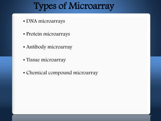
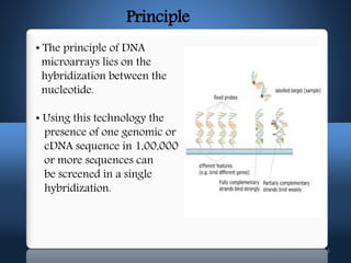
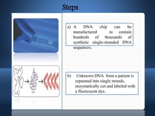
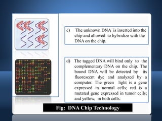
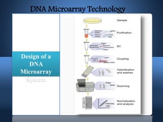
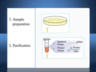
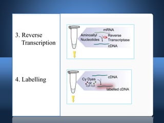
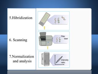
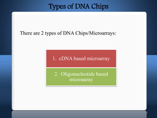
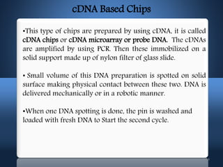
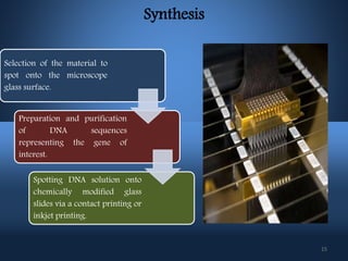
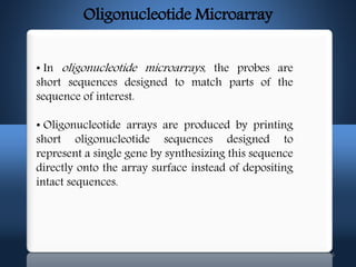
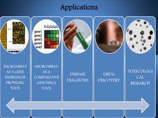
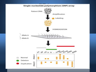
DNA microarrays, also known as DNA chips, allow researchers to analyze large numbers of DNA sequences simultaneously. They contain thousands of unique DNA probes immobilized on a solid surface in an organized grid pattern. Unknown DNA samples are labeled with fluorescent dyes and hybridized to the probes on the chip. The bound DNA is then detected using lasers and analyzed by a computer to determine which genes are present or expressed in the sample. There are two main types of DNA chips - cDNA-based chips containing amplified cDNA probes, and oligonucleotide-based chips containing short, synthesized DNA sequences. DNA microarrays have many applications including gene expression profiling, comparative genomics, disease diagnosis, and drug discovery.
Microarray -types, DNA chip, Principle and application of microarray, Preparation of DNA Chip, Affymetrix chip, microarray in genomics and proteomics, advantages and limitations of microarray
Next Generation Sequencing (NGS) Is A Modern And Cost Effective Sequencing Technology Which Enables Scientists To Sequence Nucleic Acids At Much Faster Rate. In This Presentation, You Will Learn About What is NGS, Idea Behind NGS, Methodology And Protocol, Widely Adapted NGS Protocols, Applications And References For Further Study.
STS stands for sequence tagged site which is short DNA sequence, generally between 100 and 500 bp in length, that is easily recognizable and occurs only once in the chromosome or genome being studied.
Comparative genomic hybridization is a molecular cytogenetic method for analysing copy number variations (CNVs) relative to ploidy level in the DNA of a test sample compared to a reference sample, without the need for culturing cells
whole genome analysis
history
needs
steps involved
human genome data
NGS
pyrosequencing
illumina
SOLiD
Ion torrent
PacBio
applications
problems
benefits
DNA microarray:
A DNA microarray (also commonly known as gene or genome chip, DNA chip, or gene array) is a collection of microscopic DNA spots, commonly representing single genes, arrayed on a solid surface by covalent attachment to a chemical matrix. DNA arrays are different from other types of microarray only in that they either measure DNA or use DNA as part of its detection system. Qualitative or quantitative measurements with DNA microarrays utilize the selective nature of DNA-DNA or DNA-RNA hybridization under high-stringency conditions and fluorophore-based detection. DNA arrays are commonly used for expression profiling, i.e., monitoring expression levels of thousands of genes simultaneously.
complete Single Nucleotide Polymorphiitsm Detection methods with Advance techniques with its applications
Single nucleotide polymorphisms are single base variations between genomes within a species.
There are at least 10 million polymorphic sites in the human genome.
SNPs can distinguish individuals from one another
Denaturing Gradient Gel Electrophoresis
Chemical Cleavage Of Mismatch
Single-stranded Conformation Polymorphism (SSCP)
MutS Protein-binding Assays
Mismatch Repair Detection (MRD)
Heteroduplex Analysis (HA)
Denaturing High Performance Liquid Chromatography (DHPLC)
UNG-Mediated T-Sequencing
RNA-Mediated Finger printing with MALDI MS Detection
Sequencing by Hybridization
Direct DNA Sequencing
Single-feature polymorphism (SFP)
Invader probe
Allele-specific oligonucleotide probes
PCR-based methods
Allele specific primers
Sequence Polymorphism-Derived (SPD) markers
Targeting induced local lesions in genomes (TILLinG)
Minisequencing primers
Allele-specific ligation probes
The DNA microarray is a tool used to determine whether the DNA from a particular individual contains a mutation in genes like BRCA1 and BRCA2. The chip consists of a small glass plate encased in plastic. Some companies manufacture microarrays using methods similar to those used to make computer microchips.
A DNA microarray is a collection of microscopic DNA spots attached to a solid surface. Scientists use DNA microarrays to measure the expression levels of large numbers of genes simultaneously or to genotype multiple regions of a genome. Each DNA spot contains picomoles of a specific DNA sequence, known as probes.
This chapter provides an overview of DNA microarrays. Microarrays are a technology in which 1000’s of nucleic acids are bound to a surface and are used to measure the relative concentration of nucleic acid sequences in a mixture via hybridization and subsequent detection of the hybridization events. We first cover the history of microarrays and the antecedent technologies that led to their development. We then discuss the methods of manufacture of microarrays and the most common biological applications. The chapter ends with a brief discussion of the limitations of microarrays and discusses how microarrays are being rapidly replaced by DNA sequencing technologies.
DNA microarrays
– DNA molecules printed at high density used to determine
the level of RNA or DNA in a sample.
– Can be thought of a “reverse Northern blots”
Earliest Galaxies in the JADES Origins Field: Luminosity Function and Cosmic ...Sérgio Sacani
We characterize the earliest galaxy population in the JADES Origins Field (JOF), the deepest
imaging field observed with JWST. We make use of the ancillary Hubble optical images (5 filters
spanning 0.4−0.9µm) and novel JWST images with 14 filters spanning 0.8−5µm, including 7 mediumband filters, and reaching total exposure times of up to 46 hours per filter. We combine all our data
at > 2.3µm to construct an ultradeep image, reaching as deep as ≈ 31.4 AB mag in the stack and
30.3-31.0 AB mag (5σ, r = 0.1” circular aperture) in individual filters. We measure photometric
redshifts and use robust selection criteria to identify a sample of eight galaxy candidates at redshifts
z = 11.5 − 15. These objects show compact half-light radii of R1/2 ∼ 50 − 200pc, stellar masses of
M⋆ ∼ 107−108M⊙, and star-formation rates of SFR ∼ 0.1−1 M⊙ yr−1
. Our search finds no candidates
at 15 < z < 20, placing upper limits at these redshifts. We develop a forward modeling approach to
infer the properties of the evolving luminosity function without binning in redshift or luminosity that
marginalizes over the photometric redshift uncertainty of our candidate galaxies and incorporates the
impact of non-detections. We find a z = 12 luminosity function in good agreement with prior results,
and that the luminosity function normalization and UV luminosity density decline by a factor of ∼ 2.5
from z = 12 to z = 14. We discuss the possible implications of our results in the context of theoretical
models for evolution of the dark matter halo mass function.
Cancer cell metabolism: special Reference to Lactate PathwayAADYARAJPANDEY1
Normal Cell Metabolism:
Cellular respiration describes the series of steps that cells use to break down sugar and other chemicals to get the energy we need to function.
Energy is stored in the bonds of glucose and when glucose is broken down, much of that energy is released.
Cell utilize energy in the form of ATP.
The first step of respiration is called glycolysis. In a series of steps, glycolysis breaks glucose into two smaller molecules - a chemical called pyruvate. A small amount of ATP is formed during this process.
Most healthy cells continue the breakdown in a second process, called the Kreb's cycle. The Kreb's cycle allows cells to “burn” the pyruvates made in glycolysis to get more ATP.
The last step in the breakdown of glucose is called oxidative phosphorylation (Ox-Phos).
It takes place in specialized cell structures called mitochondria. This process produces a large amount of ATP. Importantly, cells need oxygen to complete oxidative phosphorylation.
If a cell completes only glycolysis, only 2 molecules of ATP are made per glucose. However, if the cell completes the entire respiration process (glycolysis - Kreb's - oxidative phosphorylation), about 36 molecules of ATP are created, giving it much more energy to use.
IN CANCER CELL:
Unlike healthy cells that "burn" the entire molecule of sugar to capture a large amount of energy as ATP, cancer cells are wasteful.
Cancer cells only partially break down sugar molecules. They overuse the first step of respiration, glycolysis. They frequently do not complete the second step, oxidative phosphorylation.
This results in only 2 molecules of ATP per each glucose molecule instead of the 36 or so ATPs healthy cells gain. As a result, cancer cells need to use a lot more sugar molecules to get enough energy to survive.
introduction to WARBERG PHENOMENA:
WARBURG EFFECT Usually, cancer cells are highly glycolytic (glucose addiction) and take up more glucose than do normal cells from outside.
Otto Heinrich Warburg (; 8 October 1883 – 1 August 1970) In 1931 was awarded the Nobel Prize in Physiology for his "discovery of the nature and mode of action of the respiratory enzyme.
WARNBURG EFFECT : cancer cells under aerobic (well-oxygenated) conditions to metabolize glucose to lactate (aerobic glycolysis) is known as the Warburg effect. Warburg made the observation that tumor slices consume glucose and secrete lactate at a higher rate than normal tissues.
Observation of Io’s Resurfacing via Plume Deposition Using Ground-based Adapt...Sérgio Sacani
Since volcanic activity was first discovered on Io from Voyager images in 1979, changes
on Io’s surface have been monitored from both spacecraft and ground-based telescopes.
Here, we present the highest spatial resolution images of Io ever obtained from a groundbased telescope. These images, acquired by the SHARK-VIS instrument on the Large
Binocular Telescope, show evidence of a major resurfacing event on Io’s trailing hemisphere. When compared to the most recent spacecraft images, the SHARK-VIS images
show that a plume deposit from a powerful eruption at Pillan Patera has covered part
of the long-lived Pele plume deposit. Although this type of resurfacing event may be common on Io, few have been detected due to the rarity of spacecraft visits and the previously low spatial resolution available from Earth-based telescopes. The SHARK-VIS instrument ushers in a new era of high resolution imaging of Io’s surface using adaptive
optics at visible wavelengths.
Introduction:
RNA interference (RNAi) or Post-Transcriptional Gene Silencing (PTGS) is an important biological process for modulating eukaryotic gene expression.
It is highly conserved process of posttranscriptional gene silencing by which double stranded RNA (dsRNA) causes sequence-specific degradation of mRNA sequences.
dsRNA-induced gene silencing (RNAi) is reported in a wide range of eukaryotes ranging from worms, insects, mammals and plants.
This process mediates resistance to both endogenous parasitic and exogenous pathogenic nucleic acids, and regulates the expression of protein-coding genes.
What are small ncRNAs?
micro RNA (miRNA)
short interfering RNA (siRNA)
Properties of small non-coding RNA:
Involved in silencing mRNA transcripts.
Called “small” because they are usually only about 21-24 nucleotides long.
Synthesized by first cutting up longer precursor sequences (like the 61nt one that Lee discovered).
Silence an mRNA by base pairing with some sequence on the mRNA.
Discovery of siRNA?
The first small RNA:
In 1993 Rosalind Lee (Victor Ambros lab) was studying a non- coding gene in C. elegans, lin-4, that was involved in silencing of another gene, lin-14, at the appropriate time in the
development of the worm C. elegans.
Two small transcripts of lin-4 (22nt and 61nt) were found to be complementary to a sequence in the 3' UTR of lin-14.
Because lin-4 encoded no protein, she deduced that it must be these transcripts that are causing the silencing by RNA-RNA interactions.
Types of RNAi ( non coding RNA)
MiRNA
Length (23-25 nt)
Trans acting
Binds with target MRNA in mismatch
Translation inhibition
Si RNA
Length 21 nt.
Cis acting
Bind with target Mrna in perfect complementary sequence
Piwi-RNA
Length ; 25 to 36 nt.
Expressed in Germ Cells
Regulates trnasposomes activity
MECHANISM OF RNAI:
First the double-stranded RNA teams up with a protein complex named Dicer, which cuts the long RNA into short pieces.
Then another protein complex called RISC (RNA-induced silencing complex) discards one of the two RNA strands.
The RISC-docked, single-stranded RNA then pairs with the homologous mRNA and destroys it.
THE RISC COMPLEX:
RISC is large(>500kD) RNA multi- protein Binding complex which triggers MRNA degradation in response to MRNA
Unwinding of double stranded Si RNA by ATP independent Helicase
Active component of RISC is Ago proteins( ENDONUCLEASE) which cleave target MRNA.
DICER: endonuclease (RNase Family III)
Argonaute: Central Component of the RNA-Induced Silencing Complex (RISC)
One strand of the dsRNA produced by Dicer is retained in the RISC complex in association with Argonaute
ARGONAUTE PROTEIN :
1.PAZ(PIWI/Argonaute/ Zwille)- Recognition of target MRNA
2.PIWI (p-element induced wimpy Testis)- breaks Phosphodiester bond of mRNA.)RNAse H activity.
MiRNA:
The Double-stranded RNAs are naturally produced in eukaryotic cells during development, and they have a key role in regulating gene expression .
Richard's entangled aventures in wonderlandRichard Gill
Since the loophole-free Bell experiments of 2020 and the Nobel prizes in physics of 2022, critics of Bell's work have retreated to the fortress of super-determinism. Now, super-determinism is a derogatory word - it just means "determinism". Palmer, Hance and Hossenfelder argue that quantum mechanics and determinism are not incompatible, using a sophisticated mathematical construction based on a subtle thinning of allowed states and measurements in quantum mechanics, such that what is left appears to make Bell's argument fail, without altering the empirical predictions of quantum mechanics. I think however that it is a smoke screen, and the slogan "lost in math" comes to my mind. I will discuss some other recent disproofs of Bell's theorem using the language of causality based on causal graphs. Causal thinking is also central to law and justice. I will mention surprising connections to my work on serial killer nurse cases, in particular the Dutch case of Lucia de Berk and the current UK case of Lucy Letby.
The increased availability of biomedical data, particularly in the public domain, offers the opportunity to better understand human health and to develop effective therapeutics for a wide range of unmet medical needs. However, data scientists remain stymied by the fact that data remain hard to find and to productively reuse because data and their metadata i) are wholly inaccessible, ii) are in non-standard or incompatible representations, iii) do not conform to community standards, and iv) have unclear or highly restricted terms and conditions that preclude legitimate reuse. These limitations require a rethink on data can be made machine and AI-ready - the key motivation behind the FAIR Guiding Principles. Concurrently, while recent efforts have explored the use of deep learning to fuse disparate data into predictive models for a wide range of biomedical applications, these models often fail even when the correct answer is already known, and fail to explain individual predictions in terms that data scientists can appreciate. These limitations suggest that new methods to produce practical artificial intelligence are still needed.
In this talk, I will discuss our work in (1) building an integrative knowledge infrastructure to prepare FAIR and "AI-ready" data and services along with (2) neurosymbolic AI methods to improve the quality of predictions and to generate plausible explanations. Attention is given to standards, platforms, and methods to wrangle knowledge into simple, but effective semantic and latent representations, and to make these available into standards-compliant and discoverable interfaces that can be used in model building, validation, and explanation. Our work, and those of others in the field, creates a baseline for building trustworthy and easy to deploy AI models in biomedicine.
Bio
Dr. Michel Dumontier is the Distinguished Professor of Data Science at Maastricht University, founder and executive director of the Institute of Data Science, and co-founder of the FAIR (Findable, Accessible, Interoperable and Reusable) data principles. His research explores socio-technological approaches for responsible discovery science, which includes collaborative multi-modal knowledge graphs, privacy-preserving distributed data mining, and AI methods for drug discovery and personalized medicine. His work is supported through the Dutch National Research Agenda, the Netherlands Organisation for Scientific Research, Horizon Europe, the European Open Science Cloud, the US National Institutes of Health, and a Marie-Curie Innovative Training Network. He is the editor-in-chief for the journal Data Science and is internationally recognized for his contributions in bioinformatics, biomedical informatics, and semantic technologies including ontologies and linked data.
2. 2
• Introduction
•Types of Microarray
•DNA Microarray
• Principle
Steps involved in DNA microarray
• Types of DNA Chips
cDNA - based chips
Oligonucleotide - based chips
• Applications
• Advantages and disadvantages
• SNP Array
CONTENTS
3. 3
Introduction
An array is an orderly arrangement of samples where
matching of known and unknown DNA samples is done
based on base pairing rules.
A microarray is a pattern of ssDNA probes which are
immobilized on a surface called a chip or a slide.
Microarray technology evolved from Southern blotting
The concept of microarrays was first proposed in the
late 1980s by Augenlicht and his colleagues.
4. 4
Types of Microarray
• DNA microarrays
• Protein microarrays
• Antibody microarray
• Tissue microarray
• Chemical compound microarray
5. 5
DNA Microarray
• DNA microarrays are solid supports, usually of glass or
silicon, upon which DNA is attached in an organized grid
fashion.
• Each spot of DNA, called a probe, represents a single
gene.
• There are several synonyms of DNA microarrays such as
DNA chips, gene chips, DNA arrays, gene arrays and
biochips.
6. 6
Principle
• The principle of DNA
microarrays lies on the
hybridization between the
nucleotide.
• Using this technology the
presence of one genomic or
cDNA sequence in 1,00,000
or more sequences can
be screened in a single
hybridization.
7. 7
a) A DNA chip can be
manufactured to contain
hundreds of thousands of
synthetic single-stranded DNA
sequences.
b) Unknown DNA from a patient is
separated into single strands,
enzymatically cut and labeled with
a fluorescent dye.
Steps
8. 8
c) The unknown DNA is inserted into the
chip and allowed to hybridize with the
DNA on the chip.
d) The tagged DNA will bind only to the
complementary DNA on the chip. The
bound DNA will be detected by its
fluorescent dye and analyzed by a
computer. The green light is a gene
expressed in normal cells; red is a
mutated gene expressed in tumor cells;
and yellow, in both cells.
Fig: DNA Chip Technology
13. 13
There are 2 types of DNA Chips/Microarrays:
Types of DNA Chips
1. cDNA based microarray
2. Oligonucleotide based
microaaray
14. 14
•This type of chips are prepared by using cDNA, it is called
cDNA chips or cDNA microarray or probe DNA. The cDNAs
are amplified by using PCR. Then these immobilized on a
solid support made up of nylon filter of glass slide.
• Small volume of this DNA preparation is spotted on solid
surface making physical contact between these two. DNA is
delivered mechanically or in a robotic manner.
•When one DNA spotting is done, the pin is washed and
loaded with fresh DNA to Start the second cycle.
cDNA Based Chips
15. 15
Synthesis
Selection of the material to
spot onto the microscope
glass surface.
Preparation and purification
of DNA sequences
representing the gene of
interest.
Spotting DNA solution onto
chemically modified glass
slides via a contact printing or
inkjet printing.
16. 16
Oligonucleotide Microarray
• In oligonucleotide microarrays, the probes are
short sequences designed to match parts of the
sequence of interest.
• Oligonucleotide arrays are produced by printing
short oligonucleotide sequences designed to
represent a single gene by synthesizing this sequence
directly onto the array surface instead of depositing
intact sequences.
17. 17
Synthesis
•Light is directed through a photolithographic mask to
specific areas of array surface.
•Activation of areas for chemical coupling. Attachment
of A nucleotide containing
photolabile protecting
group X (MeNPOC).
•Addition of 2nd
building block T containing
a photolabile protecting
group X. This process is
repeated until the desired
product is obtained.
19. 19
• Provides data for thousands of genes.
• One experiment instead of many.
• Fast and easy to obtain results.
• Huge step closer to discovering cures for diseases
and cancer.
• Different parts of DNA can be used to study gene
expression.
Advantages
20. 20
Disadvantages
• The biggest disadvantage of DNA chips is that
they are expensive to create.
• The production of too many results at a time
requires long time for analysis, which is quite
complex in nature.
• The DNA chips do not have very long shelf life,
which proves to be another major disadvantage of
the technology.
21. 21
• A SNP is defined as a single base change in a DNA
sequence that occurs in a significant proportion (more
than 1 percent) of a large population
• SNPs are found in coding and (mostly) noncoding regions.
• Occur with a very high
frequency about 1 in 1000
bases to 1 in 100 to 300 bases.
• SNPs in coding regions
may alter the protein
structure made by that
coding region.
SNPs (Single Nucleotide Polymorphisms)
22. 22
SNP Array
• SNP array is a type of DNA Microarray which is
used to detect polymorphism within a population
• The basic principles of SNP array are the same as
the DNA microarray
• The three mandatory components of the SNP
arrays are:
• An array containing immobilized allele-
specific oligonucleotide (ASO) probes.
•Fragmented nucleic acid sequences of target,
labeled with fluorescent dyes.
•A detection system that records and interprets
the hybridization signal.