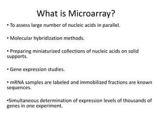
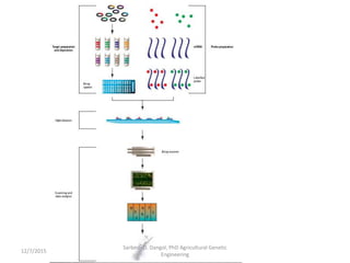
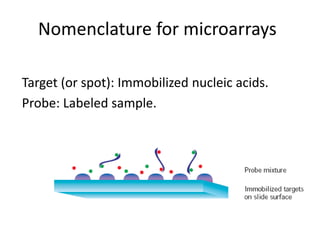
Introduction to Microarray
By: Sarbesh D. Dangol
December 7, 2015.
What is gene expression?
 The process by which a gene's information is
converted into RNA and eventually proteins.
 Various factors control at various points in the gene
sequence leading to protein synthesis.
Sarbesh D. Dangol, PhD Agricultural Genetic
Engineering
12/7/2015
Sarbesh D. Dangol, PhD Agricultural Genetic
Engineering
12/7/2015
Sarbesh D. Dangol, PhD Agricultural Genetic
Engineering
12/7/2015
General structure of a gene
Sarbesh D. Dangol, PhD Agricultural Genetic
Engineering
12/7/2015
What is Microarray?
• To assess large number of nucleic acids in parallel.
• Molecular hybridization methods.
• Preparing miniaturized collections of nucleic acids on solid
supports.
• Gene expression studies.
• mRNA samples are labeled and immobilized fractions are known
sequences.
•Simultaneous determination of expression levels of thousands of
genes in one experiment.
Sarbesh D. Dangol, PhD Agricultural Genetic
Engineering
12/7/2015
Nomenclature for microarrays
Target (or spot): Immobilized nucleic acids.
Probe: Labeled sample.
General Overview of Microarray
Sarbesh D. Dangol, PhD Agricultural Genetic
Engineering
12/7/2015
Introduction to Microarrays
• Collection of nucleic acid sequences immobilized onto
a solid support called a ‘spot’ or ‘target’.
• Varied sizes of spots different systems, but usually less
than 200 µm in diameter.
• A glass slide or glass wafer acts as the solid support.
• Tens of thousands of spots can be arrayed in a total
area of a few square centimeters.
Sarbesh D. Dangol, PhD Agricultural Genetic
Engineering
12/7/2015
• mRNA or DNA converted to a labeled
population of nucleic acids, the probe.
• The probe consists of several thousands of
different labeled nucleic acid fragments.
• 10,000 different labeled fragments
interrogating up to 100,000 different
immobilized sequences.
Sarbesh D. Dangol, PhD Agricultural Genetic
Engineering
12/7/2015
• Fluorescent dyes, cyanine dyes Cy3 (green)
and Cy5 (Red) are predominantly used.
• Detection of two or more different signals in
one experiment.
• Comparative analysis of two or more samples
on one microarray.
• Increased accuracy and throughput of
microarray.
Sarbesh D. Dangol, PhD Agricultural Genetic
Engineering
12/7/2015
• The number of duplexes formed reflects the
relative number of each specific fragment in
the probe, as long as the amount of
immobilized target nucleic acid is in excess
and not limiting the kinetics of hybridization.
Sarbesh D. Dangol, PhD Agricultural Genetic
Engineering
12/7/2015
• Two or more samples labeled with different
fluorescent dyes can be hybridized
simultaneously.
• Simultaneous hybridization taking place at
each target spot.
• By measuring different fluorescent signals
associated with each spot, relative abundance
of specific sequences in each of the samples
can be determined.
Sarbesh D. Dangol, PhD Agricultural Genetic
Engineering
12/7/2015
Manufacturing of microarray slides
• Microarray manufacture requires three
distinct components:
■ Production method
■ Microarray slide
■ Target genetic content
Sarbesh D. Dangol, PhD Agricultural Genetic
Engineering
12/7/2015
Production:
A) Oligonucleotide synthesis
• Targets synthesized directly onto slide, or purified
targets deposited onto a solid surface capable of
binding nucleic acids.
• Photolithographic masking method: attach
chemically modified linker groups, which contain
photochemically removable protective groups,
onto the glass surface.
• By masking predefined positions, it is possible to
synthesize different oligonucleotides at different
locations.
Sarbesh D. Dangol, PhD Agricultural Genetic
Engineering
12/7/2015
Microarray manufacturing by
photolithography
High Density: An area of 1.6 cm2 can contain up to 400,000 features.
Sarbesh D. Dangol, PhD Agricultural Genetic
Engineering
12/7/2015
Production:
B) Deposition
• Purified nucleic acids are attached to a modified glass
slide.
• Typically, small volumes of nucleic acid solution—
nanoliters or picoliters—are transferred onto the glass
slide.
• For preparing microarrays containing oligonucleotides,
cDNA sequences, as well as genomic DNA.
• Covalent chemical reaction between molecular groups
on the glass surface and the oligonucleotides:
-eg: amine-modified oligonucleotides to aldehyde
slides
Sarbesh D. Dangol, PhD Agricultural Genetic
Engineering
12/7/2015
Non-contact deposition
Sarbesh D. Dangol, PhD Agricultural Genetic
Engineering
12/7/2015
Contact deposition
• Solid, hollow, or split-open pen designs are used to
transfer target nucleic acid onto the slide surface.
• These pens are dipped into the target solution, a small
volume of which adheres to the pen.
• Pen comes into contact with the slide surface, a
fraction of the nucleic acid solution on the pen is
deposited onto the glass surface.
• Requires less target nucleic acid solution than the non-
contact methods.
• Results in smaller spots that can be packed more
densely on the microarray surface.
Sarbesh D. Dangol, PhD Agricultural Genetic
Engineering
12/7/2015
Sarbesh D. Dangol, PhD Agricultural Genetic
Engineering
12/7/2015
Sources of microarray target sequences
Sarbesh D. Dangol, PhD Agricultural Genetic
Engineering
12/7/2015
Printed oligonucleotide arrays
• Depositing modified oligos onto a specially
treated glass surface.
• Generally 50–70 nucleotides.
• Can be prepared in a researcher’s own laboratory
using microarray spotter equipment.
• Nucleotide sequences of the intended targets
must be known.
• Errors in sequence entries can result in non-
function in hybridization or non-complementary
target strands are used by mistake.
Sarbesh D. Dangol, PhD Agricultural Genetic
Engineering
12/7/2015
Benefits of oligonucleotide arrays
over cDNA targets
• Judicious choice of oligos makes it possible to
discriminate between related gene sequences
and study different members of gene families
simultaneously.
• Different parts of the same gene can be
represented on the array.
• Oligonucleotide targets are readily available from
commercial manufacturers or synthesized by
researchers.
• Less time constraint.
• Avoid use of 3’ UTR.
Sarbesh D. Dangol, PhD Agricultural Genetic
Engineering
12/7/2015
Design of oligonucleotide targets
• Influence the specificity and strength of hybridization with labeled
probes.
• Computer algorithms have been developed for selecting target
oligonucleotides.
• Repeat sequences should be avoided (polynucleotide stretches,
repetitive genomic elements, and palindromic sequences).
• The chosen sequences should not be homologous to other genes.
• Choose a fairly even distribution of all four nucleotides in the
sequence.
• Test them prior to use.
• mSpacer sequences can be used to increase hybridization efficiency.
Sarbesh D. Dangol, PhD Agricultural Genetic
Engineering
12/7/2015
• DNA targets do not need to be single-stranded.
• Spotting from denaturing solutions is enough to
render even double-stranded targets available for
hybridization.
• However, single-stranded DNA, which can be
generated by asymmetric PCR or by exonuclease
digestion of partially protected fragments, can
also be used as targets in microarray analysis.
Sarbesh D. Dangol, PhD Agricultural Genetic
Engineering
12/7/2015
Control DNA
• Use of housekeeping genes
• Negative controls: Blank spots (Black).
Sarbesh D. Dangol, PhD Agricultural Genetic
Engineering
12/7/2015
Methods to prepare cDNAs
Sarbesh D. Dangol, PhD Agricultural Genetic
Engineering
12/7/2015
Sarbesh D. Dangol, PhD Agricultural Genetic
Engineering
12/7/2015
Dye attachment and detection
• Use of fluorophores (Cy2, Cy3, Cy3.5, Cy5, Cy5.5, Cy7)
• Cy3 and Cy5 are bright dyes that give strong fluorescent signals.
• Alexa Fluor® 647 produces approximately 2-fold higher
signal/background ratios than Cy®5 detection (ThermoScientific)
Sarbesh D. Dangol, PhD Agricultural Genetic
Engineering
12/7/2015
Reflective slides
• Reflective surface below the spotting surface.
• Significant amount of this scattered output to
be directed towards the detector.
• Hence increasing the amount of signal
detected by the system.
Sarbesh D. Dangol, PhD Agricultural Genetic
Engineering
12/7/2015
Microarray scanners
• Lasers emit light at wavelengths that are suitable for
exciting the fluorescent dyes used as labels.
• A confocal microscope attached to a detector system
records emitted light.
• High-resolution detection of the hybridization signals.
• Specialized software extracts primary data from
scanned microarray slide images
• Normalizes this data to remove the influence of
experimental variation converting to biologically
meaningful conclusions.
Sarbesh D. Dangol, PhD Agricultural Genetic
Engineering
12/7/2015
Gene Expression analysis with
microarrays
• Compares relative expression levels of specific transcripts in two
samples.
• Control sample and the other sample is derived from cells whose
response or status is being investigated.
• Each of these samples is labeled with a different fluorescent dye,
and equal amounts of the labeled samples are combined and
hybridized with the microarray.
• The fluorescent signals corresponding to the two dyes are
measured independently from each spot after hybridization.
• After normalization, the intensity of the two hybridization signals
can be compared.
• Equal signal from both samples suggests equal expression in both
samples.
Sarbesh D. Dangol, PhD Agricultural Genetic
Engineering
12/7/2015
• Signals from one dye are presented in red (Cy5 in
this case).
• Signals from the other dye in green (Cy3).
• If equal signal is obtained from a spot, it will
appear yellow.
• Shades of green and red denote differences in
relative abundance in favor of one or the other
sample.
• As the screen appearance of microarray images
can be easily manipulated, information gained
from such images can be misleading.
Sarbesh D. Dangol, PhD Agricultural Genetic
Engineering
12/7/2015
Analysis of data
Sarbesh D. Dangol, PhD Agricultural Genetic
Engineering
12/7/2015
Sarbesh D. Dangol, PhD Agricultural Genetic
Engineering
12/7/2015
Applications of Microarray
• Increasing numbers of researchers within academic
institutions and industrial laboratories are now exploiting
this technology in diverse biomedical disciplines.
• Microarrays have not become a replacement to established
techniques, but more a novel, high-power approach to
perform analyses that were previously time consuming.
• Demonstrated for S. cerevisiae where all the expressed
genes are known.
• Microarrays can contain thousands of targets.
• Global analysis of biological processes.
• Gene expression analysis, genome analysis, and drug
discovery.
Sarbesh D. Dangol, PhD Agricultural Genetic
Engineering
12/7/2015
Genome Analysis
• Identification of new genes by examining nucleic acid
sequences derived from open reading frames.
• Understanding of gene regulation is advanced by
elucidation of transcription factor gene interactions
(immunoprecipitation of transcription factors; eg- to
identify functional regulatory elements in the yeast
genome).
• Predicting splice variants of transcripts.
• Analyzing genomic fragments derived from genetic analysis
methods, such as genomic mismatch scanning and
representational difference analysis.
• Single nucleotide polymorphisms (SNPs) and mutations.
Sarbesh D. Dangol, PhD Agricultural Genetic
Engineering
12/7/2015
Drug discovery
• Elucidating metabolic pathways by looking for
co-expressed genes.
• Define toxic properties of drugs.
Sarbesh D. Dangol, PhD Agricultural Genetic
Engineering
12/7/2015
Advantages
• Acquiring and maintaining large collections of
nucleic acid fragments is labor-intensive and
expensive.
• Adoption of microarray technology.
• The availability of ready-printed microarray
slides from both commercial companies and
academic consortiums has helped alleviate
this problem.
Sarbesh D. Dangol, PhD Agricultural Genetic
Engineering
12/7/2015
Disadvantages
• May not give information about absolute gene expression
levels in the samples.
• Intensity of the fluorescent signals is not only proportional
to the number of hybridized fragments but also to the
length of these fragments and the number of fluorescent
labels each fragment carries, i.e. labeling density.
• A strong hybridization signal from microarray analysis does
not necessarily correspond to a highly expressed gene.
• It could be derived, for example, from a gene that is
expressed at a relatively low level but yields long, highly-
labeled probe fragments.
Sarbesh D. Dangol, PhD Agricultural Genetic
Engineering
12/7/2015
Thank you. 
Sarbesh D. Dangol, PhD Agricultural Genetic
Engineering
12/7/2015

Introduction to Microarray in Gene Expression studies

  • 1.
    Introduction to Microarray By:Sarbesh D. Dangol December 7, 2015.
  • 2.
    What is geneexpression?  The process by which a gene's information is converted into RNA and eventually proteins.  Various factors control at various points in the gene sequence leading to protein synthesis. Sarbesh D. Dangol, PhD Agricultural Genetic Engineering 12/7/2015
  • 3.
    Sarbesh D. Dangol,PhD Agricultural Genetic Engineering 12/7/2015
  • 4.
    Sarbesh D. Dangol,PhD Agricultural Genetic Engineering 12/7/2015
  • 5.
    General structure ofa gene Sarbesh D. Dangol, PhD Agricultural Genetic Engineering 12/7/2015
  • 6.
    What is Microarray? •To assess large number of nucleic acids in parallel. • Molecular hybridization methods. • Preparing miniaturized collections of nucleic acids on solid supports. • Gene expression studies. • mRNA samples are labeled and immobilized fractions are known sequences. •Simultaneous determination of expression levels of thousands of genes in one experiment.
  • 7.
    Sarbesh D. Dangol,PhD Agricultural Genetic Engineering 12/7/2015
  • 8.
    Nomenclature for microarrays Target(or spot): Immobilized nucleic acids. Probe: Labeled sample.
  • 9.
    General Overview ofMicroarray Sarbesh D. Dangol, PhD Agricultural Genetic Engineering 12/7/2015
  • 10.
    Introduction to Microarrays •Collection of nucleic acid sequences immobilized onto a solid support called a ‘spot’ or ‘target’. • Varied sizes of spots different systems, but usually less than 200 µm in diameter. • A glass slide or glass wafer acts as the solid support. • Tens of thousands of spots can be arrayed in a total area of a few square centimeters. Sarbesh D. Dangol, PhD Agricultural Genetic Engineering 12/7/2015
  • 11.
    • mRNA orDNA converted to a labeled population of nucleic acids, the probe. • The probe consists of several thousands of different labeled nucleic acid fragments. • 10,000 different labeled fragments interrogating up to 100,000 different immobilized sequences. Sarbesh D. Dangol, PhD Agricultural Genetic Engineering 12/7/2015
  • 12.
    • Fluorescent dyes,cyanine dyes Cy3 (green) and Cy5 (Red) are predominantly used. • Detection of two or more different signals in one experiment. • Comparative analysis of two or more samples on one microarray. • Increased accuracy and throughput of microarray. Sarbesh D. Dangol, PhD Agricultural Genetic Engineering 12/7/2015
  • 13.
    • The numberof duplexes formed reflects the relative number of each specific fragment in the probe, as long as the amount of immobilized target nucleic acid is in excess and not limiting the kinetics of hybridization. Sarbesh D. Dangol, PhD Agricultural Genetic Engineering 12/7/2015
  • 14.
    • Two ormore samples labeled with different fluorescent dyes can be hybridized simultaneously. • Simultaneous hybridization taking place at each target spot. • By measuring different fluorescent signals associated with each spot, relative abundance of specific sequences in each of the samples can be determined. Sarbesh D. Dangol, PhD Agricultural Genetic Engineering 12/7/2015
  • 15.
    Manufacturing of microarrayslides • Microarray manufacture requires three distinct components: ■ Production method ■ Microarray slide ■ Target genetic content Sarbesh D. Dangol, PhD Agricultural Genetic Engineering 12/7/2015
  • 16.
    Production: A) Oligonucleotide synthesis •Targets synthesized directly onto slide, or purified targets deposited onto a solid surface capable of binding nucleic acids. • Photolithographic masking method: attach chemically modified linker groups, which contain photochemically removable protective groups, onto the glass surface. • By masking predefined positions, it is possible to synthesize different oligonucleotides at different locations. Sarbesh D. Dangol, PhD Agricultural Genetic Engineering 12/7/2015
  • 17.
    Microarray manufacturing by photolithography HighDensity: An area of 1.6 cm2 can contain up to 400,000 features. Sarbesh D. Dangol, PhD Agricultural Genetic Engineering 12/7/2015
  • 18.
    Production: B) Deposition • Purifiednucleic acids are attached to a modified glass slide. • Typically, small volumes of nucleic acid solution— nanoliters or picoliters—are transferred onto the glass slide. • For preparing microarrays containing oligonucleotides, cDNA sequences, as well as genomic DNA. • Covalent chemical reaction between molecular groups on the glass surface and the oligonucleotides: -eg: amine-modified oligonucleotides to aldehyde slides Sarbesh D. Dangol, PhD Agricultural Genetic Engineering 12/7/2015
  • 19.
    Non-contact deposition Sarbesh D.Dangol, PhD Agricultural Genetic Engineering 12/7/2015
  • 20.
    Contact deposition • Solid,hollow, or split-open pen designs are used to transfer target nucleic acid onto the slide surface. • These pens are dipped into the target solution, a small volume of which adheres to the pen. • Pen comes into contact with the slide surface, a fraction of the nucleic acid solution on the pen is deposited onto the glass surface. • Requires less target nucleic acid solution than the non- contact methods. • Results in smaller spots that can be packed more densely on the microarray surface. Sarbesh D. Dangol, PhD Agricultural Genetic Engineering 12/7/2015
  • 21.
    Sarbesh D. Dangol,PhD Agricultural Genetic Engineering 12/7/2015
  • 22.
    Sources of microarraytarget sequences Sarbesh D. Dangol, PhD Agricultural Genetic Engineering 12/7/2015
  • 23.
    Printed oligonucleotide arrays •Depositing modified oligos onto a specially treated glass surface. • Generally 50–70 nucleotides. • Can be prepared in a researcher’s own laboratory using microarray spotter equipment. • Nucleotide sequences of the intended targets must be known. • Errors in sequence entries can result in non- function in hybridization or non-complementary target strands are used by mistake. Sarbesh D. Dangol, PhD Agricultural Genetic Engineering 12/7/2015
  • 24.
    Benefits of oligonucleotidearrays over cDNA targets • Judicious choice of oligos makes it possible to discriminate between related gene sequences and study different members of gene families simultaneously. • Different parts of the same gene can be represented on the array. • Oligonucleotide targets are readily available from commercial manufacturers or synthesized by researchers. • Less time constraint. • Avoid use of 3’ UTR. Sarbesh D. Dangol, PhD Agricultural Genetic Engineering 12/7/2015
  • 25.
    Design of oligonucleotidetargets • Influence the specificity and strength of hybridization with labeled probes. • Computer algorithms have been developed for selecting target oligonucleotides. • Repeat sequences should be avoided (polynucleotide stretches, repetitive genomic elements, and palindromic sequences). • The chosen sequences should not be homologous to other genes. • Choose a fairly even distribution of all four nucleotides in the sequence. • Test them prior to use. • mSpacer sequences can be used to increase hybridization efficiency. Sarbesh D. Dangol, PhD Agricultural Genetic Engineering 12/7/2015
  • 26.
    • DNA targetsdo not need to be single-stranded. • Spotting from denaturing solutions is enough to render even double-stranded targets available for hybridization. • However, single-stranded DNA, which can be generated by asymmetric PCR or by exonuclease digestion of partially protected fragments, can also be used as targets in microarray analysis. Sarbesh D. Dangol, PhD Agricultural Genetic Engineering 12/7/2015
  • 27.
    Control DNA • Useof housekeeping genes • Negative controls: Blank spots (Black). Sarbesh D. Dangol, PhD Agricultural Genetic Engineering 12/7/2015
  • 28.
    Methods to preparecDNAs Sarbesh D. Dangol, PhD Agricultural Genetic Engineering 12/7/2015
  • 29.
    Sarbesh D. Dangol,PhD Agricultural Genetic Engineering 12/7/2015
  • 30.
    Dye attachment anddetection • Use of fluorophores (Cy2, Cy3, Cy3.5, Cy5, Cy5.5, Cy7) • Cy3 and Cy5 are bright dyes that give strong fluorescent signals. • Alexa Fluor® 647 produces approximately 2-fold higher signal/background ratios than Cy®5 detection (ThermoScientific) Sarbesh D. Dangol, PhD Agricultural Genetic Engineering 12/7/2015
  • 31.
    Reflective slides • Reflectivesurface below the spotting surface. • Significant amount of this scattered output to be directed towards the detector. • Hence increasing the amount of signal detected by the system. Sarbesh D. Dangol, PhD Agricultural Genetic Engineering 12/7/2015
  • 32.
    Microarray scanners • Lasersemit light at wavelengths that are suitable for exciting the fluorescent dyes used as labels. • A confocal microscope attached to a detector system records emitted light. • High-resolution detection of the hybridization signals. • Specialized software extracts primary data from scanned microarray slide images • Normalizes this data to remove the influence of experimental variation converting to biologically meaningful conclusions. Sarbesh D. Dangol, PhD Agricultural Genetic Engineering 12/7/2015
  • 33.
    Gene Expression analysiswith microarrays • Compares relative expression levels of specific transcripts in two samples. • Control sample and the other sample is derived from cells whose response or status is being investigated. • Each of these samples is labeled with a different fluorescent dye, and equal amounts of the labeled samples are combined and hybridized with the microarray. • The fluorescent signals corresponding to the two dyes are measured independently from each spot after hybridization. • After normalization, the intensity of the two hybridization signals can be compared. • Equal signal from both samples suggests equal expression in both samples. Sarbesh D. Dangol, PhD Agricultural Genetic Engineering 12/7/2015
  • 34.
    • Signals fromone dye are presented in red (Cy5 in this case). • Signals from the other dye in green (Cy3). • If equal signal is obtained from a spot, it will appear yellow. • Shades of green and red denote differences in relative abundance in favor of one or the other sample. • As the screen appearance of microarray images can be easily manipulated, information gained from such images can be misleading. Sarbesh D. Dangol, PhD Agricultural Genetic Engineering 12/7/2015
  • 35.
    Analysis of data SarbeshD. Dangol, PhD Agricultural Genetic Engineering 12/7/2015
  • 36.
    Sarbesh D. Dangol,PhD Agricultural Genetic Engineering 12/7/2015
  • 37.
    Applications of Microarray •Increasing numbers of researchers within academic institutions and industrial laboratories are now exploiting this technology in diverse biomedical disciplines. • Microarrays have not become a replacement to established techniques, but more a novel, high-power approach to perform analyses that were previously time consuming. • Demonstrated for S. cerevisiae where all the expressed genes are known. • Microarrays can contain thousands of targets. • Global analysis of biological processes. • Gene expression analysis, genome analysis, and drug discovery. Sarbesh D. Dangol, PhD Agricultural Genetic Engineering 12/7/2015
  • 38.
    Genome Analysis • Identificationof new genes by examining nucleic acid sequences derived from open reading frames. • Understanding of gene regulation is advanced by elucidation of transcription factor gene interactions (immunoprecipitation of transcription factors; eg- to identify functional regulatory elements in the yeast genome). • Predicting splice variants of transcripts. • Analyzing genomic fragments derived from genetic analysis methods, such as genomic mismatch scanning and representational difference analysis. • Single nucleotide polymorphisms (SNPs) and mutations. Sarbesh D. Dangol, PhD Agricultural Genetic Engineering 12/7/2015
  • 39.
    Drug discovery • Elucidatingmetabolic pathways by looking for co-expressed genes. • Define toxic properties of drugs. Sarbesh D. Dangol, PhD Agricultural Genetic Engineering 12/7/2015
  • 40.
    Advantages • Acquiring andmaintaining large collections of nucleic acid fragments is labor-intensive and expensive. • Adoption of microarray technology. • The availability of ready-printed microarray slides from both commercial companies and academic consortiums has helped alleviate this problem. Sarbesh D. Dangol, PhD Agricultural Genetic Engineering 12/7/2015
  • 41.
    Disadvantages • May notgive information about absolute gene expression levels in the samples. • Intensity of the fluorescent signals is not only proportional to the number of hybridized fragments but also to the length of these fragments and the number of fluorescent labels each fragment carries, i.e. labeling density. • A strong hybridization signal from microarray analysis does not necessarily correspond to a highly expressed gene. • It could be derived, for example, from a gene that is expressed at a relatively low level but yields long, highly- labeled probe fragments. Sarbesh D. Dangol, PhD Agricultural Genetic Engineering 12/7/2015
  • 42.
    Thank you.  SarbeshD. Dangol, PhD Agricultural Genetic Engineering 12/7/2015