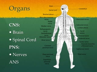
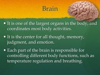
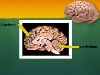
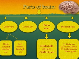
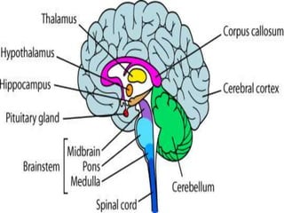
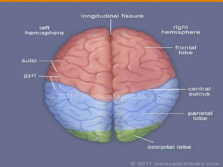
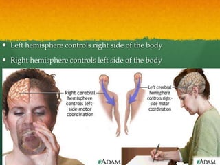
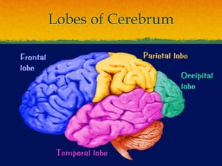
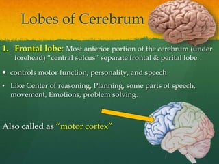
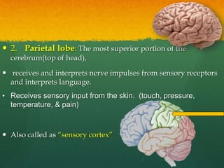
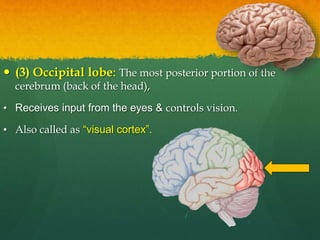
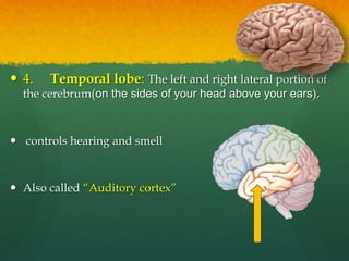
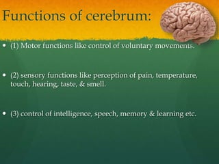
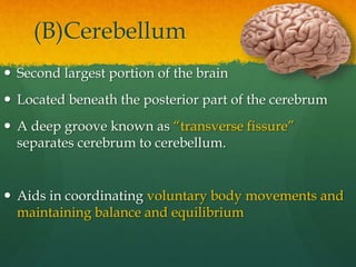
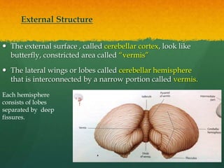
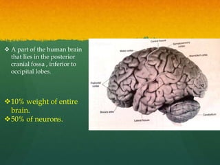
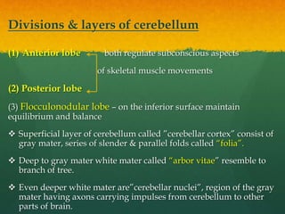
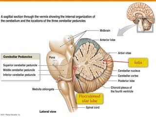
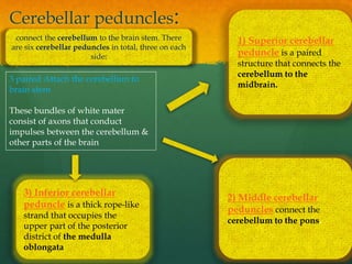
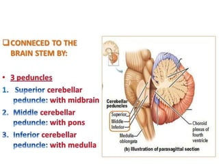
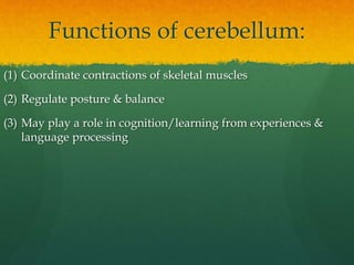
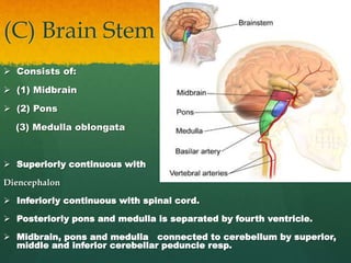
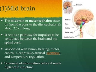
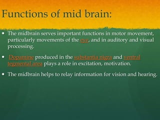
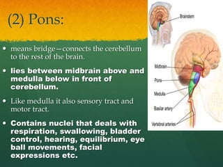
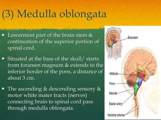
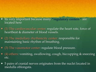
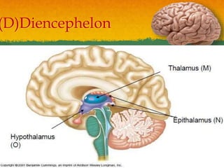
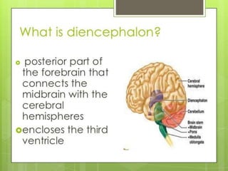
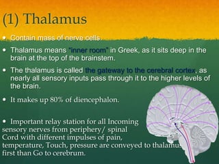
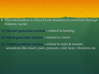
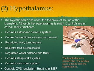
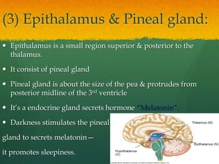
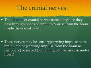
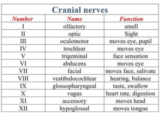
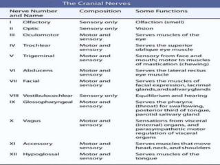
The Anatomy and Physiology of
Brain Sarita Sharma
Associate Professor
MMCP, MMDU
Organs
CNS:
 Brain
 Spinal Cord
PNS:
 Nerves
ANS
Brain
 It is one of the largest organs in the body, and
coordinates most body activities.
 It is the center for all thought, memory,
judgment, and emotion.
 Each part of the brain is responsible for
controlling different body functions, such as
temperature regulation and breathing.
 The brain is contained in skull & weighs 1300 - 1400 g
 made up of about 1000 billion neurons & each neuron is
surrounded by about 10 glial cells (neuroglia ).
 Neurons cannot multiply & many neurons are lost everyday
in life but glial cells can multiply throughout the life.
 Brain is also covered by “Meninges” like spinal cord---outer
duramater, middle arachnoidmater & inner piamater
Cerebrum
cerebellum
Ventricles of the brain
 There are certain cavities in the brain called as “ventricles”.
 These ventricles are filled with CSF.
 There are 4 ventricles of brain
2 lateral ventricles
in two hemisphere
of the brain
3rd lies between
hypothalamus &
thalamus
4th between brain stem &
cerebellum
& is continuous with the
central cavity of spinal
cord
Parts of brain:
Cerebrum Cerebellum
Brain
stem
Diencephelon
(1)Medulla
(2)Pons
(3)Mid brain
(1) Thalamus
(2) Hypothalamus
(3) Epithalamus &
Pineal gland
Right
cerebral
hemisph
ere
Left
cerebral
hemisph
ere
(A) Cerebrum
 It is the largest section of the brain
 It is located in the upper portion of the brain and is the area
that processes thoughts, judgment, memory, problem solving,
and language, imaginations.
 Cerebral cortex – the superficial layer of the cerebrum is “gray
mater” & this is 2-4 mm thick called “Cerebral cortex ” contains
billions neurons.
 During embryonic development when the brain size increases
rapidly, the gray mater of the cortex enlarges much faster than
deeper white mater so as result cortical region roles & fold
upon itself.
 The folds are called “gyri” or “convolutions”
 The deepest grooves between the folds are called “fissures”
 The shallower grooves between folds are called “sulci”
 The most prominent fissure, the longitudinal fissure,
separates the cerebrum into right & left halves called
“cerebral hemispheres”. Each hemisphere has 4 lobes.
 The cerebrum is subdivided into the left and right Both
hemisphere are connected by a bridge of nerve fibers that
relay information between 2 hemisphere called “corpus
callosum”
 Left hemisphere controls right side of the body
 Right hemisphere controls left side of the body
Lobes of Cerebrum
Lobes of Cerebrum
1. Frontal lobe: Most anterior portion of the cerebrum (under
forehead) “central sulcus” separate frontal & perital lobe.
 controls motor function, personality, and speech
• Like Center of reasoning, Planning, some parts of speech,
movement, Emotions, problem solving.
Also called as “motor cortex”
 2. Parietal lobe: The most superior portion of the
cerebrum(top of head),
 receives and interprets nerve impulses from sensory receptors
and interprets language.
• Receives sensory input from the skin. (touch, pressure,
temperature, & pain)
 Also called as “sensory cortex”
 (3) Occipital lobe: The most posterior portion of the
cerebrum (back of the head),
• Receives input from the eyes & controls vision.
• Also called as “visual cortex”.
 4. Temporal lobe: The left and right lateral portion of
the cerebrum(on the sides of your head above your ears),
 controls hearing and smell
 Also called “Auditory cortex”
Functions of cerebrum:
 (1) Motor functions like control of voluntary movements.
 (2) sensory functions like perception of pain, temperature,
touch, hearing, taste, & smell.
 (3) control of intelligence, speech, memory & learning etc.
(B)Cerebellum
 Second largest portion of the brain
 Located beneath the posterior part of the cerebrum
 A deep groove known as “transverse fissure”
separates cerebrum to cerebellum.
 Aids in coordinating voluntary body movements and
maintaining balance and equilibrium
External Structure
 The external surface , called cerebellar cortex, look like
butterfly, constricted area called “vermis”
 The lateral wings or lobes called cerebellar hemisphere
that is interconnected by a narrow portion called vermis.
Each hemisphere
consists of lobes
separated by deep
fissures.
 A part of the human brain
that lies in the posterior
cranial fossa , inferior to
occipital lobes.
10% weight of entire
brain.
50% of neurons.
Divisions & layers of cerebellum
(1) Anterior lobe both regulate subconscious aspects
of skeletal muscle movements
(2) Posterior lobe
(3) Flocculonodular lobe – on the inferior surface maintain
equilibrium and balance
 Superficial layer of cerebellum called ”cerebellar cortex” consist of
gray mater, series of slender & parallel folds called “folia”.
 Deep to gray mater white mater called “arbor vitae” resemble to
branch of tree.
 Even deeper white mater are”cerebellar nuclei”, region of the gray
mater having axons carrying impulses from cerebellum to other
parts of brain.
Flocculonod
ular lobe
folia
Cerebellar peduncles:
3) Inferior cerebellar
peduncle is a thick rope-like
strand that occupies the
upper part of the posterior
district of the medulla
oblongata
2) Middle cerebellar
peduncles connect the
cerebellum to the pons.
1) Superior cerebellar
peduncle is a paired
structure that connects the
cerebellum to the
midbrain.
3 paired Attach the cerebellum to
brain stem
These bundles of white mater
consist of axons that conduct
impulses between the cerebellum &
other parts of the brain
connect the cerebellum to the brain stem. There
are six cerebellar peduncles in total, three on each
side:
Functions of cerebellum:
(1) Coordinate contractions of skeletal muscles
(2) Regulate posture & balance
(3) May play a role in cognition/learning from experiences &
language processing
(C) Brain Stem
 Consists of:
 (1) Midbrain
 (2) Pons
(3) Medulla oblongata
 Superiorly continuous with
Diencephalon
 Inferiorly continuous with spinal cord.
 Posteriorly pons and medulla is separated by fourth ventricle.
 Midbrain, pons and medulla connected to cerebellum by superior,
middle and inferior cerebellar peduncle resp.
(1)Mid brain
 The midbrain or mesencephalon exten
ds from the pons to the diencephalon &
about 2.5 cm long.
 It acts as a pathway for impulses to be
conducted between the brain and the
spinal cord.
 associated with vision, hearing, motor
control, sleep/wake, arousal (alertness),
and temperature regulation.
 Screening of information before it reach
high brain structure
Functions of mid brain:
 The midbrain serves important functions in motor movement,
particularly movements of the eye, and in auditory and visual
processing.
 Dopamine produced in the substantia nigra and ventral
tegmental area plays a role in excitation, motivation.
 The midbrain helps to relay information for vision and hearing.
(2) Pons:
 means bridge—connects the cerebellum
to the rest of the brain.
 lies between midbrain above and
medulla below in front of
cerebellum.
 Like medulla it also sensory tract and
motor tract.
 Contains nuclei that deals with
respiration, swallowing, bladder
control, hearing, equilibrium, eye
ball movements, facial
expressions etc.
(3) Medulla oblongata
 Lowermost part of the brain stem &
continuation of the superior portion of
spinal cord.
 Situated at the base of the skull/ starts
from foramen magnum & extends to the
inferior border of the pons, a distance of
about 3 cm.
 The ascending & descending sensory &
motor white mater tracts (nerves)
connecting brain to spinal cord pass
through medulla oblongata.
 Its very important because many “regulatory centers ” are
located here
 (1) The cardiovascular center: regulate the heart rate, force of
heartbeat & diameter of blood vessels.
 (2) The medullary rhythmicity center: responsible for
maintaining basic rhythm of breathing.
 (3) The vasomotor center: regulate blood pressure.
 (4) others: vomiting, swallowing, cough, hiccupping & sneezing
etc.
 5 pairs of cranial nerves originates from the nuclei located in
medulla oblongata.
(D)Diencephelon
(1) Thalamus
 Contain mass of nerve cells.
 Thalamus means “inner room” in Greek, as it sits deep in the
brain at the top of the brainstem.
 The thalamus is called the gateway to the cerebral cortex, as
nearly all sensory inputs pass through it to the higher levels of
the brain.
 It makes up 80% of diencephalon.
 Important relay station for all Incoming
sensory nerves from periphery/ spinal
Cord with different impulses of pain,
temperature, Touch, pressure are conveyed to thalamus
first than Go to cerebrum.
 This information is relayed from thalamus to cerebrum through
thalamic nuclei:
(1) Median geniculate nucleus—related to hearing
(2) lateral geniculate nucleus—related to vision
(3) Ventral posterior nucleus—related to taste & somatic
sensations like touch, pain, pressure, cold, heat, vibrations etc.
(2) Hypothalamus:
 The hypothalamus sits under the thalamus at the top of the
brainstem. Although the hypothalamus is small, it controls many
critical bodily functions:
• Controls autonomic nervous system
• Center for emotional response and behavior
• Regulates body temperature
• Regulates food intake(appetite)
• Regulates water balance and thirst
• Controls sleep-wake cycles
• Controls endocrine system
• Controls CVS regulation- Heart rate & BP
The hypothalamus is
shaded blue. The pituitary
gland extends from the
hypothalamus.
(3) Epithalamus & Pineal gland:
 Epithalamus is a small region superior & posterior to the
thalamus.
 It consist of pineal gland
 Pineal gland is about the size of the pea & protrudes from
posterior midline of the 3rd ventricle
 It’s a endocrine gland secrets hormone “Melatonin”.
 Darkness stimulates the pineal
gland to secrets melatonin—
it promotes sleepiness.
The cranial nerves:
 The 12 pairs of cranial nerves named because they
pass through bones of cranium & arise from the brain
inside the cranial cavity.
 These nerves may be sensory(carrying impulse to the
brain), motor (carrying impulse from the brain to
periphery) or mixed (containing both sensory & motor
fibers)
Thank you

The human brain.pptx

  • 1.
    The Anatomy andPhysiology of Brain Sarita Sharma Associate Professor MMCP, MMDU
  • 2.
    Organs CNS:  Brain  SpinalCord PNS:  Nerves ANS
  • 3.
    Brain  It isone of the largest organs in the body, and coordinates most body activities.  It is the center for all thought, memory, judgment, and emotion.  Each part of the brain is responsible for controlling different body functions, such as temperature regulation and breathing.
  • 4.
     The brainis contained in skull & weighs 1300 - 1400 g  made up of about 1000 billion neurons & each neuron is surrounded by about 10 glial cells (neuroglia ).  Neurons cannot multiply & many neurons are lost everyday in life but glial cells can multiply throughout the life.  Brain is also covered by “Meninges” like spinal cord---outer duramater, middle arachnoidmater & inner piamater
  • 5.
  • 6.
    Ventricles of thebrain  There are certain cavities in the brain called as “ventricles”.  These ventricles are filled with CSF.  There are 4 ventricles of brain 2 lateral ventricles in two hemisphere of the brain 3rd lies between hypothalamus & thalamus 4th between brain stem & cerebellum & is continuous with the central cavity of spinal cord
  • 8.
    Parts of brain: CerebrumCerebellum Brain stem Diencephelon (1)Medulla (2)Pons (3)Mid brain (1) Thalamus (2) Hypothalamus (3) Epithalamus & Pineal gland Right cerebral hemisph ere Left cerebral hemisph ere
  • 10.
    (A) Cerebrum  Itis the largest section of the brain  It is located in the upper portion of the brain and is the area that processes thoughts, judgment, memory, problem solving, and language, imaginations.  Cerebral cortex – the superficial layer of the cerebrum is “gray mater” & this is 2-4 mm thick called “Cerebral cortex ” contains billions neurons.  During embryonic development when the brain size increases rapidly, the gray mater of the cortex enlarges much faster than deeper white mater so as result cortical region roles & fold upon itself.
  • 11.
     The foldsare called “gyri” or “convolutions”  The deepest grooves between the folds are called “fissures”  The shallower grooves between folds are called “sulci”  The most prominent fissure, the longitudinal fissure, separates the cerebrum into right & left halves called “cerebral hemispheres”. Each hemisphere has 4 lobes.  The cerebrum is subdivided into the left and right Both hemisphere are connected by a bridge of nerve fibers that relay information between 2 hemisphere called “corpus callosum”
  • 13.
     Left hemispherecontrols right side of the body  Right hemisphere controls left side of the body
  • 14.
  • 15.
    Lobes of Cerebrum 1.Frontal lobe: Most anterior portion of the cerebrum (under forehead) “central sulcus” separate frontal & perital lobe.  controls motor function, personality, and speech • Like Center of reasoning, Planning, some parts of speech, movement, Emotions, problem solving. Also called as “motor cortex”
  • 16.
     2. Parietallobe: The most superior portion of the cerebrum(top of head),  receives and interprets nerve impulses from sensory receptors and interprets language. • Receives sensory input from the skin. (touch, pressure, temperature, & pain)  Also called as “sensory cortex”
  • 17.
     (3) Occipitallobe: The most posterior portion of the cerebrum (back of the head), • Receives input from the eyes & controls vision. • Also called as “visual cortex”.
  • 18.
     4. Temporallobe: The left and right lateral portion of the cerebrum(on the sides of your head above your ears),  controls hearing and smell  Also called “Auditory cortex”
  • 20.
    Functions of cerebrum: (1) Motor functions like control of voluntary movements.  (2) sensory functions like perception of pain, temperature, touch, hearing, taste, & smell.  (3) control of intelligence, speech, memory & learning etc.
  • 21.
    (B)Cerebellum  Second largestportion of the brain  Located beneath the posterior part of the cerebrum  A deep groove known as “transverse fissure” separates cerebrum to cerebellum.  Aids in coordinating voluntary body movements and maintaining balance and equilibrium
  • 22.
    External Structure  Theexternal surface , called cerebellar cortex, look like butterfly, constricted area called “vermis”  The lateral wings or lobes called cerebellar hemisphere that is interconnected by a narrow portion called vermis. Each hemisphere consists of lobes separated by deep fissures.
  • 23.
     A partof the human brain that lies in the posterior cranial fossa , inferior to occipital lobes. 10% weight of entire brain. 50% of neurons.
  • 24.
    Divisions & layersof cerebellum (1) Anterior lobe both regulate subconscious aspects of skeletal muscle movements (2) Posterior lobe (3) Flocculonodular lobe – on the inferior surface maintain equilibrium and balance  Superficial layer of cerebellum called ”cerebellar cortex” consist of gray mater, series of slender & parallel folds called “folia”.  Deep to gray mater white mater called “arbor vitae” resemble to branch of tree.  Even deeper white mater are”cerebellar nuclei”, region of the gray mater having axons carrying impulses from cerebellum to other parts of brain.
  • 25.
  • 26.
    Cerebellar peduncles: 3) Inferiorcerebellar peduncle is a thick rope-like strand that occupies the upper part of the posterior district of the medulla oblongata 2) Middle cerebellar peduncles connect the cerebellum to the pons. 1) Superior cerebellar peduncle is a paired structure that connects the cerebellum to the midbrain. 3 paired Attach the cerebellum to brain stem These bundles of white mater consist of axons that conduct impulses between the cerebellum & other parts of the brain connect the cerebellum to the brain stem. There are six cerebellar peduncles in total, three on each side:
  • 28.
    Functions of cerebellum: (1)Coordinate contractions of skeletal muscles (2) Regulate posture & balance (3) May play a role in cognition/learning from experiences & language processing
  • 29.
    (C) Brain Stem Consists of:  (1) Midbrain  (2) Pons (3) Medulla oblongata  Superiorly continuous with Diencephalon  Inferiorly continuous with spinal cord.  Posteriorly pons and medulla is separated by fourth ventricle.  Midbrain, pons and medulla connected to cerebellum by superior, middle and inferior cerebellar peduncle resp.
  • 30.
    (1)Mid brain  Themidbrain or mesencephalon exten ds from the pons to the diencephalon & about 2.5 cm long.  It acts as a pathway for impulses to be conducted between the brain and the spinal cord.  associated with vision, hearing, motor control, sleep/wake, arousal (alertness), and temperature regulation.  Screening of information before it reach high brain structure
  • 31.
    Functions of midbrain:  The midbrain serves important functions in motor movement, particularly movements of the eye, and in auditory and visual processing.  Dopamine produced in the substantia nigra and ventral tegmental area plays a role in excitation, motivation.  The midbrain helps to relay information for vision and hearing.
  • 32.
    (2) Pons:  meansbridge—connects the cerebellum to the rest of the brain.  lies between midbrain above and medulla below in front of cerebellum.  Like medulla it also sensory tract and motor tract.  Contains nuclei that deals with respiration, swallowing, bladder control, hearing, equilibrium, eye ball movements, facial expressions etc.
  • 33.
    (3) Medulla oblongata Lowermost part of the brain stem & continuation of the superior portion of spinal cord.  Situated at the base of the skull/ starts from foramen magnum & extends to the inferior border of the pons, a distance of about 3 cm.  The ascending & descending sensory & motor white mater tracts (nerves) connecting brain to spinal cord pass through medulla oblongata.
  • 34.
     Its veryimportant because many “regulatory centers ” are located here  (1) The cardiovascular center: regulate the heart rate, force of heartbeat & diameter of blood vessels.  (2) The medullary rhythmicity center: responsible for maintaining basic rhythm of breathing.  (3) The vasomotor center: regulate blood pressure.  (4) others: vomiting, swallowing, cough, hiccupping & sneezing etc.  5 pairs of cranial nerves originates from the nuclei located in medulla oblongata.
  • 35.
  • 37.
    (1) Thalamus  Containmass of nerve cells.  Thalamus means “inner room” in Greek, as it sits deep in the brain at the top of the brainstem.  The thalamus is called the gateway to the cerebral cortex, as nearly all sensory inputs pass through it to the higher levels of the brain.  It makes up 80% of diencephalon.  Important relay station for all Incoming sensory nerves from periphery/ spinal Cord with different impulses of pain, temperature, Touch, pressure are conveyed to thalamus first than Go to cerebrum.
  • 38.
     This informationis relayed from thalamus to cerebrum through thalamic nuclei: (1) Median geniculate nucleus—related to hearing (2) lateral geniculate nucleus—related to vision (3) Ventral posterior nucleus—related to taste & somatic sensations like touch, pain, pressure, cold, heat, vibrations etc.
  • 39.
    (2) Hypothalamus:  Thehypothalamus sits under the thalamus at the top of the brainstem. Although the hypothalamus is small, it controls many critical bodily functions: • Controls autonomic nervous system • Center for emotional response and behavior • Regulates body temperature • Regulates food intake(appetite) • Regulates water balance and thirst • Controls sleep-wake cycles • Controls endocrine system • Controls CVS regulation- Heart rate & BP The hypothalamus is shaded blue. The pituitary gland extends from the hypothalamus.
  • 40.
    (3) Epithalamus &Pineal gland:  Epithalamus is a small region superior & posterior to the thalamus.  It consist of pineal gland  Pineal gland is about the size of the pea & protrudes from posterior midline of the 3rd ventricle  It’s a endocrine gland secrets hormone “Melatonin”.  Darkness stimulates the pineal gland to secrets melatonin— it promotes sleepiness.
  • 41.
    The cranial nerves: The 12 pairs of cranial nerves named because they pass through bones of cranium & arise from the brain inside the cranial cavity.  These nerves may be sensory(carrying impulse to the brain), motor (carrying impulse from the brain to periphery) or mixed (containing both sensory & motor fibers)
  • 44.