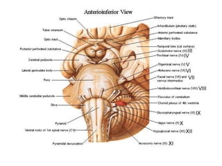
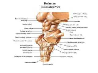
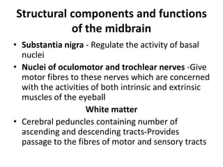
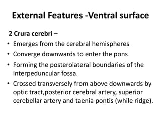
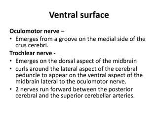
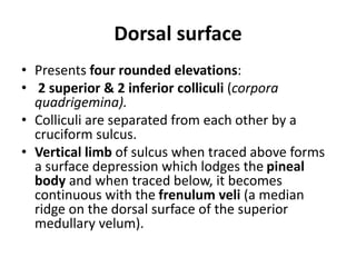
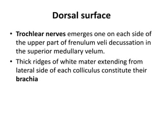
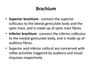
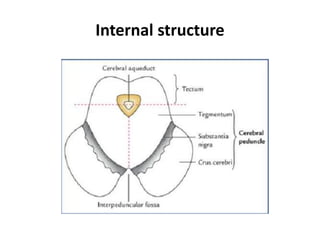
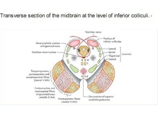
Midbrain
DR.DEEPAK KHEDEKAR
DEPT.OF ANATOMY
LTMMC &GH.
Introduction
• Upper and shortest part of the brain-stem.
• 2.5 cm long and 2.5 cm wide.
• Connects the hindbrain with the forebrain.
• Its cavity, the cerebral aqueduct (aqueduct of
Sylvius) connects the third ventricle with the
fourth ventricle.
• Passes through the tentorial notch.
Relation
• On each side - optic tract, Parahippocampal
gyrus, Posterior cerebral artery, Basal vein;
• Anteriorly -interpeduncular structures, viz.
mammillary bodies,tuber cinereum, etc.;
• Posteriorly - splenium of corpus callosum,
great cerebral vein, pineal body and posterior
ends of right and left thalami.
MIDBRAIN
Structural components and functions
of the midbrain
• Superior colliculi -Reflex centres for visual
reflexes
• Inferior colliculi - Lower auditory centres,
probably concerned with reflexes involving
auditory stimuli
• Red nuclei - Involuntary control of muscle
tone and posture
Structural components and functions
of the midbrain
• Substantia nigra - Regulate the activity of basal
nuclei
• Nuclei of oculomotor and trochlear nerves -Give
motor fibres to these nerves which are concerned
with the activities of both intrinsic and extrinsic
muscles of the eyeball
White matter
• Cerebral peduncles containing number of
ascending and descending tracts-Provides
passage to the fibres of motor and sensory tracts
External Features -Ventral surface
2 Crura cerebri –
• Emerges from the cerebral hemispheres
• Converge downwards to enter the pons
• Forming the posterolateral boundaries of the
interpeduncular fossa.
• Crossed transversely from above downwards by
optic tract,posterior cerebral artery, superior
cerebellar artery and taenia pontis (while ridge).
Ventral surface
Oculomotor nerve –
• Emerges from a groove on the medial side of the
crus cerebri.
Trochlear nerve -
• Emerges on the dorsal aspect of the midbrain
• curls around the lateral aspect of the cerebral
peduncle to appear on the ventral aspect of the
midbrain lateral to the oculomotor nerve.
• 2 nerves run forward between the posterior
cerebral and the superior cerebellar arteries.
Dorsal surface
• Presents four rounded elevations:
• 2 superior & 2 inferior colliculi (corpora
quadrigemina).
• Colliculi are separated from each other by a
cruciform sulcus.
• Vertical limb of sulcus when traced above forms
a surface depression which lodges the pineal
body and when traced below, it becomes
continuous with the frenulum veli (a median
ridge on the dorsal surface of the superior
medullary velum).
Dorsal surface
• Trochlear nerves emerges one on each side of
the upper part of frenulum veli decussation in
the superior medullary velum.
• Thick ridges of white mater extending from
lateral side of each colliculus constitute their
brachia
MIDBRAIN
Brachium
• Superior brachium - connect the superior
colliculus to the lateral geniculate body and the
optic tract, and is made up of optic tract fibres.
• Inferior brachium -connect the inferior colliculus
to the medial geniculate body, and is made up of
auditory fibres.
• Superior and inferior colliculi are concerned with
reflex activities triggered by auditory and visual
impulses respectively.
Internal structure
Internal structure
• Small posterior part is called tectum and
consists of four colliculi.
• Large anterior part is divided into two equal
right and left halves by a vertical plane, the
cerebral peduncle.
• Each cerebral peduncle is further subdivided
into three parts, from dorsal to ventral these
are: (a) teg-mentum, (b) substantia nigra, and
(c) crus cerebri.
Crus cerebri (Basis pedunculi)
• Part of cerebral peduncle situated
anterolateral to the substantia nigra.
• Contains important descending tracts which
connect the cerebral cortex to the anterior
horn cells of the spinal cord, cranial nerve
nuclei, and pontine nuclei.
Cerebral Peduncles - Tract
• Corticospinal and corticonuclear fibres
(pyramidal tract) occupy the middle two-
thirds of the crus.
• Frontopontine fibres occupy the medial one-
sixth of the crus. The temporopontine,
parietopontine, and occipitopontine fibres
occupy the lateral one-sixth of the crus
Substantia nigra
• Curved (crescent-shaped) pigmented band of
grey mater (thicker medially than laterally)
situated between tegmentum and crus cerebri
• Large motor nucleus that extends throughout the
length of midbrain
• Divided into two parts:
(a) Dorsal part (pars compacta) containing
medium sized cells and
(b) Ventral part (pars reticularis) containing fewer
cells. The pars reticularis is intermingled with the
fibres of crus cerebri.
Substantia nigra
• Made up of deeply pigmented nerve cells
which contain melanin (a polymerized form of
dopamine) and iron.
• Cells synthesize dopamine which is carried
through their axons (nigrostriatal fibres) to
the corpus striatum.
THANK YOU

MIDBRAIN.DK.pptx

  • 1.
  • 2.
    Introduction • Upper andshortest part of the brain-stem. • 2.5 cm long and 2.5 cm wide. • Connects the hindbrain with the forebrain. • Its cavity, the cerebral aqueduct (aqueduct of Sylvius) connects the third ventricle with the fourth ventricle. • Passes through the tentorial notch.
  • 5.
    Relation • On eachside - optic tract, Parahippocampal gyrus, Posterior cerebral artery, Basal vein; • Anteriorly -interpeduncular structures, viz. mammillary bodies,tuber cinereum, etc.; • Posteriorly - splenium of corpus callosum, great cerebral vein, pineal body and posterior ends of right and left thalami.
  • 6.
  • 7.
    Structural components andfunctions of the midbrain • Superior colliculi -Reflex centres for visual reflexes • Inferior colliculi - Lower auditory centres, probably concerned with reflexes involving auditory stimuli • Red nuclei - Involuntary control of muscle tone and posture
  • 8.
    Structural components andfunctions of the midbrain • Substantia nigra - Regulate the activity of basal nuclei • Nuclei of oculomotor and trochlear nerves -Give motor fibres to these nerves which are concerned with the activities of both intrinsic and extrinsic muscles of the eyeball White matter • Cerebral peduncles containing number of ascending and descending tracts-Provides passage to the fibres of motor and sensory tracts
  • 9.
    External Features -Ventralsurface 2 Crura cerebri – • Emerges from the cerebral hemispheres • Converge downwards to enter the pons • Forming the posterolateral boundaries of the interpeduncular fossa. • Crossed transversely from above downwards by optic tract,posterior cerebral artery, superior cerebellar artery and taenia pontis (while ridge).
  • 10.
    Ventral surface Oculomotor nerve– • Emerges from a groove on the medial side of the crus cerebri. Trochlear nerve - • Emerges on the dorsal aspect of the midbrain • curls around the lateral aspect of the cerebral peduncle to appear on the ventral aspect of the midbrain lateral to the oculomotor nerve. • 2 nerves run forward between the posterior cerebral and the superior cerebellar arteries.
  • 11.
    Dorsal surface • Presentsfour rounded elevations: • 2 superior & 2 inferior colliculi (corpora quadrigemina). • Colliculi are separated from each other by a cruciform sulcus. • Vertical limb of sulcus when traced above forms a surface depression which lodges the pineal body and when traced below, it becomes continuous with the frenulum veli (a median ridge on the dorsal surface of the superior medullary velum).
  • 12.
    Dorsal surface • Trochlearnerves emerges one on each side of the upper part of frenulum veli decussation in the superior medullary velum. • Thick ridges of white mater extending from lateral side of each colliculus constitute their brachia
  • 13.
  • 15.
    Brachium • Superior brachium- connect the superior colliculus to the lateral geniculate body and the optic tract, and is made up of optic tract fibres. • Inferior brachium -connect the inferior colliculus to the medial geniculate body, and is made up of auditory fibres. • Superior and inferior colliculi are concerned with reflex activities triggered by auditory and visual impulses respectively.
  • 16.
  • 17.
    Internal structure • Smallposterior part is called tectum and consists of four colliculi. • Large anterior part is divided into two equal right and left halves by a vertical plane, the cerebral peduncle. • Each cerebral peduncle is further subdivided into three parts, from dorsal to ventral these are: (a) teg-mentum, (b) substantia nigra, and (c) crus cerebri.
  • 18.
    Crus cerebri (Basispedunculi) • Part of cerebral peduncle situated anterolateral to the substantia nigra. • Contains important descending tracts which connect the cerebral cortex to the anterior horn cells of the spinal cord, cranial nerve nuclei, and pontine nuclei.
  • 20.
    Cerebral Peduncles -Tract • Corticospinal and corticonuclear fibres (pyramidal tract) occupy the middle two- thirds of the crus. • Frontopontine fibres occupy the medial one- sixth of the crus. The temporopontine, parietopontine, and occipitopontine fibres occupy the lateral one-sixth of the crus
  • 21.
    Substantia nigra • Curved(crescent-shaped) pigmented band of grey mater (thicker medially than laterally) situated between tegmentum and crus cerebri • Large motor nucleus that extends throughout the length of midbrain • Divided into two parts: (a) Dorsal part (pars compacta) containing medium sized cells and (b) Ventral part (pars reticularis) containing fewer cells. The pars reticularis is intermingled with the fibres of crus cerebri.
  • 22.
    Substantia nigra • Madeup of deeply pigmented nerve cells which contain melanin (a polymerized form of dopamine) and iron. • Cells synthesize dopamine which is carried through their axons (nigrostriatal fibres) to the corpus striatum.
  • 25.