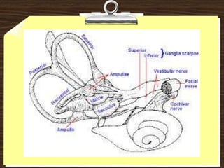
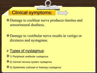
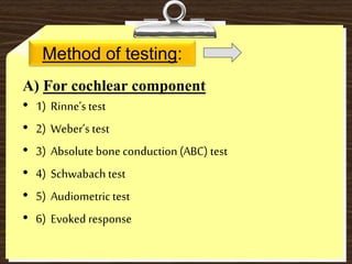
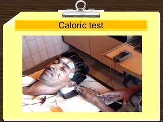
VESTIBULOCOCHLEAR NERVE
- NITHIN
NAIR
INTRODUCTION
Stato-acoustic or Auditory nerve
8th cranial nerve
2 components: a) cochlear (hearing) b) vestibular (equilibrium)
OBJECTIVES
1) Anatomy and functions.
2) Purpose of test
3) How to test
4) Interpretations
ANATOMY AND FUNCTIONS
Cochlear Nerve:
Dorsal and ventral cochlear nuclei -- situated---
inferior cerebellar peduncle.
The auditory receptors -- hair cells of the organ of
corti
Innervated – peripheral processes of bipolar neurons
of spiral ganglion
Their central processes form cochlear nerve which
ends in dorsal and ventral cochlear nuclei
AUDITORY PATHWAY
The 1st sensory neuron lies in the spiral ganglion of
bipolar cell. The peripheral processes are distributed
to the organ of corti.The central processes form
cochlear nerve –ends in dorsal and ventral cochlear
nuclei.
The 2nd neuron fibres start from the dorsal and
ventral cochlear nuclei; their axons form trapezoid
body and ends in dorsal nucleus of trapezoid body of
the same and opp. side
AUDITORY PATHWAY
The 3rd neuron fibres from dorsal nuclei of trapezoid
body ascend as lat. Lemniscus on both sides. Some of
the fibres of lat. Lemniscus end in inf. Colliculus. Rest
of the lat. Lemniscus relays in med. Geniculate body.
The 4th neuron fibres from med. Geniculate body –
auditory radiations – pass thru sublentiform part of
internal capsule – projected to auditory cortex of
temporal lobe.
Vestibular Nerve:
The 4 vestibular nuclei are situated partly in medulla
and partly in pons in relation to the floor of 4th
ventrical.
Receptors – maculae of saccule and utricle (static
balance) and cristae of the ampullae of semicircular
ducts (kinetic balance)
Innervated – peripheral processes of bipolar neuron
of vestibular ganglion (acoustic meatus)
Their central processes –form vestibular nerve –
ends in vestibular nuclei.
Vestibular pathway:
Innervates the maculae of the urticle and succule
(sensitive to static changes in equilibrium)
The ampullae of semicircular canals(sensitive to
dynamic changes in equilibrium)
Impulses pass: vestibular nerve vestibular nuclei
(pontomedullary junction)
Fibres from vestibular nuclei pass via inferior
cerebellar peduncle cerebellum
Clinical symptoms:
Damage to cochlear nerve produces tinnitus and
sensorineural deafness.
Damage to vestibular nerve results in vertigo or
dizziness and nystagmus.
 Types of nystagmus:
1) Peripheral vestibular nystagmus
2) Central nervous system nystagmus
3) Optokinetic (railroad or freeway) nystagmus
Purpose of test:
To detect whether any deafness is bilateral or
unilateral, and whether sensorineural or
conduction.
To determine whether disturbance of
vestibular function originates in the labyrinth,
the vestibular nerve, or the brain stem.
HOWTO TEST
Method of testing:
A) For cochlear component
• 1) Rinne’stest
• 2) Weber’s test
• 3) Absolute bone conduction (ABC) test
• 4) Schwabach test
• 5) Audiometric test
• 6) Evoked response
B) For vestibular component
• 1) Rotationaltest.
• 2) Caloric test.
• 3) Dix Hallpikemaneuver for nystagmus.
• 4) Electronystagmography
Rinne’s test
Weber’s test
Audiometric test
Evoked response
Rotational test
Caloric test
Dix hallpike maneuver
Electronystagmography (ENG)
INTERPRETATIONS
Interpretations
Whether hearing loss is senorineural or conductive
TEST NORMAL CONDUCTIVE
DEAFNESS
SENSORINEURAL
DEAFNESS
Rinne AC > BC BC > AC AC > BC
Weber Not lateralized Lateralized to
diseased ear
Lateralized to
better ear
ABC Equal Lengthened Shortened
Schwabach Equal Lengthened Shortened
Interpretations
Whether nystagmus is peripheral or central
FINDING PERIPHERAL CENTRAL
LATENCY Yes 3 – 10 sec No
FATIGABILITY Yes No
NYSTAGMUS DIRECTION Fixed, typically mixed
rotational
Changing, variable and
pure vertical or pure
horizontal
SUPPRESSION BY VISUAL
FIXATION
Yes No
SEVERITY Markedly severe Mild to moderate
CONSISTENCY Less consistent More consistent
PAST POINTING In direction of slow
phase
In direction of fast
phase
References...
 BD CHAURASIA’S HUMAN ANATOMY
 NEUROLOGICAL EXAMINATION BY
NAVNEET KUMAR
 BICKERSTAFF’S NEUROLOGICAL
EXAMINATION
 NEUROLOGICAL EXAMINATION MADE
EASY – GERAINT FULLER
 PJ MEHTA’S PRACTICAL MEDICINE
 PRACTICAL PHYSIOLOGY – VD JOSHI
 WWW.GOOGLE.COM

Vestibulocochlear nerve 8

  • 1.
  • 2.
    INTRODUCTION Stato-acoustic or Auditorynerve 8th cranial nerve 2 components: a) cochlear (hearing) b) vestibular (equilibrium) OBJECTIVES 1) Anatomy and functions. 2) Purpose of test 3) How to test 4) Interpretations
  • 3.
    ANATOMY AND FUNCTIONS CochlearNerve: Dorsal and ventral cochlear nuclei -- situated--- inferior cerebellar peduncle. The auditory receptors -- hair cells of the organ of corti Innervated – peripheral processes of bipolar neurons of spiral ganglion Their central processes form cochlear nerve which ends in dorsal and ventral cochlear nuclei
  • 6.
    AUDITORY PATHWAY The 1stsensory neuron lies in the spiral ganglion of bipolar cell. The peripheral processes are distributed to the organ of corti.The central processes form cochlear nerve –ends in dorsal and ventral cochlear nuclei. The 2nd neuron fibres start from the dorsal and ventral cochlear nuclei; their axons form trapezoid body and ends in dorsal nucleus of trapezoid body of the same and opp. side
  • 7.
    AUDITORY PATHWAY The 3rdneuron fibres from dorsal nuclei of trapezoid body ascend as lat. Lemniscus on both sides. Some of the fibres of lat. Lemniscus end in inf. Colliculus. Rest of the lat. Lemniscus relays in med. Geniculate body. The 4th neuron fibres from med. Geniculate body – auditory radiations – pass thru sublentiform part of internal capsule – projected to auditory cortex of temporal lobe.
  • 9.
    Vestibular Nerve: The 4vestibular nuclei are situated partly in medulla and partly in pons in relation to the floor of 4th ventrical. Receptors – maculae of saccule and utricle (static balance) and cristae of the ampullae of semicircular ducts (kinetic balance) Innervated – peripheral processes of bipolar neuron of vestibular ganglion (acoustic meatus) Their central processes –form vestibular nerve – ends in vestibular nuclei.
  • 11.
    Vestibular pathway: Innervates themaculae of the urticle and succule (sensitive to static changes in equilibrium) The ampullae of semicircular canals(sensitive to dynamic changes in equilibrium) Impulses pass: vestibular nerve vestibular nuclei (pontomedullary junction) Fibres from vestibular nuclei pass via inferior cerebellar peduncle cerebellum
  • 12.
    Clinical symptoms: Damage tocochlear nerve produces tinnitus and sensorineural deafness. Damage to vestibular nerve results in vertigo or dizziness and nystagmus.  Types of nystagmus: 1) Peripheral vestibular nystagmus 2) Central nervous system nystagmus 3) Optokinetic (railroad or freeway) nystagmus
  • 13.
    Purpose of test: Todetect whether any deafness is bilateral or unilateral, and whether sensorineural or conduction. To determine whether disturbance of vestibular function originates in the labyrinth, the vestibular nerve, or the brain stem.
  • 14.
  • 15.
    Method of testing: A)For cochlear component • 1) Rinne’stest • 2) Weber’s test • 3) Absolute bone conduction (ABC) test • 4) Schwabach test • 5) Audiometric test • 6) Evoked response
  • 16.
    B) For vestibularcomponent • 1) Rotationaltest. • 2) Caloric test. • 3) Dix Hallpikemaneuver for nystagmus. • 4) Electronystagmography
  • 17.
  • 18.
  • 19.
  • 20.
  • 21.
  • 22.
  • 23.
  • 24.
  • 25.
  • 26.
    Interpretations Whether hearing lossis senorineural or conductive TEST NORMAL CONDUCTIVE DEAFNESS SENSORINEURAL DEAFNESS Rinne AC > BC BC > AC AC > BC Weber Not lateralized Lateralized to diseased ear Lateralized to better ear ABC Equal Lengthened Shortened Schwabach Equal Lengthened Shortened
  • 27.
    Interpretations Whether nystagmus isperipheral or central FINDING PERIPHERAL CENTRAL LATENCY Yes 3 – 10 sec No FATIGABILITY Yes No NYSTAGMUS DIRECTION Fixed, typically mixed rotational Changing, variable and pure vertical or pure horizontal SUPPRESSION BY VISUAL FIXATION Yes No SEVERITY Markedly severe Mild to moderate CONSISTENCY Less consistent More consistent PAST POINTING In direction of slow phase In direction of fast phase
  • 28.
    References...  BD CHAURASIA’SHUMAN ANATOMY  NEUROLOGICAL EXAMINATION BY NAVNEET KUMAR  BICKERSTAFF’S NEUROLOGICAL EXAMINATION  NEUROLOGICAL EXAMINATION MADE EASY – GERAINT FULLER  PJ MEHTA’S PRACTICAL MEDICINE  PRACTICAL PHYSIOLOGY – VD JOSHI  WWW.GOOGLE.COM