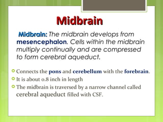
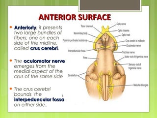
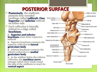
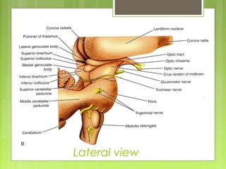
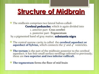
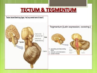
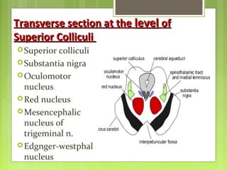
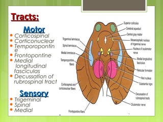
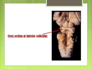
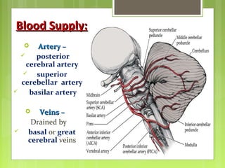
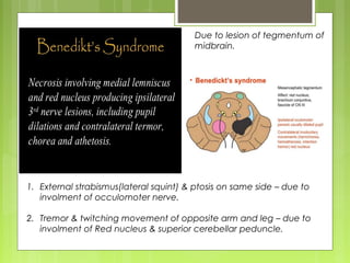
Basic Anatomy
&
Applied aspects
By: Pulak Agrawal
Roll No.: 001
Midbrain:Midbrain: The midbrain develops from
mesencephalon. Cells within the midbrain
multiply continually and are compressed
to form cerebral aqueduct.
MidbrainMidbrain
 Connects the pons and cerebellum with the forebrain.
 It is about 0.8 inch in length
 The midbrain is traversed by a narrow channel called
cerebral aqueduct filled with CSF.
 AnteriorlyAnteriorly, it presents
two large bundles of
fibers, one on each
side of the midline,
called crus cerebricrus cerebri..
 The oculomotor nerveoculomotor nerve
emerges from the
medial aspect of the
crus of the same side
 The crus cerebri
bounds the
interpeduncular fossainterpeduncular fossa
on either side.
ANTERIOR SURFACEANTERIOR SURFACE
POSTERIOR SURFACEPOSTERIOR SURFACE
 PosteriorlyPosteriorly, the midbrain
presents four rounded
swellings called colliculi- Onecolliculi- One
SuperiorSuperior and inferiorinferior colliculi
on either side.
 Each colliculus is laterally
related to a ridge called
brachium.brachium.
o Superior and inferiorSuperior and inferior
brachiumbrachium arises from respective
colliculi
o Superior brachium connects the
superior colliculus to laterallateral
geniculate bodygeniculate body
o Inferior brachium connects the
inferior colluculus to medialmedial
geniculate bodygeniculate body
o In the midline below the inferior
colliculus, the trochlear nervetrochlear nerve
emerges which then winds round
the side of the midbrain to reach its
ventral aspectventral aspect
Lateral view
Structure of MidbrainStructure of Midbrain
 The midbrain comprises two lateral halves called-
Cerebral pedunclesCerebral peduncles; which is again divided into
1. anterior part- Crus cerebri
2. posterior part -Tegmentum
by a pigmented band of gray matter, substantia nigrasubstantia nigra
 The central narrow cavity is called the cerebral aqueduct or
aqueduct of Sylvius, which connects the 3rd
and 4th
ventricles.
 The tectum is the part of the midbrain posterior to the cerebral
aqueduct; it has four small surface swellings referred to previously;
these are two superior and two inferior colliculi.
 The tegmentum forms the floor of mid brain
TECTUM & TEGMENTUMTECTUM & TEGMENTUM
Transverse section at theTransverse section at the levellevel ofof
Superior ColliculiSuperior Colliculi
Superior colliculi
Substantia nigra
Oculomotor
nucleus
Red nucleus
Mesencephalic
nucleus of
trigeminal n.
Edgnger-westphal
nucleus
Tracts:Tracts:
MotorMotor
 Corticospinal
 Corticonuclear
 Temporopontin
e
 Frontopontine
 Medial
longitudinal
fasciculas
 Decussation of
rubrospinal tract
SensorySensory
 Trigeminal
 Spinal
 Medial
TransverseTransverse Section at the level ofSection at the level of
Inferior ColliculiInferior Colliculi
 Substantia
nigra
 Troclear
nucleus
 Mesencephal
ic nucleus of
trigeminal
nerve
Tracts:Tracts:
MotorMotor
 Corticospinal
 Corticonuclear
 Temporopontine
 Frontopontine
 Medial longitudinal
fasciculas
SensorySensory
 Lateral
 Trigeminal
 Spinal
 Medial lemnisci
 Decussation of
superior cerebellar
peduncles
Blood Supply:Blood Supply:
 Artery –Artery –
 posterior
cerebral artery
 superior
cerebellar artery
 basilar artery
 Veins –Veins –
Drained by
 basal or great
cerebral veins
Due to lesion of tegmentum of
midbrain.
1. External strabismus(lateral squint) & ptosis on same side – due to
involment of occulomoter nerve.
2. Tremor & twitching movement of opposite arm and leg – due to
involment of Red nucleus & superior cerebellar peduncle.
 Rubral tremor (Holme’s tremor):
A coarse, slow (4Hz) tremor, especially present in the
upper extremities, that is found at rest, postural and
intention.
Claude Syndrome:
Ipsi CN III, contra tremor, ataxia and incoordination
Parinaud’s Syndrome:
Due to lesion of superior colliculi as occurs when
this area becomes compressed by tumour of
pineal gland.
Weakness of upward gaze& vertical nystagmus
Refrences:
Snell's Clinical Neuroanatomy
IB Singh - Textbook of Human Neuroanatomy
internet
MIDBRAIN basic anatomy and applied aspects.

MIDBRAIN basic anatomy and applied aspects.

  • 1.
    Basic Anatomy & Applied aspects By:Pulak Agrawal Roll No.: 001
  • 2.
    Midbrain:Midbrain: The midbraindevelops from mesencephalon. Cells within the midbrain multiply continually and are compressed to form cerebral aqueduct. MidbrainMidbrain  Connects the pons and cerebellum with the forebrain.  It is about 0.8 inch in length  The midbrain is traversed by a narrow channel called cerebral aqueduct filled with CSF.
  • 3.
     AnteriorlyAnteriorly, itpresents two large bundles of fibers, one on each side of the midline, called crus cerebricrus cerebri..  The oculomotor nerveoculomotor nerve emerges from the medial aspect of the crus of the same side  The crus cerebri bounds the interpeduncular fossainterpeduncular fossa on either side. ANTERIOR SURFACEANTERIOR SURFACE
  • 4.
    POSTERIOR SURFACEPOSTERIOR SURFACE PosteriorlyPosteriorly, the midbrain presents four rounded swellings called colliculi- Onecolliculi- One SuperiorSuperior and inferiorinferior colliculi on either side.  Each colliculus is laterally related to a ridge called brachium.brachium. o Superior and inferiorSuperior and inferior brachiumbrachium arises from respective colliculi o Superior brachium connects the superior colliculus to laterallateral geniculate bodygeniculate body o Inferior brachium connects the inferior colluculus to medialmedial geniculate bodygeniculate body o In the midline below the inferior colliculus, the trochlear nervetrochlear nerve emerges which then winds round the side of the midbrain to reach its ventral aspectventral aspect
  • 5.
  • 6.
    Structure of MidbrainStructureof Midbrain  The midbrain comprises two lateral halves called- Cerebral pedunclesCerebral peduncles; which is again divided into 1. anterior part- Crus cerebri 2. posterior part -Tegmentum by a pigmented band of gray matter, substantia nigrasubstantia nigra  The central narrow cavity is called the cerebral aqueduct or aqueduct of Sylvius, which connects the 3rd and 4th ventricles.  The tectum is the part of the midbrain posterior to the cerebral aqueduct; it has four small surface swellings referred to previously; these are two superior and two inferior colliculi.  The tegmentum forms the floor of mid brain
  • 7.
  • 9.
    Transverse section attheTransverse section at the levellevel ofof Superior ColliculiSuperior Colliculi Superior colliculi Substantia nigra Oculomotor nucleus Red nucleus Mesencephalic nucleus of trigeminal n. Edgnger-westphal nucleus
  • 10.
    Tracts:Tracts: MotorMotor  Corticospinal  Corticonuclear Temporopontin e  Frontopontine  Medial longitudinal fasciculas  Decussation of rubrospinal tract SensorySensory  Trigeminal  Spinal  Medial
  • 12.
    TransverseTransverse Section atthe level ofSection at the level of Inferior ColliculiInferior Colliculi  Substantia nigra  Troclear nucleus  Mesencephal ic nucleus of trigeminal nerve
  • 13.
    Tracts:Tracts: MotorMotor  Corticospinal  Corticonuclear Temporopontine  Frontopontine  Medial longitudinal fasciculas SensorySensory  Lateral  Trigeminal  Spinal  Medial lemnisci  Decussation of superior cerebellar peduncles
  • 14.
    Blood Supply:Blood Supply: Artery –Artery –  posterior cerebral artery  superior cerebellar artery  basilar artery  Veins –Veins – Drained by  basal or great cerebral veins
  • 19.
    Due to lesionof tegmentum of midbrain. 1. External strabismus(lateral squint) & ptosis on same side – due to involment of occulomoter nerve. 2. Tremor & twitching movement of opposite arm and leg – due to involment of Red nucleus & superior cerebellar peduncle.
  • 20.
     Rubral tremor(Holme’s tremor): A coarse, slow (4Hz) tremor, especially present in the upper extremities, that is found at rest, postural and intention. Claude Syndrome: Ipsi CN III, contra tremor, ataxia and incoordination Parinaud’s Syndrome: Due to lesion of superior colliculi as occurs when this area becomes compressed by tumour of pineal gland. Weakness of upward gaze& vertical nystagmus
  • 21.
    Refrences: Snell's Clinical Neuroanatomy IBSingh - Textbook of Human Neuroanatomy internet