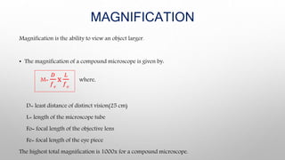
Download to read offline


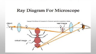
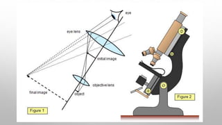


This document describes the components and workings of a compound microscope. A compound microscope uses multiple lenses, including an objective lens and ocular lens, to magnify specimens. The objective lens collects light from the specimen and forms a primary magnified image, which is further magnified by the ocular lens for viewing. Key parts include the mechanical base and arm, illuminating mirrors and condenser, and magnifying objective and ocular lenses. Compound microscopes can magnify specimens up to 1000x and are useful laboratory and educational tools for examining biological and mineral samples.


