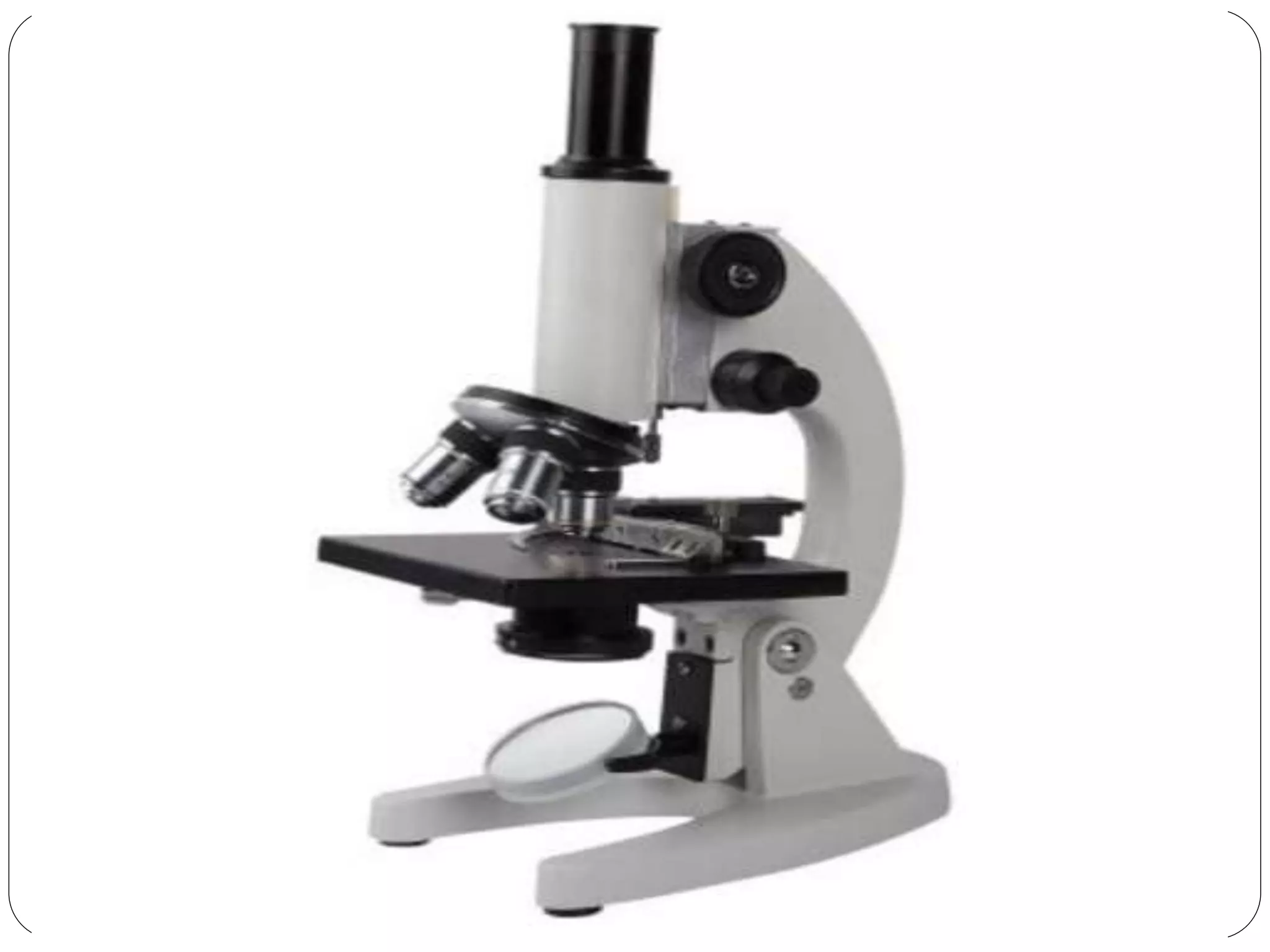
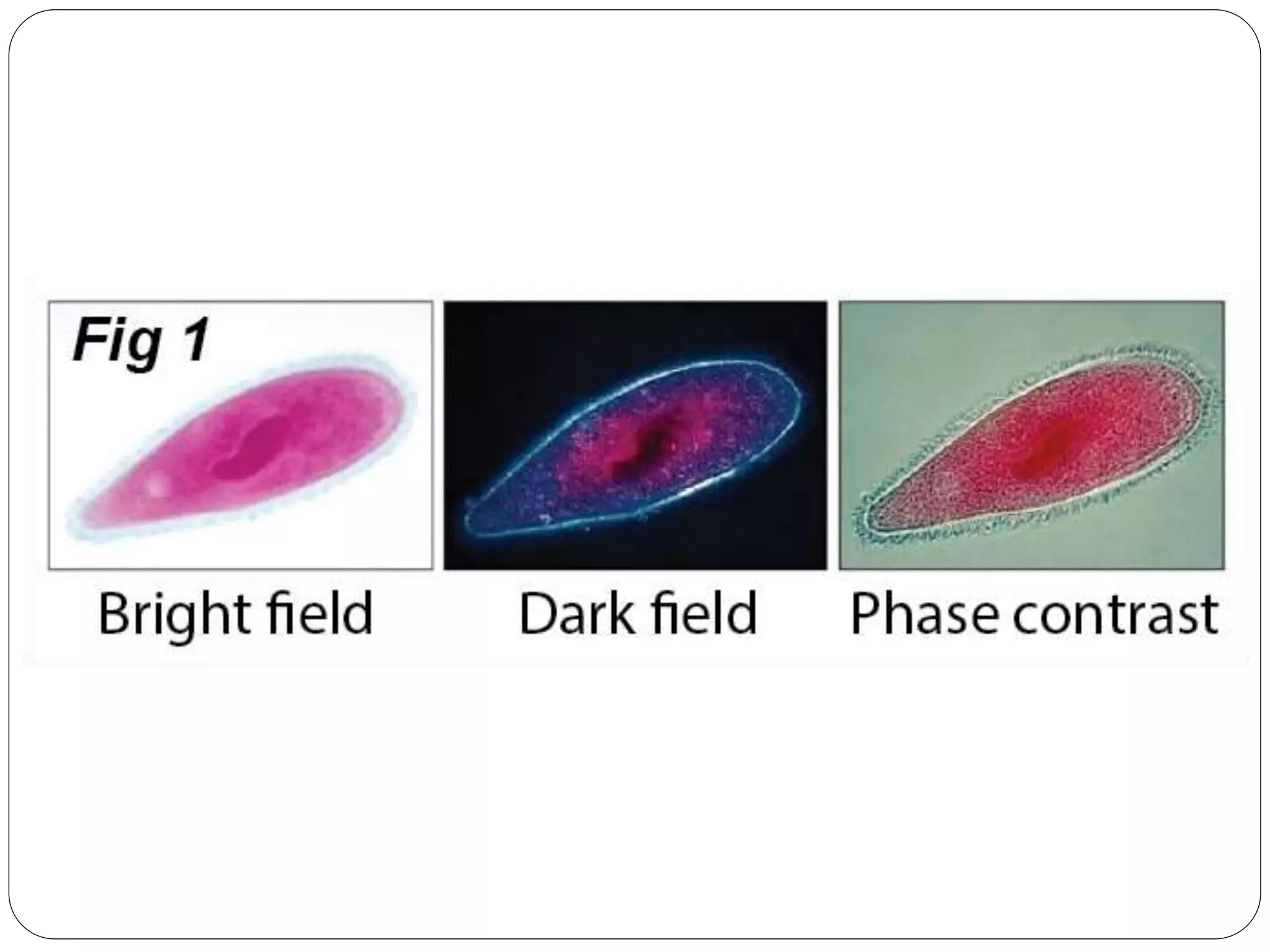
Ankur vashishtha
Microscopy
Why we do microscopy?
 Bacteria of medical importance measure 2-5 µm.
 The resolution power of necked eye is about 200 µm.
 Bacteria being much smaller than the resolution limit, can
be visualized only under the magnification.
 Hence, the study of Microorganism requires the use of
Microscope.
Introduction
 Microscopy is defend as the use of a Microscope (i.e.
visually enlarged) objects too small to be visualized with
the naked eye so that their characteristics are readily
observable.
What is microscope?
 Microscope is an
instrument, which
produce greatly enlarged
image of minute objects.
Applications of Microscopy in Diagnostic
Microbiology
 Rapid organism identification by direct visualization in patient
specimens.
 Rapid final identification of certain organisms by direct
visualization in patient specimens.
 Detection of different organisms present in the same specimen.
 Detection of organisms not easily cultivated in the laboratory.
Properties of a microscope
1. Good resolution
2. Good contrast
3. Good magnification
Good resolution
Resolution power is the ability to produce separate images of closely
placed objects so that they can be distinguished as two separate
entities.
o Oil has a higher refractive index than air; hence, enhances the
resolution power.
Good contrast
Can be improved by staining the specimen. When the stains
bind to the cells, the contrast is increased.
Good magnification
Achieved by use of lenses.
o Ocular lens with a magnification power of
10x, Objective lens-scanning (4x), low power
(10x), high power (40x) and oil immersion
(100x).
o Total magnification = magnification of
objective lens * ocular lens. Magnification of
scanning field, low power, high power and oil
immersion field is 40x, 100x, 400x and
1000x respectively.
Types of Microscopes
1. Bright field or light microscope.
2. Dark field (dark ground) microscope.
3. Phase contrast microscope.
4. Fluorescent microscope.
5. Electron microscope.
Bright field or light microscope.
 The bright-field or light microscope forms a dark
image against a brighter background, hence the
name.
 Parts in a bright-field microscope are divided into
three groups.
1. Mechanical part 2. Magnifying parts 3. Illuminating part
 Base
 C-shaped arm
 Mechanical
stage
 Ocular lens
 Objective lens
 Condenser
 Iris diaphragm
 Light source
 Fine & coarse
adjustment
1.
Working Principle
Dark field (dark ground) microscope
 Working principle - Object appears bright against a dark
background- by use of a special dark field condenser.
 The condenser is blackened in the centre and light passes
through its periphery illuminating the living microorganism
on a glass slide.
 To identify the living, unstained cells and thin bacteria
like spirochetes which cannot be visualized by light
Working Principle
Phase contrast microscope
o The condenser is similar to
that of dark field microscope.
o Cone of light passes through
a cell - some light rays are
bent due to variations in
density and refractive index
within the specimen and are
retarded by about one-fourth
of a wavelength.
Light rays go through →
condenser → specimen →
phase ring → objective lens
→ ocular lens.
Working Principle
Use of phase contrast microscope
 Studying microbial motility.
 Determining the shape of living cells.
 Detecting bacterial components, such as
endospores and inclusion bodies (have refractive
indices markedly different from that of water).
Fluorescence microscope
 By the use of dyes, some microorganisms and particles
having low wavelength which is invisible to eye are
converted into longer wavelength which is visible to the
eyes.
 UV light is used for illumination.
 This method is used for demonstration of microorganisms ;
naturally occurring fluorescent material or particles after
staining with some fluorescent dyes eg. M. tuberculosis.
Working Principle
 Exciting rays get
reflected by a
dichromatic mirror -
fall on the specimen
stained by
fluorescent dye.
 Barrier filter –
removes remaining
ultraviolet light
(damage the
viewer’s eyes), or
blue and violet light
(reduce contrast).
Electron microscope
 The electron microscope uses an electron beam
to create an image, with electromagnets acting as
lenses.
 The limit of resolution is improved by a factor of
1000 (theoretically down to 0.1 nm, but more
realistically down to 2 nm) over the light
microscope.
 Electron microscopes are of two types:
1. Transmission electron microscope
2. Scanning electron microscope
Transmission Electron Microscope. Scanning Electron Microscope.
THANK
YOU

Microscope

  • 1.
  • 2.
    Why we domicroscopy?  Bacteria of medical importance measure 2-5 µm.  The resolution power of necked eye is about 200 µm.  Bacteria being much smaller than the resolution limit, can be visualized only under the magnification.  Hence, the study of Microorganism requires the use of Microscope.
  • 3.
    Introduction  Microscopy isdefend as the use of a Microscope (i.e. visually enlarged) objects too small to be visualized with the naked eye so that their characteristics are readily observable.
  • 4.
    What is microscope? Microscope is an instrument, which produce greatly enlarged image of minute objects.
  • 5.
    Applications of Microscopyin Diagnostic Microbiology  Rapid organism identification by direct visualization in patient specimens.  Rapid final identification of certain organisms by direct visualization in patient specimens.  Detection of different organisms present in the same specimen.  Detection of organisms not easily cultivated in the laboratory.
  • 6.
    Properties of amicroscope 1. Good resolution 2. Good contrast 3. Good magnification
  • 7.
    Good resolution Resolution poweris the ability to produce separate images of closely placed objects so that they can be distinguished as two separate entities. o Oil has a higher refractive index than air; hence, enhances the resolution power.
  • 8.
    Good contrast Can beimproved by staining the specimen. When the stains bind to the cells, the contrast is increased.
  • 9.
    Good magnification Achieved byuse of lenses. o Ocular lens with a magnification power of 10x, Objective lens-scanning (4x), low power (10x), high power (40x) and oil immersion (100x). o Total magnification = magnification of objective lens * ocular lens. Magnification of scanning field, low power, high power and oil immersion field is 40x, 100x, 400x and 1000x respectively.
  • 10.
    Types of Microscopes 1.Bright field or light microscope. 2. Dark field (dark ground) microscope. 3. Phase contrast microscope. 4. Fluorescent microscope. 5. Electron microscope.
  • 11.
    Bright field orlight microscope.  The bright-field or light microscope forms a dark image against a brighter background, hence the name.  Parts in a bright-field microscope are divided into three groups. 1. Mechanical part 2. Magnifying parts 3. Illuminating part  Base  C-shaped arm  Mechanical stage  Ocular lens  Objective lens  Condenser  Iris diaphragm  Light source  Fine & coarse adjustment
  • 12.
  • 15.
  • 16.
    Dark field (darkground) microscope  Working principle - Object appears bright against a dark background- by use of a special dark field condenser.  The condenser is blackened in the centre and light passes through its periphery illuminating the living microorganism on a glass slide.  To identify the living, unstained cells and thin bacteria like spirochetes which cannot be visualized by light
  • 17.
  • 18.
    Phase contrast microscope oThe condenser is similar to that of dark field microscope. o Cone of light passes through a cell - some light rays are bent due to variations in density and refractive index within the specimen and are retarded by about one-fourth of a wavelength.
  • 19.
    Light rays gothrough → condenser → specimen → phase ring → objective lens → ocular lens. Working Principle
  • 20.
    Use of phasecontrast microscope  Studying microbial motility.  Determining the shape of living cells.  Detecting bacterial components, such as endospores and inclusion bodies (have refractive indices markedly different from that of water).
  • 21.
    Fluorescence microscope  Bythe use of dyes, some microorganisms and particles having low wavelength which is invisible to eye are converted into longer wavelength which is visible to the eyes.  UV light is used for illumination.  This method is used for demonstration of microorganisms ; naturally occurring fluorescent material or particles after staining with some fluorescent dyes eg. M. tuberculosis.
  • 22.
    Working Principle  Excitingrays get reflected by a dichromatic mirror - fall on the specimen stained by fluorescent dye.  Barrier filter – removes remaining ultraviolet light (damage the viewer’s eyes), or blue and violet light (reduce contrast).
  • 23.
    Electron microscope  Theelectron microscope uses an electron beam to create an image, with electromagnets acting as lenses.  The limit of resolution is improved by a factor of 1000 (theoretically down to 0.1 nm, but more realistically down to 2 nm) over the light microscope.
  • 24.
     Electron microscopesare of two types: 1. Transmission electron microscope 2. Scanning electron microscope Transmission Electron Microscope. Scanning Electron Microscope.
  • 25.