Recommended
PPT
PPTX
PPTX
PPTX
Study of microscope in pharmacy degree.pptx
PPTX
PDF
🧫 Compound Microscope microbiology pdf notes
PPT
Q2 module-1-2 -microscope
PPT
Q2 module-1-2 -microscope
PDF
General knowledge about compound microscopes
PPTX
PPTX
PPT on Microscopy - Compound Microscope, History, Parts and Function
PPTX
Using_Compofbdcbdcc cndund_Microscope.pptx
PPTX
W1 - MICROSCOPE.pptx for grade 7 students
PPTX
sci 7 q2 1 Identify the parts and functions, and demonstrate proper handling ...
PPTX
THE PARTS OF THE MICROSCOPE: UNDERSTANDING MICROSCOPY
PPTX
INTRODUCTION TO MICROSCOPE- SIMPLE & COMPOUND
PPTX
10 DEV SINGH.pptxxcvbnm,xcvbnmsdfcgvhbnZ
PPTX
Science Subject_ Lesson on Compound Microscope
PPTX
compoundmicroscope-201031072503.pptx....
PPTX
learn the history, parts, and functions of microscope
PPT
Introduction To Microscopes History & Parts
PPTX
PPTX
Clinical microscopefor biomedical hag) .pptx
PDF
PPTX
microscope thelat help to see tissue under slide
PPTX
compound microscope (basic)
PPTX
Temporary Compound microscope slide .pptx
PPTX
Compound-Microscope-Unveiling-the-Unseen.pptx
PDF
19 January 2026 Andreas Schleicher Digital Education Outlook 2026.pdf
DOCX
COMMUNITY HEALTH NURSING OSCE CHECKLIST.docx
More Related Content
PPT
PPTX
PPTX
PPTX
Study of microscope in pharmacy degree.pptx
PPTX
PDF
🧫 Compound Microscope microbiology pdf notes
PPT
Q2 module-1-2 -microscope
PPT
Q2 module-1-2 -microscope
Similar to Compound Microscope microbiology slide.pptx
PDF
General knowledge about compound microscopes
PPTX
PPTX
PPT on Microscopy - Compound Microscope, History, Parts and Function
PPTX
Using_Compofbdcbdcc cndund_Microscope.pptx
PPTX
W1 - MICROSCOPE.pptx for grade 7 students
PPTX
sci 7 q2 1 Identify the parts and functions, and demonstrate proper handling ...
PPTX
THE PARTS OF THE MICROSCOPE: UNDERSTANDING MICROSCOPY
PPTX
INTRODUCTION TO MICROSCOPE- SIMPLE & COMPOUND
PPTX
10 DEV SINGH.pptxxcvbnm,xcvbnmsdfcgvhbnZ
PPTX
Science Subject_ Lesson on Compound Microscope
PPTX
compoundmicroscope-201031072503.pptx....
PPTX
learn the history, parts, and functions of microscope
PPT
Introduction To Microscopes History & Parts
PPTX
PPTX
Clinical microscopefor biomedical hag) .pptx
PDF
PPTX
microscope thelat help to see tissue under slide
PPTX
compound microscope (basic)
PPTX
Temporary Compound microscope slide .pptx
PPTX
Compound-Microscope-Unveiling-the-Unseen.pptx
Recently uploaded
PDF
19 January 2026 Andreas Schleicher Digital Education Outlook 2026.pdf
DOCX
COMMUNITY HEALTH NURSING OSCE CHECKLIST.docx
PDF
GIÁO ÁN KẾ HOẠCH BÀI DẠY NĂNG LỰC SỐ MÔN TIẾNG ANH LỚP 11 CẢ NĂM - GLOBAL SUC...
PDF
Caribbean Examinations Council Literacy and Numeracy Standards
PPTX
Statistical Data Analysis using R Programming.pptx
PPTX
Burnout_among_medical teachers_&_mentoring .pptx
PDF
GIÁO ÁN KẾ HOẠCH BÀI DẠY NĂNG LỰC SỐ MÔN TIẾNG ANH LỚP 12 CẢ NĂM - GLOBAL SUC...
PDF
ĐỀ MINH HỌA KỲ THI TỐT NGHIỆP TRUNG HỌC PHỔ THÔNG NĂM 2026 MÔN TIẾNG ANH 2026...
PPTX
Overview of Unapproved Timeoff in Odoo 19
PDF
To synthesis and submit Benzamide synthesis.pdf
PPTX
Accounting Theory Group Presentation - Why Study Accounting Theory
PPTX
Setting Up and Processing Down Payments in Odoo 18 Sales
PPTX
How to Add or Remove Multiple Followers in a Records
PPTX
How to Manage Product Types in Odoo 18 Sales
PPTX
Introduction of Carbohydrates - Dr.M.Jothimuniyandi
PPTX
OXYGEN ADMINISTRATION/THERAPY ......pptx
PDF
The Unique Wildlife of Ethiopia: From the Simien to the Bale Mountains
PPTX
How to Manage Empty Location in Odoo 18 Inventory
PPTX
Activity on Job position in Odoo 19 Recruitment
PPTX
NMR Spectroscopy: Principles and Applications
Compound Microscope microbiology slide.pptx 1. 2. Introduction
• A microscope is an instrument used to see objects that are too small for the nak
• Microscopes are vital in biology, medicine, and material science.
• Types include light, electron, and compound microscopes.
3. What is a Compound Microscope?
• A compound microscope uses two sets of lenses to magnify objects.
• It provides high magnification and better resolution than simple microscopes.
4. History of the Compound Microscope
• Invented in the late 16th century.
• Zacharias Janssen credited with early development.
• Antonie van Leeuwenhoek improved lens quality.
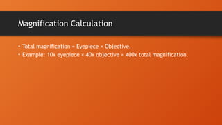
5. Working Principle
• Light passes through the specimen.
• Objective lens creates a magnified image.
• Eyepiece lens further magnifies the image.
6. 7. Structural Parts
• Base: Supports microscope.
• Arm: Holds body tube and connects to base.
• Stage: Platform where slides are placed.
8. Eyepiece (Ocular Lens)
• Top lens you look through.
• Common magnification is 10x.
• May have pointer or measurement scale.
9. 10. 11. Light Source and Condenser
• Mirror or electric light source.
• Condenser focuses light onto the slide.
12. Diaphragm and Iris
• Regulates amount of light reaching the specimen.
• Improves contrast and clarity.
13. Stage Clips and Nosepiece
• Clips hold slide steady.
• Nosepiece rotates objective lenses.
14. 15. Proper Usage Steps
• Carry with two hands.
• Start with lowest objective.
• Adjust light and focus carefully.
16. Care and Maintenance
• Clean lenses with lens paper.
• Cover when not in use.
• Avoid touching lenses with fingers.
17. 18. 19. 20. Conclusion & Q&A
• Compound microscopes are essential tools in science.
• Understanding their parts and usage enhances learning and research.