Download to read offline
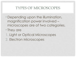
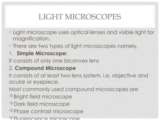


The document provides an overview of different types of microscopes, primarily categorizing them into light (optical) and electron microscopes. Light microscopes are further divided into simple and compound microscopes, detailing their components and functions, including illumination and imaging systems. It also addresses the limitations of compound microscopes in terms of magnification power and resolution.