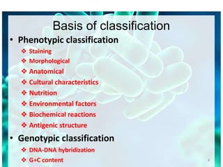
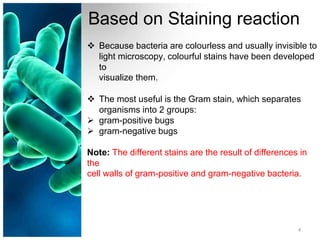
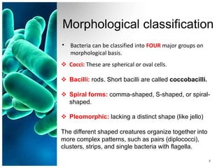
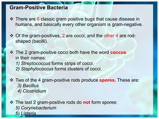
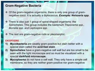
CLASSIFICATION OF
BACTERIA
1
1
BACTERIAL TAXONOMY
 All organisms have a name consisting of two parts:
the
genus followed by the species (i.e., Homo sapiens).
 This is no different from bacteria
Genus species
Bacillus subtilis
Clostridium tetani
Staphylococcus aureus
2
Basis of classification
• Phenotypic classification
❖ Staining
❖ Morphological
❖ Anatomical
❖ Cultural characteristics
❖ Nutrition
❖ Environmental factors
❖ Biochemical reactions
❖ Antigenic structure
• Genotypic classification
❖ DNA-DNA hybridization
❖ G+C content 2
3
Based on Staining reaction
 Because bacteria are colourless and usually invisible to
light microscopy, colourful stains have been developed
to
visualize them.
 The most useful is the Gram stain, which separates
organisms into 2 groups:
 gram-positive bugs
 gram-negative bugs
Note: The different stains are the result of differences in
the
cell walls of gram-positive and gram-negative bacteria.
4
Based on Staining reaction
5
• GRAM’S STAIN
– Gram-positive cocci – Staphylococcus aureus
– Gram-negative cocci – Neisseria gonorrhoeae
– Gram-positive rods – Clostridium spp.
– Gram-negative rods – E. coli
• ACID FAST STAIN
– Acid-fast bacilli –Mycobacterium tuberculosis
– Non-acid-fast bacilli – Staphylococcus aureus
Morphological classification
• Bacteria can be classified into FOUR major groups on
morphological basis.
 Cocci: These are spherical or oval cells.
 Bacilli: rods. Short bacilli are called coccobacilli.
 Spiral forms: comma-shaped, S-shaped, or spiral-
shaped.
 Pleomorphic: lacking a distinct shape (like jello)
The different shaped creatures organize together into
more complex patterns, such as pairs (diplococci),
clusters, strips, and single bacteria with flagella.
3
6
7
8
Gram-Positive Bacteria
 There are 6 classic gram positive bugs that cause disease in
humans, and basically every other organism is gram-negative.
 Of the gram-positives, 2 are cocci, and the other 4 are rod-
shaped (bacilli).
 The 2 gram-positive cocci both have the word coccus
in their names:
1) Streptococcus forms strips of cocci.
2) Staphylococcus forms clusters of cocci.
 Two of the 4 gram-positive rods produce spores. These are:
3) Bacillus
4) Clostridium
 The last 2 gram-positive rods do not form spores:
5) Corynebacterium
6) Listeria 9
Gram-Negative Bacteria
 Of the gram-negative organisms, there is only one group of gram-
negative cocci. It is actually a diplococcus. Example: Neisseria spp.
 There is also just 1 group of spiral-shaped organisms: the
Spirochetes. This group includes the bacterium Treponema spp.,
Borrelia spp. and Leptospira spp.
 The rest are gram-negative rods or pleomorphic.
EXCEPTIONS
1) Mycobacteria are weakly gram-positive but stain better with a
special stain called the acid-fast stain
2) Spirochetes have a gram-negative cell wall but are too small to be
seen with the light microscope and so must be visualized with a
special darkfield microscope.
3) Mycoplasma do not have a cell wall. They only have a simple cell
membrane, so they are neither gram-positive nor gram-negative
1
0
Based on Anatomical features
• Capsule
– Capsulate– Streptococcus pneumoniae
– Non-capsulate – Viridans streptococci
Flagella
– Flagellate – vibro cholerae
– Aflagellate – Shigella spp.
Spore
– Spore-forming – Bacillus spp.
– Non-sporing – Escherichia coli
•
10
1
1
Based on Cultural characteristics
12
• Extra growth factors requirements
– Fastidious – Hemophilus influenzae
– Non-fastidious – Escherichia coli
• Hemolysis on Sheep Blood Agar
– Alpha-hemolysis – Streptococcus pneumoniae
– Beta-hemolysis – Streptococcus pyogenes
• Utilization of carbohydrates
– Oxidative - Micrococcus
– Fermentative – Escherichia coli
Based on Cultural characteristics
13
• Growth rate
– Rapid growers– Vibrio cholerae
– Slow growers – Mycobacterium tuberculosis
• Pigment production
– Pigment producer – Staphylococcus aureus
– Pigment non-producer – Escherichia coli
Based on Nutrition
14
• Autotrophs: use inorganic sources, such
as ammonium and sulphide as energy
sources.
Heterotrophs: use organic carbon
sources as energy sources
Based on environmental factors
15
• Temperature
• Oxygen dependence
• pH
• Salt concentration
• Atmospheric pressure
Temperature
• Psychrophiles (15-200
C) – Pseudomonas
fluorescens
• Mesophiles (20-400
C) – Escherichia coli,
Salmonella enterica, Staphylococcus aureus
• Thermophiles (50-600
C)- Bacillus
stearothermophilus
• Extremely thermophiles (as high as 2500
C)
16
Oxygen dependence
• Aerobe (grow in ambient temperature, which
contains 21% O2 and a small amount of CO2
,
0.03%)
• Obligate aerobes – Strictly require O2for their
growth (Pseudomonas aeruginosa)
• Microaerophilic (grow under reduced O2
, 5-
10% and increased CO2
, 8-10%)- Campylobacter
jejuni, Helicobacter pylori
17
Oxygen dependence
• Facultative anaerobe (capable of growing
either in presence or absence of O2
)- E. coli
• Obligate anaerobe – Clostridium spp.
• Capnophilic (require increased concentration
of CO2
, i.e., 5-10%) –
H. influenzae,
N. gonorrhoeae
• Aerotolerant 18
pH
• Acidophiles (Lactobacillus acidophilus)
• Alkaliphiles (Vibrio)
• Neutralophiles (pH 6-8)
Majority of the medically important bacteria grow
best at neutral or slightly alkaline reaction (pH
7.2-7.6)
19
Salt concentration
• Halophiles
• Non-halophiles
20
Other ways of classification
21
•
•
•
•
Motile/Non-motile
Pathogenic/Non-pathogenic
Sensitive/Resistant (to particular antibiotic/ chemicals)
Lactose fermenter/Lactose non-fermenter
5
Time (Minutes)
Bacterial Growth Curve
1 10
Lag phase
Stationary phase
Death
phase
Log phase
22

classification of bacteria.pptx

  • 1.
  • 2.
    BACTERIAL TAXONOMY  Allorganisms have a name consisting of two parts: the genus followed by the species (i.e., Homo sapiens).  This is no different from bacteria Genus species Bacillus subtilis Clostridium tetani Staphylococcus aureus 2
  • 3.
    Basis of classification •Phenotypic classification ❖ Staining ❖ Morphological ❖ Anatomical ❖ Cultural characteristics ❖ Nutrition ❖ Environmental factors ❖ Biochemical reactions ❖ Antigenic structure • Genotypic classification ❖ DNA-DNA hybridization ❖ G+C content 2 3
  • 4.
    Based on Stainingreaction  Because bacteria are colourless and usually invisible to light microscopy, colourful stains have been developed to visualize them.  The most useful is the Gram stain, which separates organisms into 2 groups:  gram-positive bugs  gram-negative bugs Note: The different stains are the result of differences in the cell walls of gram-positive and gram-negative bacteria. 4
  • 5.
    Based on Stainingreaction 5 • GRAM’S STAIN – Gram-positive cocci – Staphylococcus aureus – Gram-negative cocci – Neisseria gonorrhoeae – Gram-positive rods – Clostridium spp. – Gram-negative rods – E. coli • ACID FAST STAIN – Acid-fast bacilli –Mycobacterium tuberculosis – Non-acid-fast bacilli – Staphylococcus aureus
  • 6.
    Morphological classification • Bacteriacan be classified into FOUR major groups on morphological basis.  Cocci: These are spherical or oval cells.  Bacilli: rods. Short bacilli are called coccobacilli.  Spiral forms: comma-shaped, S-shaped, or spiral- shaped.  Pleomorphic: lacking a distinct shape (like jello) The different shaped creatures organize together into more complex patterns, such as pairs (diplococci), clusters, strips, and single bacteria with flagella. 3 6
  • 7.
  • 8.
  • 9.
    Gram-Positive Bacteria  Thereare 6 classic gram positive bugs that cause disease in humans, and basically every other organism is gram-negative.  Of the gram-positives, 2 are cocci, and the other 4 are rod- shaped (bacilli).  The 2 gram-positive cocci both have the word coccus in their names: 1) Streptococcus forms strips of cocci. 2) Staphylococcus forms clusters of cocci.  Two of the 4 gram-positive rods produce spores. These are: 3) Bacillus 4) Clostridium  The last 2 gram-positive rods do not form spores: 5) Corynebacterium 6) Listeria 9
  • 10.
    Gram-Negative Bacteria  Ofthe gram-negative organisms, there is only one group of gram- negative cocci. It is actually a diplococcus. Example: Neisseria spp.  There is also just 1 group of spiral-shaped organisms: the Spirochetes. This group includes the bacterium Treponema spp., Borrelia spp. and Leptospira spp.  The rest are gram-negative rods or pleomorphic. EXCEPTIONS 1) Mycobacteria are weakly gram-positive but stain better with a special stain called the acid-fast stain 2) Spirochetes have a gram-negative cell wall but are too small to be seen with the light microscope and so must be visualized with a special darkfield microscope. 3) Mycoplasma do not have a cell wall. They only have a simple cell membrane, so they are neither gram-positive nor gram-negative 1 0
  • 11.
    Based on Anatomicalfeatures • Capsule – Capsulate– Streptococcus pneumoniae – Non-capsulate – Viridans streptococci Flagella – Flagellate – vibro cholerae – Aflagellate – Shigella spp. Spore – Spore-forming – Bacillus spp. – Non-sporing – Escherichia coli • 10 1 1
  • 12.
    Based on Culturalcharacteristics 12 • Extra growth factors requirements – Fastidious – Hemophilus influenzae – Non-fastidious – Escherichia coli • Hemolysis on Sheep Blood Agar – Alpha-hemolysis – Streptococcus pneumoniae – Beta-hemolysis – Streptococcus pyogenes • Utilization of carbohydrates – Oxidative - Micrococcus – Fermentative – Escherichia coli
  • 13.
    Based on Culturalcharacteristics 13 • Growth rate – Rapid growers– Vibrio cholerae – Slow growers – Mycobacterium tuberculosis • Pigment production – Pigment producer – Staphylococcus aureus – Pigment non-producer – Escherichia coli
  • 14.
    Based on Nutrition 14 •Autotrophs: use inorganic sources, such as ammonium and sulphide as energy sources. Heterotrophs: use organic carbon sources as energy sources
  • 15.
    Based on environmentalfactors 15 • Temperature • Oxygen dependence • pH • Salt concentration • Atmospheric pressure
  • 16.
    Temperature • Psychrophiles (15-200 C)– Pseudomonas fluorescens • Mesophiles (20-400 C) – Escherichia coli, Salmonella enterica, Staphylococcus aureus • Thermophiles (50-600 C)- Bacillus stearothermophilus • Extremely thermophiles (as high as 2500 C) 16
  • 17.
    Oxygen dependence • Aerobe(grow in ambient temperature, which contains 21% O2 and a small amount of CO2 , 0.03%) • Obligate aerobes – Strictly require O2for their growth (Pseudomonas aeruginosa) • Microaerophilic (grow under reduced O2 , 5- 10% and increased CO2 , 8-10%)- Campylobacter jejuni, Helicobacter pylori 17
  • 18.
    Oxygen dependence • Facultativeanaerobe (capable of growing either in presence or absence of O2 )- E. coli • Obligate anaerobe – Clostridium spp. • Capnophilic (require increased concentration of CO2 , i.e., 5-10%) – H. influenzae, N. gonorrhoeae • Aerotolerant 18
  • 19.
    pH • Acidophiles (Lactobacillusacidophilus) • Alkaliphiles (Vibrio) • Neutralophiles (pH 6-8) Majority of the medically important bacteria grow best at neutral or slightly alkaline reaction (pH 7.2-7.6) 19
  • 20.
  • 21.
    Other ways ofclassification 21 • • • • Motile/Non-motile Pathogenic/Non-pathogenic Sensitive/Resistant (to particular antibiotic/ chemicals) Lactose fermenter/Lactose non-fermenter
  • 22.
    5 Time (Minutes) Bacterial GrowthCurve 1 10 Lag phase Stationary phase Death phase Log phase 22