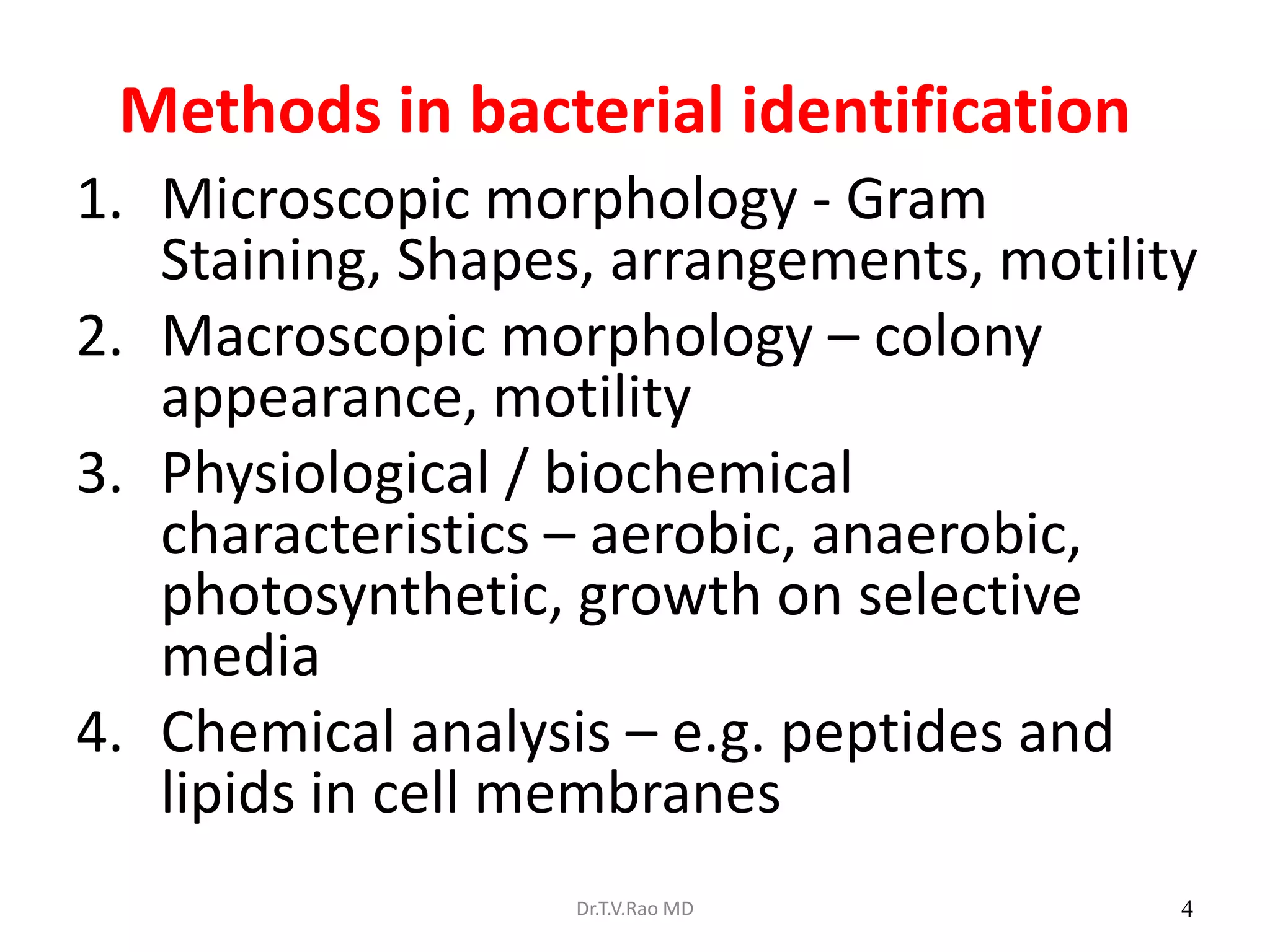
This document provides information on identifying bacteria. It discusses the hierarchy of biological classification and describes methods for bacterial identification including microscopic morphology, macroscopic morphology, physiological/biochemical characteristics, and genetic/molecular analysis. Steps for diagnostic isolation and identification are outlined beginning with streaking samples on culture plates and observing colony characteristics. Methods for examining bacterial cells like Gram stain, flagella, capsules, and spores are covered. Biochemical tests for identification like indole, methyl red, Voges-Proskauer, citrate, and lactose fermentation are also discussed.


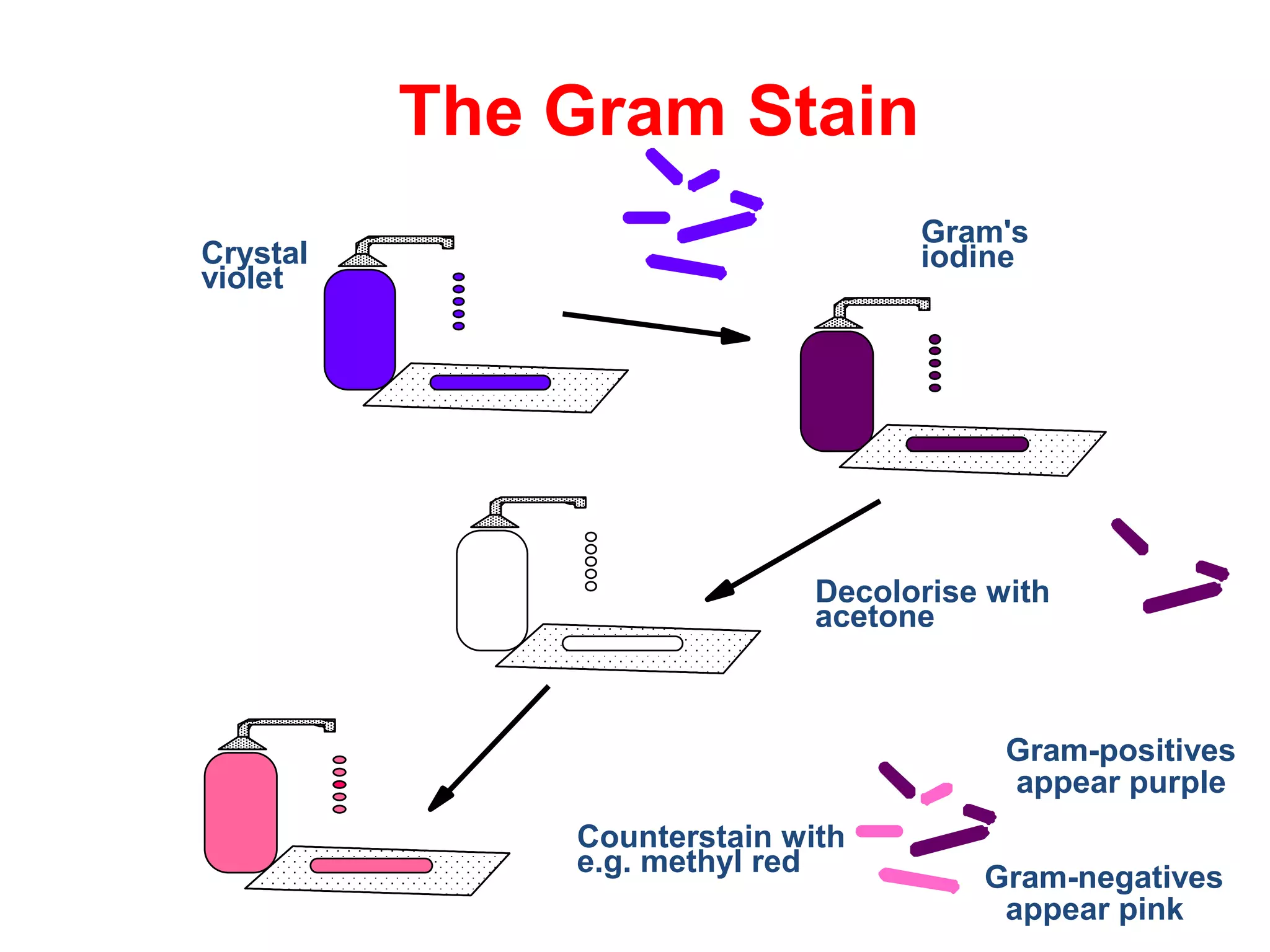
![Gram stain
a. Gram stain divides the bacteria into Gram positive
& Gram negative.
The basic procedure :
i. Take a heat fixed bacterial smear.
ii. Flood the smear with CRYSTAL VIOLET or Methyl violet for 1
minute, then wash with water. [PRIMARY STAIN]
iii. Flood the smear with IODINE for 1 minute, then wash with water.
iv. Flood the smear with ETHANOL-ACETONE, quickly, then wash with
water. [DECOLORI
v. Flood the smear with SAFRANIN for 1 minute, then wash with
water. [COUNTERSTAIN]
vi. Blot the smear, air dry and observe.
Dr.T.V.Rao MD 12](https://image.slidesharecdn.com/identificationofbacteria-180120102225-211202091130/75/Identificationofbacteria-180120102225-12-2048.jpg)