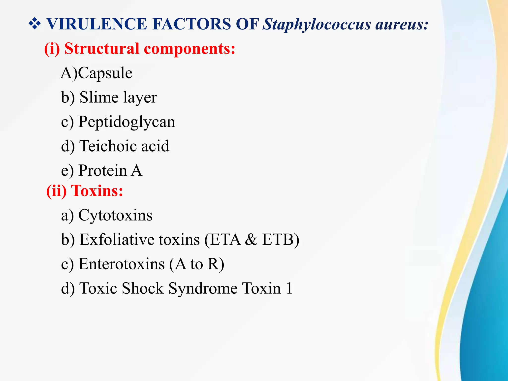
Staphylococcus aureus is a gram-positive, spherical bacterium that can cause several diseases in humans. It is a normal member of the skin and nasal flora but can become pathogenic. S. aureus produces several toxins and enzymes that allow it to infect the skin, blood, lungs, heart, bones and joints. Diseases include impetigo, cellulitis, abscesses, pneumonia, osteomyelitis, endocarditis and toxic shock syndrome. Laboratory diagnosis involves culturing and identifying its characteristic gram-positive cocci in clusters and positive tests for catalase, coagulase and DNase. Treatment involves antibiotics like oxacillin or vancomycin depending on antibiotic resistance.
The genus Shigella exclusively infects human intestine.
Shigella dysenteriae is the causative agent of bacillary dysentery or shigellosis in humans.
It is a diarrheal illness which is characterized by frequent passage of blood stained mucopurulent stools.
The four important species of the genus Shigella are:
Shigella dysenteriae
Shigella flexneri
Shigella sonnei
Shigella boydii.
Staphylococcus aureus is a bacterium that causes staphylococcal food poisoning, a form of gastroenteritis with rapid onset of symptoms. S. aureus is commonly found in the environment (soil, water and air) and is also found in the nose and on the skin of humans.
Microbiology of E coli giving basic of Escherichia coli, its morphology, cultural and biochemical characteristics, Antigenic character, pathogenesis, laboratory diagnosis, prevention and control
Lecture notes of Staphylococcus. A detailed account on the morphology, culture characteristics, biochemical characteristics, pathogenesis, laboratory diagnosis of S. aureus.
one of the bacteria named staphylococcus which causes infection in human, from mild to severe.
It is useful for all medical students and paramedical students.
Microbial corrosion, also called microbiologically influenced corrosion, microbially induced corrosion or biocorrosion, is "corrosion affected by the presence or activity of microorganisms in biofilms on the surface of the corroding material."
The increased availability of biomedical data, particularly in the public domain, offers the opportunity to better understand human health and to develop effective therapeutics for a wide range of unmet medical needs. However, data scientists remain stymied by the fact that data remain hard to find and to productively reuse because data and their metadata i) are wholly inaccessible, ii) are in non-standard or incompatible representations, iii) do not conform to community standards, and iv) have unclear or highly restricted terms and conditions that preclude legitimate reuse. These limitations require a rethink on data can be made machine and AI-ready - the key motivation behind the FAIR Guiding Principles. Concurrently, while recent efforts have explored the use of deep learning to fuse disparate data into predictive models for a wide range of biomedical applications, these models often fail even when the correct answer is already known, and fail to explain individual predictions in terms that data scientists can appreciate. These limitations suggest that new methods to produce practical artificial intelligence are still needed.
In this talk, I will discuss our work in (1) building an integrative knowledge infrastructure to prepare FAIR and "AI-ready" data and services along with (2) neurosymbolic AI methods to improve the quality of predictions and to generate plausible explanations. Attention is given to standards, platforms, and methods to wrangle knowledge into simple, but effective semantic and latent representations, and to make these available into standards-compliant and discoverable interfaces that can be used in model building, validation, and explanation. Our work, and those of others in the field, creates a baseline for building trustworthy and easy to deploy AI models in biomedicine.
Bio
Dr. Michel Dumontier is the Distinguished Professor of Data Science at Maastricht University, founder and executive director of the Institute of Data Science, and co-founder of the FAIR (Findable, Accessible, Interoperable and Reusable) data principles. His research explores socio-technological approaches for responsible discovery science, which includes collaborative multi-modal knowledge graphs, privacy-preserving distributed data mining, and AI methods for drug discovery and personalized medicine. His work is supported through the Dutch National Research Agenda, the Netherlands Organisation for Scientific Research, Horizon Europe, the European Open Science Cloud, the US National Institutes of Health, and a Marie-Curie Innovative Training Network. He is the editor-in-chief for the journal Data Science and is internationally recognized for his contributions in bioinformatics, biomedical informatics, and semantic technologies including ontologies and linked data.
Earliest Galaxies in the JADES Origins Field: Luminosity Function and Cosmic ...Sérgio Sacani
We characterize the earliest galaxy population in the JADES Origins Field (JOF), the deepest
imaging field observed with JWST. We make use of the ancillary Hubble optical images (5 filters
spanning 0.4−0.9µm) and novel JWST images with 14 filters spanning 0.8−5µm, including 7 mediumband filters, and reaching total exposure times of up to 46 hours per filter. We combine all our data
at > 2.3µm to construct an ultradeep image, reaching as deep as ≈ 31.4 AB mag in the stack and
30.3-31.0 AB mag (5σ, r = 0.1” circular aperture) in individual filters. We measure photometric
redshifts and use robust selection criteria to identify a sample of eight galaxy candidates at redshifts
z = 11.5 − 15. These objects show compact half-light radii of R1/2 ∼ 50 − 200pc, stellar masses of
M⋆ ∼ 107−108M⊙, and star-formation rates of SFR ∼ 0.1−1 M⊙ yr−1
. Our search finds no candidates
at 15 < z < 20, placing upper limits at these redshifts. We develop a forward modeling approach to
infer the properties of the evolving luminosity function without binning in redshift or luminosity that
marginalizes over the photometric redshift uncertainty of our candidate galaxies and incorporates the
impact of non-detections. We find a z = 12 luminosity function in good agreement with prior results,
and that the luminosity function normalization and UV luminosity density decline by a factor of ∼ 2.5
from z = 12 to z = 14. We discuss the possible implications of our results in the context of theoretical
models for evolution of the dark matter halo mass function.
Seminar of U.V. Spectroscopy by SAMIR PANDASAMIR PANDA
Spectroscopy is a branch of science dealing the study of interaction of electromagnetic radiation with matter.
Ultraviolet-visible spectroscopy refers to absorption spectroscopy or reflect spectroscopy in the UV-VIS spectral region.
Ultraviolet-visible spectroscopy is an analytical method that can measure the amount of light received by the analyte.
(May 29th, 2024) Advancements in Intravital Microscopy- Insights for Preclini...Scintica Instrumentation
Intravital microscopy (IVM) is a powerful tool utilized to study cellular behavior over time and space in vivo. Much of our understanding of cell biology has been accomplished using various in vitro and ex vivo methods; however, these studies do not necessarily reflect the natural dynamics of biological processes. Unlike traditional cell culture or fixed tissue imaging, IVM allows for the ultra-fast high-resolution imaging of cellular processes over time and space and were studied in its natural environment. Real-time visualization of biological processes in the context of an intact organism helps maintain physiological relevance and provide insights into the progression of disease, response to treatments or developmental processes.
In this webinar we give an overview of advanced applications of the IVM system in preclinical research. IVIM technology is a provider of all-in-one intravital microscopy systems and solutions optimized for in vivo imaging of live animal models at sub-micron resolution. The system’s unique features and user-friendly software enables researchers to probe fast dynamic biological processes such as immune cell tracking, cell-cell interaction as well as vascularization and tumor metastasis with exceptional detail. This webinar will also give an overview of IVM being utilized in drug development, offering a view into the intricate interaction between drugs/nanoparticles and tissues in vivo and allows for the evaluation of therapeutic intervention in a variety of tissues and organs. This interdisciplinary collaboration continues to drive the advancements of novel therapeutic strategies.
Slide 1: Title Slide
Extrachromosomal Inheritance
Slide 2: Introduction to Extrachromosomal Inheritance
Definition: Extrachromosomal inheritance refers to the transmission of genetic material that is not found within the nucleus.
Key Components: Involves genes located in mitochondria, chloroplasts, and plasmids.
Slide 3: Mitochondrial Inheritance
Mitochondria: Organelles responsible for energy production.
Mitochondrial DNA (mtDNA): Circular DNA molecule found in mitochondria.
Inheritance Pattern: Maternally inherited, meaning it is passed from mothers to all their offspring.
Diseases: Examples include Leber’s hereditary optic neuropathy (LHON) and mitochondrial myopathy.
Slide 4: Chloroplast Inheritance
Chloroplasts: Organelles responsible for photosynthesis in plants.
Chloroplast DNA (cpDNA): Circular DNA molecule found in chloroplasts.
Inheritance Pattern: Often maternally inherited in most plants, but can vary in some species.
Examples: Variegation in plants, where leaf color patterns are determined by chloroplast DNA.
Slide 5: Plasmid Inheritance
Plasmids: Small, circular DNA molecules found in bacteria and some eukaryotes.
Features: Can carry antibiotic resistance genes and can be transferred between cells through processes like conjugation.
Significance: Important in biotechnology for gene cloning and genetic engineering.
Slide 6: Mechanisms of Extrachromosomal Inheritance
Non-Mendelian Patterns: Do not follow Mendel’s laws of inheritance.
Cytoplasmic Segregation: During cell division, organelles like mitochondria and chloroplasts are randomly distributed to daughter cells.
Heteroplasmy: Presence of more than one type of organellar genome within a cell, leading to variation in expression.
Slide 7: Examples of Extrachromosomal Inheritance
Four O’clock Plant (Mirabilis jalapa): Shows variegated leaves due to different cpDNA in leaf cells.
Petite Mutants in Yeast: Result from mutations in mitochondrial DNA affecting respiration.
Slide 8: Importance of Extrachromosomal Inheritance
Evolution: Provides insight into the evolution of eukaryotic cells.
Medicine: Understanding mitochondrial inheritance helps in diagnosing and treating mitochondrial diseases.
Agriculture: Chloroplast inheritance can be used in plant breeding and genetic modification.
Slide 9: Recent Research and Advances
Gene Editing: Techniques like CRISPR-Cas9 are being used to edit mitochondrial and chloroplast DNA.
Therapies: Development of mitochondrial replacement therapy (MRT) for preventing mitochondrial diseases.
Slide 10: Conclusion
Summary: Extrachromosomal inheritance involves the transmission of genetic material outside the nucleus and plays a crucial role in genetics, medicine, and biotechnology.
Future Directions: Continued research and technological advancements hold promise for new treatments and applications.
Slide 11: Questions and Discussion
Invite Audience: Open the floor for any questions or further discussion on the topic.
2. GENERAL CHARACTERISTICS
• Gram’s classification – Gram positive bacteria
• Shape – Spherical (Cocci)
• Arrangement – Bunched or Grape like clusters
• Motility – Non-Motile
• Capsule - Absent
• Endospores – Absent
• Respiration – Aerobic/Facultative anaerobic
• Optimum Temperature - 37 °C
• Optimum pH – 7.0 to 7.5
• Normal human flora on skin and mucosal surfaces.
• The organisms are ubiquitous, so there are no Geographic or seasonal limitations.
• Discovered by Anton J. Rosenbach (German Surgeon) in 1884 .
3. PATHOGENICITY OF Staphylococcus
aureus:
Staphylococcus aureus Infections are mainly transmitted by
a) Direct contact with an infected person
b) By using a contaminated object
c) By inhaling infected droplets dispersed by sneezing or
coughing.
1 to 6 Hours
4. (i) Structural components:
A)Capsule
b) Slime layer
c) Peptidoglycan
d) Teichoic acid
e) Protein A
(ii) Toxins:
a) Cytotoxins
b) Exfoliative toxins (ETA & ETB)
c) Enterotoxins (A to R)
d) Toxic Shock Syndrome Toxin 1
5. (iii) Enzymes:
a) Coagulase
b) Hyaluronic acid
c) Fibrinolysin
d) Lipases
e) Nucleases
f) Beta Lactamase
g) Staphylokinase
6. PATHOGENESIS OF Staphylococcus aureus:
Staphylococcus aureus enters into the surface of the
Skin, grows into Hair follicles and invadesSebaceous
glands.
After that, it triggers Fever and Inflammation, which
are natural responses against infection, and causes the
follicle to enlarge and fill with pus composed of
leukocytes, dead cells, and bacteria.
The infection may spread into the Hypodermis to form
a Furuncle or into neighboring hair follicles to form a
Carbuncle.
7. Staphylococcus aureus may also spread into the
blood - a condition called Bacteremia and is carried to
the lining of the heart, lungs, and bones, causing
Endocarditis, Pneumonia and Osteomyelitis,
respectively.
8. CLINICAL DISEASES CAUSED BY
Staphylococcus aureus:
I) TOXIN – MEDIATED DISEASES :
Also known as Ritter von Ritterschein disease ,
Ritter disease, and Staphylococcal epidermal
necrolysis.
Superficial blistering skin disorders caused by the
Exfoliative toxins of some strains of Staphylococcus
aureus.
Exfoliative toxin that causes the outer layers of Skin to
Blister within the upper layers of the skin) and Peel.
9. Staphylococcal Food Poisoning results after
consumption of food contaminated with heat-stable
Enterotoxin.
Symptoms include Nausea,Vomiting, Abdominal
cramping and Diarrhea.
In more severe cases - Dehydration, Headache, Muscle
cramping, and changes in Blood pressure and Pulse
rate may occur.
The condition is typically over in 2 days. But it is not
unusual for complete recovery to take 3 days and
sometimes longer in severe cases.
10. Toxic Shock Syndrome (TSS) is a rare but serious
medical condition caused by a bacterial infection. The
bacterium Staphylococcus aureus gets into the blood
stream and produces Toxins.
Sudden high fever; Low blood pressure ;Vomiting;
Diarrhea; Rash resembling a Sunburn; Confusion;
Muscle aches; Redness of eyes, mouth & throat;
Seizures; Headaches; Shock; Renal failure and
sometimes Death.
11. (II) SUPPURATIVE INFECTIONS:
The most common cause of impetigo is bacteria called
Staphylococcus aureus. Another bacteria source is group A
Streptococcus.
Impetigo is a common and highly contagious skin infection
that mainly affects infants and children.
Impetigo usually appears as red sores on the face,
especially around a child's nose and mouth, and on hands
and feet.
- Impetigo involving hair follicles
12. - Large, painful, pus-filled
cutaneous nodules.
– Coalescence of Furuncles with
extension into the subcutaneous tissues and evidence of
systemic disease
- Spread of bacteria into the blood from a
focus of infection.
– Endocarditis characterized by damage
to the endothelial lining of the heart.
13. - Consolidation and
abscess formation in the lungs; seen in the very young
and elderly and in patients with underlying or recent
pulmonary disease; a severe form of necrotizing
pneumonia with septic shock and high mortality is
recognized.
- Destruction of bones,
particularly the metaphyseal area of long bones.
Painful erythematous joint with
collection of purulent material in the joint space.
14. LABORATORY DIAGNOSIS OF Staphylococcus aureus:
Gram stating – Violet coloured Gram positive cocci
arranged in form of clusters.
Motility test – Non-Motile.
Nutrient agar - Smooth, golden yellow colonies
Blood agar – Beta Hemolysis.
MacConkey agar – Pink coloured Lactose fermenting
colonies.
Selective medium - Mannitol Salt Agar (MSA) –
15. BIOCHEMICAL TESTS:
a) Catalase test - Positive
b) Oxidase test - Negative
c) Urease test – Negative
d) Indole test - Negative
e) Methyl Red (MR) test – Negative
f) Voges Proskauer (VP) test - Positive
g) Citrate utilization test – Positive
h) Starch Hydrolysis – Negative
i) Casein Hydrolysis - Negative
k) Tellurite Reduction Test – Positive
l) DNase Test - Positive
16. ANTIBIOTIC THERAPY AND TREATMENT:
Oxacillin (or other Penicillinase - resistant Penicillin)
Vancomycin for Oxacillin-resistant strains.
Removal of the foreign body is often required for
successful treatment.