Chlamydiae
Chlamydiae thatinfect humans are divided into three species, Chlamydia
trachomatis, Chlamydia (Chlamydophila) pneumoniae, and Chlamydia
(Chlamydophila) psittaci, on the basis of antigenic composition, intracellular
inclusions, sulfonamide susceptibility, and disease production.
Chlamydiae possess shared genus-specific antigens. These are heat-stable
lipopolysaccharides with 2-keto-3-deoxyoctanoic acid. Antibody to these
genus-specific antigens can be detected by immunofluorescence.
All chlamydiae exhibit similar morphologic features and multiply in the
cytoplasm of their host cells by a distinctive developmental cycle. The
chlamydiae can be viewed as gram-negative bacteria that lack mechanisms
for the production of metabolic energy and cannot synthesize ATP. This restricts
them to an intracellular existence. Thus, chlamydiae are obligate intracellular
parasites.
3.
Developmental Cycle
Allchlamydiae have a common reproductive cycle. The environmentally
stable infectious particle is a small cell called the elementary body or EB
(0.3 µm) in diameter. The EBs have a high affinity for host epithelial cells and
rapidly enter them through Heparan sulfate-like proteoglycans on the
surface of C trachomatis mediates the initial interaction between EBs and
host cells, or through other potential adhesins outer membrane protein and
other surface proteins. EBs then subsequently engulfed by the host cell.
4.
Developmental Cycle
Shortlyafter entry into the host cell, the disulfide bonds of the EB membrane
proteins are no longer cross-linked and the EB is reorganized into a larger
structure called a reticulate body or RB measuring about 0.5-1 µm. Within
the membrane-bound vacuole, the RB grows in size and divides repeatedly
by binary fission. Eventually, the entire vacuole becomes filled with
elementary bodies derived from the reticulate bodies to form a
cytoplasmic inclusion. The newly formed EBs may be liberated from the host
cell to infect new cells. The developmental cycle takes 24–48 hours.
6.
Staining Properties
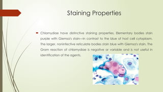
Chlamydiaehave distinctive staining properties. Elementary bodies stain
purple with Giemsa's stain—in contrast to the blue of host cell cytoplasm.
The larger, noninfective reticulate bodies stain blue with Giemsa's stain. The
Gram reaction of chlamydiae is negative or variable and is not useful in
identification of the agents.
7.
Chlamydia trachomatis Ocular
Infections
Humans are the natural host for C trachomatis. Intracytoplasmic replication
results in the formation of compact inclusions with a glycogen matrix in
which elementary bodies are embedded.
Trachoma is an ancient eye disease, it is a chronic keratoconjunctivitis that
begins with acute inflammatory changes in the conjunctiva and cornea
and progresses to scarring and blindness. The C trachomatis serovars A, B,
and C are associated with clinical trachoma.
8.
Clinical Findings
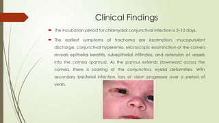
Theincubation period for chlamydial conjunctival infection is 3–10 days.
The earliest symptoms of trachoma are lacrimation, mucopurulent
discharge, conjunctival hyperemia. Microscopic examination of the cornea
reveals epithelial keratitis, subepithelial infiltrates, and extension of vessels
into the cornea (pannus). As the pannus extends downward across the
cornea, there is scarring of the conjunctiva, eyelid deformities. With
secondary bacterial infection, loss of vision progresses over a period of
years.
9.
Treatment, Prevention andControl
Clinical trials, in an endemic trachoma, using azithromycin treatment show
that infection and clinical disease are greatly decreased at 6 and 12
months post therapy. Topical therapy is of little value.
The WHO has initiated the S-A-F-E program to eliminate blinding trachoma
and at least markedly reduce clinically active disease.
The S-A-F-E program is as follows: Surgery for deformed eyelids; periodic
Azithromycin therapy; Face washing and hygiene; and, Environmental
improvement such as decreasing the number of flies that feed on
conjunctival exudates. It is clear that improved socioeconomic conditions
enhance the disappearance of endemic trachoma.
10.
Chlamydia trachomatis Genital
Infections
C trachomatis serovars D–K cause sexually transmitted diseases and may
also produce infection of the eye.
In sexually active men, C trachomatis causes nongonococcal urethritis and,
occasionally, epididymitis.
In women, C trachomatis causes urethritis, cervicitis, and pelvic
inflammatory disease, which can lead to sterility and predispose to ectopic
pregnancy.
The newborn acquires the infection during passage through an infected
birth canal. Probably 20–60% of infants of infected mothers acquire the
infection.
11.
Chlamydia trachomatis Genital
Infections
Laboratory Diagnosis
1- Specimen Collection: Proper specimen collection is the
key to the laboratory diagnosis of chlamydia infection.
Because the chlamydiae are obligate intracellular bacteria,
it is important that the specimens contain infected human
cells as well as the extracellular material where they might
also be present. Collect endocervical specimens following
removal of discharge and secretions from the cervix. A
swab or cytology brush is used to scrape epithelial cells from
1 to 2 cm deep into the endocervix.
12.
Chlamydia trachomatis Genital
Infections
2- Nucleic Acid Detection: Nucleic acid amplification tests are the tests of
choice to diagnose genital C trachomatis infections, e.g. the PCR technique.
3- Culture: Culture is generally much less sensitive than the nucleic acid
detection assays.
4- Serology: Because of the relatively great antigenic mass of chlamydiae in
genital tract infections, serum antibodies occur much more commonly than in
trachoma and are of higher titer. A titer rise occurs during and after acute
chlamydial infection.
Treatment: Azithromycin is effective and can be given to pregnant women.
Tetracyclines (eg, doxycycline) are commonly used in non gonococcal
urethritis and in non pregnant infected females.
13.
Chlamydophila pneumoniae &Respiratory
Infections
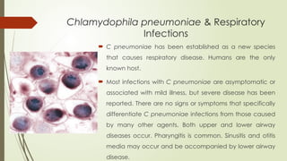
C pneumoniae has been established as a new species
that causes respiratory disease. Humans are the only
known host.
Most infections with C pneumoniae are asymptomatic or
associated with mild illness, but severe disease has been
reported. There are no signs or symptoms that specifically
differentiate C pneumoniae infections from those caused
by many other agents. Both upper and lower airway
diseases occur. Pharyngitis is common. Sinusitis and otitis
media may occur and be accompanied by lower airway
disease.
14.
Laboratory Diagnosis andtreatment
Smears: Direct detection of elementary bodies in clinical specimens using
fluorescent antibody techniques is insensitive.
Culture: Swab specimens of the pharynx should be put into a chlamydiae
transport medium and placed at 4°C; C pneumoniae is rapidly inactivated
at room temperature. It grows poorly in cell culture, forming inclusions
smaller than those formed by the other chlamydiae. C pneumoniae
Growth is better at 35°C than 37°C.
Serology: Primary infection yields IgM antibody after about 3 weeks
followed by IgG antibody at 6–8 weeks. In reinfection, the IgM response
may be absent or minimal and the IgG response occurs in 1–2 weeks.
Treatment: C pneumoniae is susceptible to the macrolides and
tetracyclines and to some fluoroquinolones.
15.
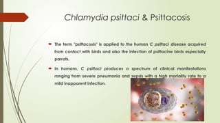
Chlamydia psittaci &Psittacosis
The term "psittacosis" is applied to the human C psittaci disease acquired
from contact with birds and also the infection of psittacine birds especially
parrots.
In humans, C psittaci produces a spectrum of clinical manifestations
ranging from severe pneumonia and sepsis with a high mortality rate to a
mild inapparent infection.
16.
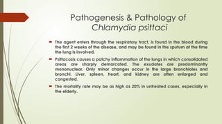
Pathogenesis & Pathologyof
Chlamydia psittaci
The agent enters through the respiratory tract, is found in the blood during
the first 2 weeks of the disease, and may be found in the sputum at the time
the lung is involved.
Psittacosis causes a patchy inflammation of the lungs in which consolidated
areas are sharply demarcated. The exudates are predominantly
mononuclear. Only minor changes occur in the large bronchioles and
bronchi. Liver, spleen, heart, and kidney are often enlarged and
congested.
The mortality rate may be as high as 20% in untreated cases, especially in
the elderly.
17.
Laboratory Diagnosis andtreatment
Culture: Culture of C psittaci can be dangerous, and detection of
the organism using immunoassays or PCR is preferred. If necessary,
C psittaci can be cultured from blood or sputum or from lung tissue
by culture in tissue culture cells, or embryonated eggs.
Because of the difficulty in obtaining laboratory confirmation of C
psittaci infection, most infections are treated based only on the
clinical diagnosis. Information on therapeutic efficacy comes from
several clinical trials. Azithromycin, clarithromycin, and
erythromycin (and doxycycline in adults) clear most, but not all,
respiratory C psittaci infections. All the patients improve clinically,
even those with persistent infection.