The document summarizes key aspects of the nervous system, including:
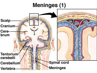
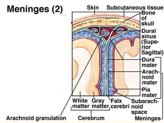
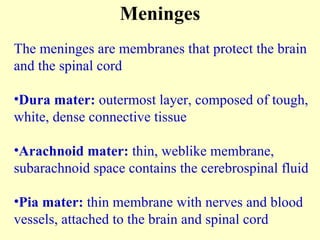
- The meninges (dura mater, arachnoid mater, pia mater) protect the brain and spinal cord.
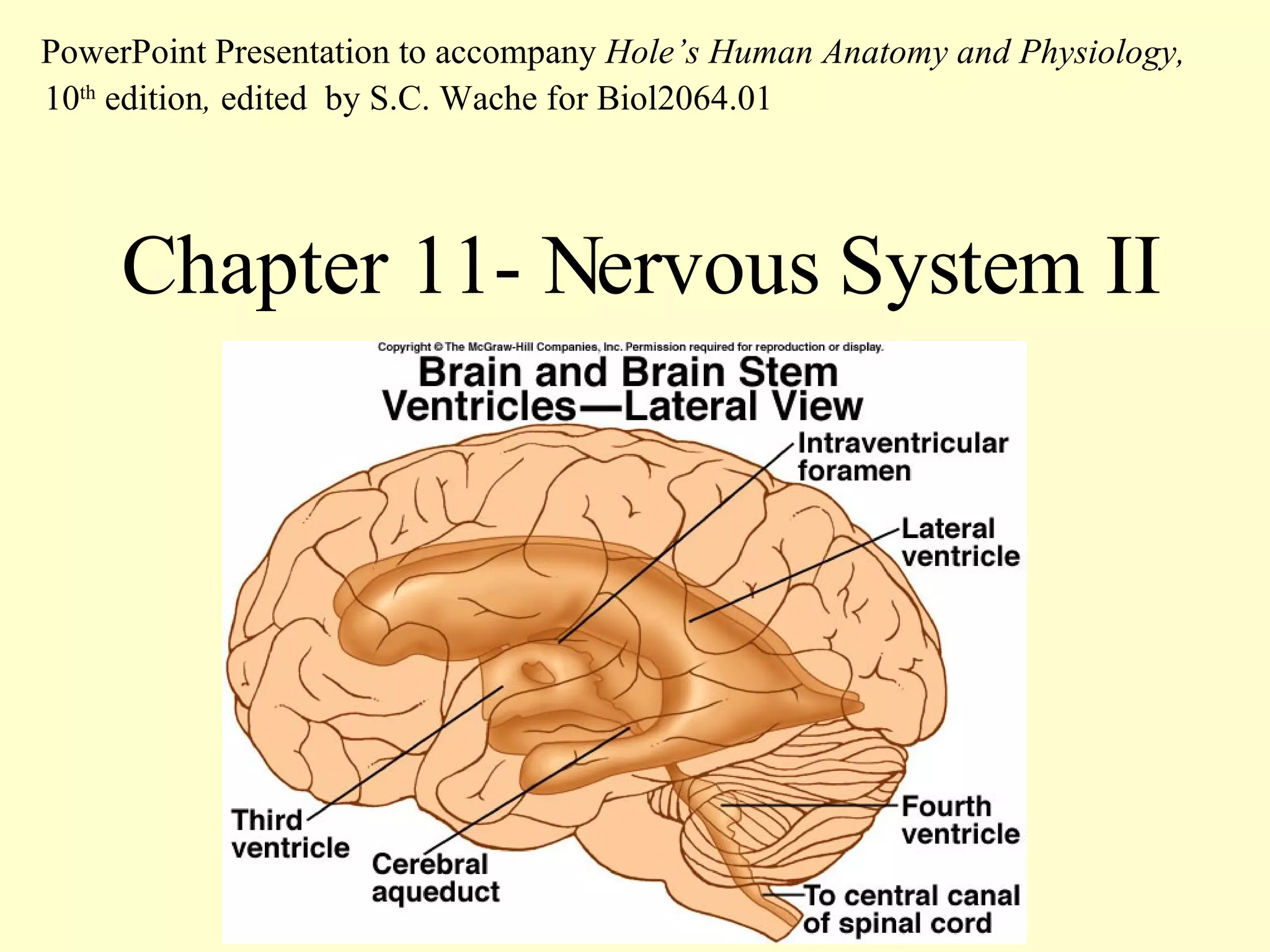
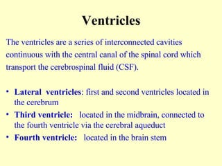
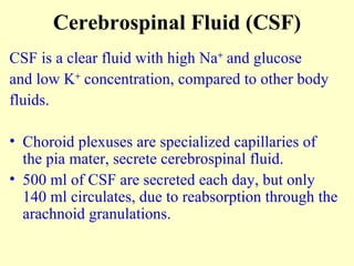
- The ventricles and cerebrospinal fluid (CSF) transport CSF within the brain and spinal cord.
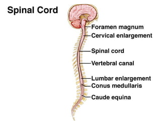
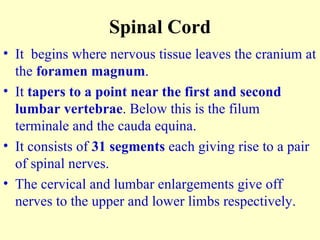
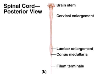
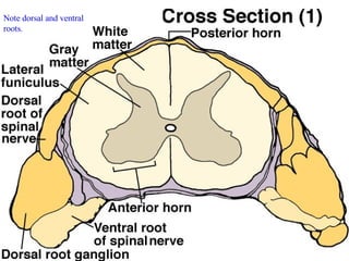
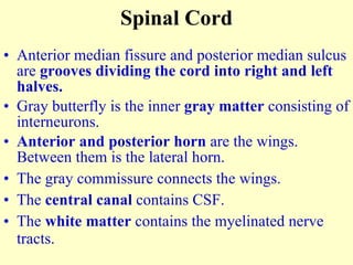
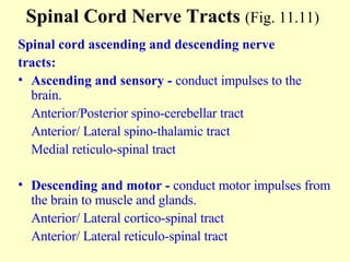
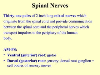
- The spinal cord contains gray matter with neurons and white matter with nerve tracts. It relays sensory and motor signals.
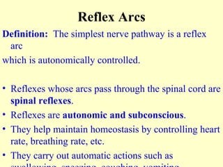
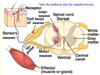
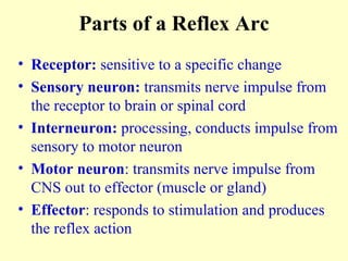
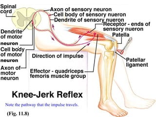
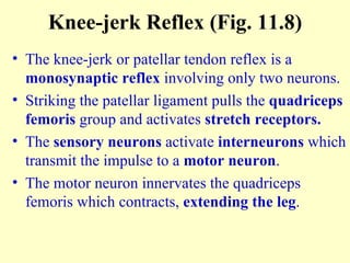
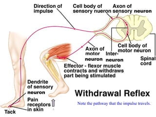
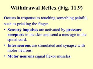
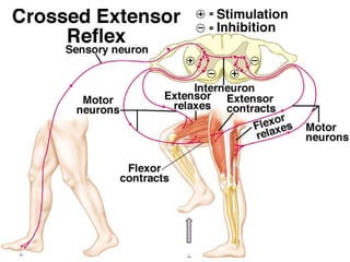
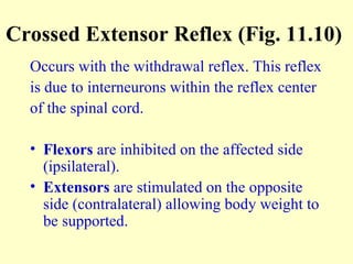
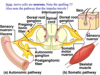
- Reflex arcs involve receptors, sensory and motor neurons, and effectors in automatic responses like withdrawal reflexes.
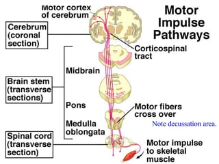
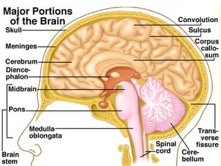
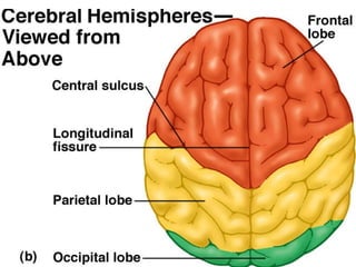
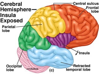
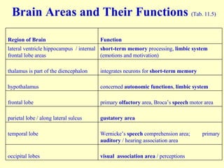
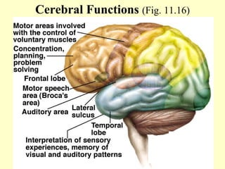
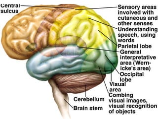
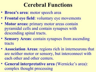
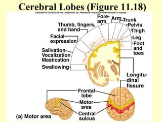
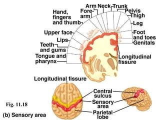
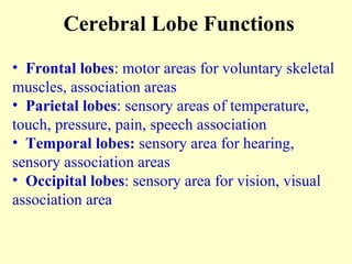
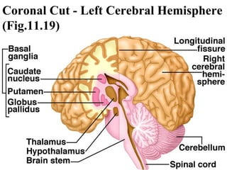
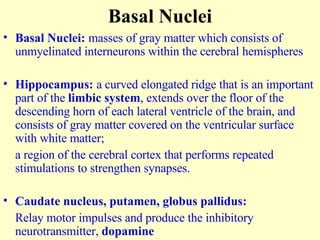
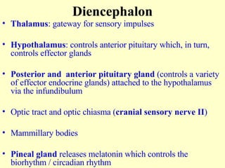
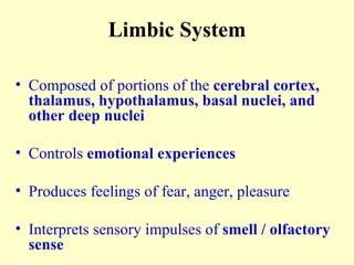
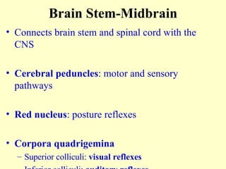
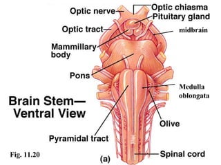
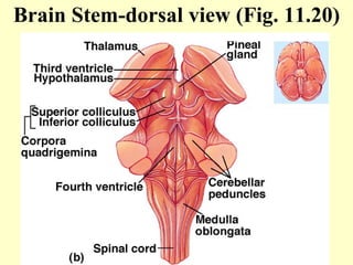
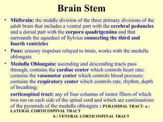
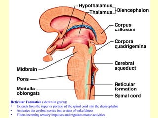
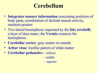
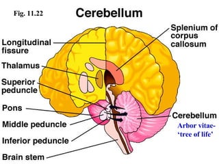
- The brain contains structures like the cerebrum, cerebellum, and brain stem that control functions like memory, movement, and homeostasis.

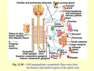
![Plexuses Cervical : first four cervical nerves [C1-C4] Brachial : lower four cervical nerves and first thoracic nerve [C5-C8 and T1] Lumbosacral : last thoracic nerve and lumbar, sacral, and coccygeal nerves [T12-S5] PNS - Autonomic Nervous System Functions independently, involuntary / subconscious control Controls visceral activities by regulating smooth and cardiac muscles and glands Regulates heart rate, blood pressure, breathing, body temperature and other homeostatic mechanisms Responds to stress](https://image.slidesharecdn.com/ch11-ppt-lect-1202693080414246-4/85/Ch11-Ppt-Lect-55-320.jpg)
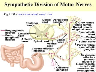
![Fig. 11.36 – note the meninges, dorsal and ventral roots. Note that the spinal nerves are mixed function nerves [AM-PS].](https://image.slidesharecdn.com/ch11-ppt-lect-1202693080414246-4/85/Ch11-Ppt-Lect-58-320.jpg)


![Chapt06 Holes Lecture Animation[1]](https://cdn.slidesharecdn.com/ss_thumbnails/chapt06holeslectureanimation1-091122122041-phpapp02-thumbnail.jpg?width=640&height=640&fit=bounds)
![Chapt05 Holes Lecture[1]](https://cdn.slidesharecdn.com/ss_thumbnails/chapt05holeslecture1-091122121913-phpapp02-thumbnail.jpg?width=640&height=640&fit=bounds)


![Chapt11 Holes Lecture[1]](https://cdn.slidesharecdn.com/ss_thumbnails/chapt11holeslecture1-091122123910-phpapp01-thumbnail.jpg?width=640&height=640&fit=bounds)