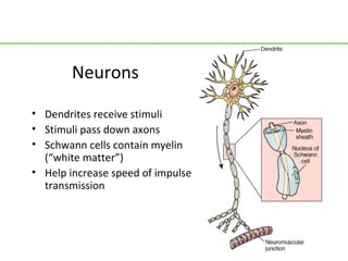
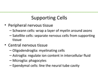
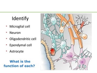
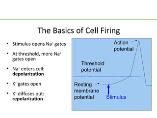
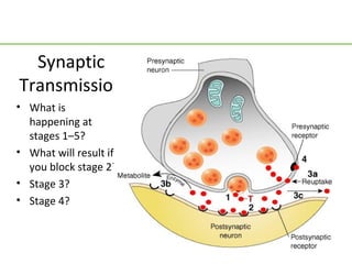
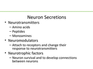
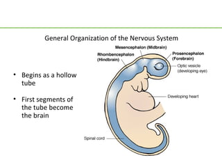
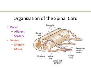
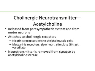
Nervous System Physiology
         Review
Neurons

• Dendrites receive stimuli
• Stimuli pass down axons
• Schwann cells contain myelin
  (“white matter”)
• Help increase speed of impulse
  transmission
Supporting Cells
• Peripheral nervous tissue
  – Schwann cells: wrap a layer of myelin around axons
  – Satellite cells: separate nervous cells from supporting
    tissue
• Central nervous tissue
  – Oligodendroglia: myelinating cells
  – Astroglia: regulate ion content in intercellular fluid
  – Microglia: phagocytes
  – Ependymal cells: line the neural tube cavity
Identify
 • Microglial cell
 • Neuron
 • Oligodendritic cell
 • Ependymal cell
 • Astrocyte

   What is the
function of each?
Question
Tell whether the following statement is true or
  false.
All neurons are myelinated.
False               Answer
Rationale: The myelin sheath increases the
  speed of impulse transmission (the impulse
  can skip over the myelinated/insulated parts
  of the neuron), but speed is not important
  everywhere (like the digestive tract). If every
  neuron was myelinated, neurons would take
  up a lot more space, too.
The Basics of Cell Firing
• Stimulus opens Na+ gates                    Action
                                              potential
• At threshold, more Na+
  gates open
                               Threshold
• Na+ enters cell:             potential
  depolarization
• K+ gates open              Resting
• K+ diffuses out:           membrane
  repolarization             potential     Stimulus
Synaptic
Transmission
• What is
  happening at
  stages 1–5?
• What will result if
  you block stage 2?
• Stage 3?
• Stage 4?
Neuron Secretions
• Neurotransmitters
  – Amino acids
  – Peptides
  – Monoamines
• Neuromodulators
  – Attach to receptors and change their
    response to neurotransmitters
• Neurotrophic factors
  – Neuron survival and to develop connections
    between neurons
General Organization of the Nervous System


• Begins as a hollow
  tube

• First segments of
  the tube become
  the brain
Organization of the Spinal Cord
• Dorsal
   – Afferent
   – Sensory
• Ventral
   – Efferent
   – Motor
Scenario
A woman developed polyneuropathy.
• Her spinal nerves were damaged
• She lost the ability to tell where her body was positioned
• She has to look every time she takes a step, to tell where
  she is moving her feet to
Question:
• What parts of her spinal nerves were damaged?
Cell Columns of the Spinal Cord
• What
  problems
  would you
  expect in              A
  someone
  who                B
  suffered
  ischemia     C
  to:
  – Area A
  – Area B
  – Area C
Question
If you place your hand on a hot surface, which
    ganglion carries the impulse to the spinal
    cord?
b. Ventral
c. Dorsal
d. Interneuron
e. Association neuron
a. Dorsal              Answer
Rationale: Afferent neurons carry sensory
   impulses to the spinal cord through the dorsal
   root ganglion; efferent neurons carry motor
   responses through the ventral root ganglion
   to effector cells in the tissue.
Layers of the White Matter
•   Archi layer
     – Connects neighboring segments
     – Contains neurons  reticular
        activating system
•   Paleo layer
     – Fibers reach to the brain stem
•   Neo layer
     – Pathways for bladder control and
        fine motor skills
     – Develop by fifth year of life
Brain Regions
          •   Cerebrum
          •   Thalamus
          •   Hypothalamus
          •   Cerebral peduncles
          •   Cerebral aqueduct
          •   Colliculi
          •   Cerebellum
          •   Pons
          •   Medulla oblongata
Functions of the Hindbrain
• Medulla oblongata, cerebellum, and pons

• Reflex centers for heart and respiration rates, coughing,
  swallowing, vomiting, etc.

• Gives rise to cranial nerves V–XII controlling viscera,
  hearing, facial, and mouth/throat functions

• Cerebellum allows fine motor coordination
Functions of the Midbrain
• Cerebral peduncles carry nerve fibers from the
  cerebrum to the hindbrain
• Cerebral aqueduct lets cerebrospinal fluid drain
  from the fourth ventricle inside the cerebrum
• Superior colliculi control reflex eye movements
• Inferior colliculi control reflex reactions to sound
• Gives rise to cranial nerves III and IV, controlling eye
  movement
Functions of the Forebrain
• Thalamus: “switchboard” or relay station for impulses going to
  and coming from the cerebrum
• Hypothalamus: homeostatic control
• Cerebrum
• Gives rise to cranial nerves I and II, for smell and sight
Cerebrum
• Frontal lobe: motor, anticipation
• Parietal lobe: somatosensory
• Temporal lobe: hearing, memory
• Occipital lobe: vision

• Limbic system: emotional
Question
Which part of the brain maintains vital functions
   like breathing, heart rate, and digestion?
b. Forebrain
c. Midbrain
d. Hindbrain
e. Cerebellum
a. Hindbrain       Answer
Rationale: Also known as the brain stem, this is
   the vasomotor center that controls
   cardiopulmonary function and digestion.
Dura Mater
• Has two layers
• Inner layer bends over
  to form a fold (falx
  cerebri) that separates
  the cerebral
  hemispheres
• It forms a second fold
  (tentorium) that holds
  the cerebrum up off
  the cerebellum
Dura Mater
      (cont.)
• Between the layers of
  the dura, at the base of
  each fold, venous blood
  drains out of the brain
  in a sinus
• Bridging veins carry
  blood from the brain
  across the inner layer of
  the dura mater to the
  sinus
• The sinus also collects
  cerebrospinal fluid
Arachnoid

• Lies just beneath the dura
  mater
• Waterproof
• Cerebrospinal fluid (CSF)
  lies under the arachnoid
  to cushion the brain
• Extensions of the
  arachnoid (villi) poke
  through the inner layer of
  the dura mater into the
  sinuses, to let CSF drain
  into the sinuses
Pia Mater
• Lies right on the surface
  of the brain
• Holds the cerebral
  arteries in place
Meninges and Meningeal Spaces
• Epidural space: meningeal arteries
• Dura mater
   – Subdural space: bridging veins
• Arachnoid
   – Subarachnoid space: cerebral arteries,
     cerebrospinal fluid
• Pia mater
Cerebrospinal Fluid
• Leaks out of capillaries inside the brain’s hollow
  ventricles
   – Composition controlled by the blood-brain barrier
• Passes out an opening below the cerebellum
• Circulates around the brain and spinal cord in the
  subarachnoid space
• Passes through arachnoid villi into blood in the dural
  sinuses and is returned to the heart
Autonomic Nervous System
• Sympathetic
   – Catecholamines
   – Epinephrine, norepinephrine, dopamine
   – Attach to adrenergic receptors
• Parasympathetic
   – Acetylcholine
   – Attaches to cholinergic receptors
Adrenergic Neurotransmitters
• Synthesized in the sympathetic system
• Attach to adrenergic receptors
  – Alpha-1 receptors: constrict blood vessels
  – Alpha-2 receptors: negative feedback to stop
    neurotransmitter release
  – Beta-1 receptors: speed and strengthen heart
  – Beta-2 receptors: bronchodilation
• Neurotransmitter is removed from synapse by
  reuptake or degraded by enzymes
Cholinergic Neurotransmitter—
               Acetylcholine
• Released from parasympathetic system and from
  motor neurons
• Attaches to cholinergic receptors
   – Nicotinic receptors: excite skeletal muscle cells
   – Muscarinic receptors: slow heart, stimulate GI tract,
     vasodilate
• Neurotransmitter is removed from synapse by
  acetylcholinesterase
Question
Tell whether the following statement is true or
  false.
The sympathetic division of the ANS is also
  known as fight-or-flight.
True                Answer
Rationale: The SNS is characterized by the
  release of adrenaline, which results in pupil
  dilation, bronchodilation, and increased HR,
  BP, and glucose production—all the things
  that come in handy when you are running
  from something!

Neuro pptrevised

  • 1.
  • 2.
    Neurons • Dendrites receivestimuli • Stimuli pass down axons • Schwann cells contain myelin (“white matter”) • Help increase speed of impulse transmission
  • 3.
    Supporting Cells • Peripheralnervous tissue – Schwann cells: wrap a layer of myelin around axons – Satellite cells: separate nervous cells from supporting tissue • Central nervous tissue – Oligodendroglia: myelinating cells – Astroglia: regulate ion content in intercellular fluid – Microglia: phagocytes – Ependymal cells: line the neural tube cavity
  • 4.
    Identify • Microglialcell • Neuron • Oligodendritic cell • Ependymal cell • Astrocyte What is the function of each?
  • 5.
    Question Tell whether thefollowing statement is true or false. All neurons are myelinated.
  • 6.
    False Answer Rationale: The myelin sheath increases the speed of impulse transmission (the impulse can skip over the myelinated/insulated parts of the neuron), but speed is not important everywhere (like the digestive tract). If every neuron was myelinated, neurons would take up a lot more space, too.
  • 7.
    The Basics ofCell Firing • Stimulus opens Na+ gates Action potential • At threshold, more Na+ gates open Threshold • Na+ enters cell: potential depolarization • K+ gates open Resting • K+ diffuses out: membrane repolarization potential Stimulus
  • 8.
    Synaptic Transmission • What is happening at stages 1–5? • What will result if you block stage 2? • Stage 3? • Stage 4?
  • 9.
    Neuron Secretions • Neurotransmitters – Amino acids – Peptides – Monoamines • Neuromodulators – Attach to receptors and change their response to neurotransmitters • Neurotrophic factors – Neuron survival and to develop connections between neurons
  • 10.
    General Organization ofthe Nervous System • Begins as a hollow tube • First segments of the tube become the brain
  • 11.
    Organization of theSpinal Cord • Dorsal – Afferent – Sensory • Ventral – Efferent – Motor
  • 12.
    Scenario A woman developedpolyneuropathy. • Her spinal nerves were damaged • She lost the ability to tell where her body was positioned • She has to look every time she takes a step, to tell where she is moving her feet to Question: • What parts of her spinal nerves were damaged?
  • 13.
    Cell Columns ofthe Spinal Cord • What problems would you expect in A someone who B suffered ischemia C to: – Area A – Area B – Area C
  • 14.
    Question If you placeyour hand on a hot surface, which ganglion carries the impulse to the spinal cord? b. Ventral c. Dorsal d. Interneuron e. Association neuron
  • 15.
    a. Dorsal Answer Rationale: Afferent neurons carry sensory impulses to the spinal cord through the dorsal root ganglion; efferent neurons carry motor responses through the ventral root ganglion to effector cells in the tissue.
  • 16.
    Layers of theWhite Matter • Archi layer – Connects neighboring segments – Contains neurons  reticular activating system • Paleo layer – Fibers reach to the brain stem • Neo layer – Pathways for bladder control and fine motor skills – Develop by fifth year of life
  • 17.
    Brain Regions • Cerebrum • Thalamus • Hypothalamus • Cerebral peduncles • Cerebral aqueduct • Colliculi • Cerebellum • Pons • Medulla oblongata
  • 18.
    Functions of theHindbrain • Medulla oblongata, cerebellum, and pons • Reflex centers for heart and respiration rates, coughing, swallowing, vomiting, etc. • Gives rise to cranial nerves V–XII controlling viscera, hearing, facial, and mouth/throat functions • Cerebellum allows fine motor coordination
  • 19.
    Functions of theMidbrain • Cerebral peduncles carry nerve fibers from the cerebrum to the hindbrain • Cerebral aqueduct lets cerebrospinal fluid drain from the fourth ventricle inside the cerebrum • Superior colliculi control reflex eye movements • Inferior colliculi control reflex reactions to sound • Gives rise to cranial nerves III and IV, controlling eye movement
  • 20.
    Functions of theForebrain • Thalamus: “switchboard” or relay station for impulses going to and coming from the cerebrum • Hypothalamus: homeostatic control • Cerebrum • Gives rise to cranial nerves I and II, for smell and sight
  • 21.
    Cerebrum • Frontal lobe:motor, anticipation • Parietal lobe: somatosensory • Temporal lobe: hearing, memory • Occipital lobe: vision • Limbic system: emotional
  • 22.
    Question Which part ofthe brain maintains vital functions like breathing, heart rate, and digestion? b. Forebrain c. Midbrain d. Hindbrain e. Cerebellum
  • 23.
    a. Hindbrain Answer Rationale: Also known as the brain stem, this is the vasomotor center that controls cardiopulmonary function and digestion.
  • 24.
    Dura Mater • Hastwo layers • Inner layer bends over to form a fold (falx cerebri) that separates the cerebral hemispheres • It forms a second fold (tentorium) that holds the cerebrum up off the cerebellum
  • 25.
    Dura Mater (cont.) • Between the layers of the dura, at the base of each fold, venous blood drains out of the brain in a sinus • Bridging veins carry blood from the brain across the inner layer of the dura mater to the sinus • The sinus also collects cerebrospinal fluid
  • 26.
    Arachnoid • Lies justbeneath the dura mater • Waterproof • Cerebrospinal fluid (CSF) lies under the arachnoid to cushion the brain • Extensions of the arachnoid (villi) poke through the inner layer of the dura mater into the sinuses, to let CSF drain into the sinuses
  • 27.
    Pia Mater • Liesright on the surface of the brain • Holds the cerebral arteries in place
  • 28.
    Meninges and MeningealSpaces • Epidural space: meningeal arteries • Dura mater – Subdural space: bridging veins • Arachnoid – Subarachnoid space: cerebral arteries, cerebrospinal fluid • Pia mater
  • 29.
    Cerebrospinal Fluid • Leaksout of capillaries inside the brain’s hollow ventricles – Composition controlled by the blood-brain barrier • Passes out an opening below the cerebellum • Circulates around the brain and spinal cord in the subarachnoid space • Passes through arachnoid villi into blood in the dural sinuses and is returned to the heart
  • 30.
    Autonomic Nervous System •Sympathetic – Catecholamines – Epinephrine, norepinephrine, dopamine – Attach to adrenergic receptors • Parasympathetic – Acetylcholine – Attaches to cholinergic receptors
  • 31.
    Adrenergic Neurotransmitters • Synthesizedin the sympathetic system • Attach to adrenergic receptors – Alpha-1 receptors: constrict blood vessels – Alpha-2 receptors: negative feedback to stop neurotransmitter release – Beta-1 receptors: speed and strengthen heart – Beta-2 receptors: bronchodilation • Neurotransmitter is removed from synapse by reuptake or degraded by enzymes
  • 32.
    Cholinergic Neurotransmitter— Acetylcholine • Released from parasympathetic system and from motor neurons • Attaches to cholinergic receptors – Nicotinic receptors: excite skeletal muscle cells – Muscarinic receptors: slow heart, stimulate GI tract, vasodilate • Neurotransmitter is removed from synapse by acetylcholinesterase
  • 33.
    Question Tell whether thefollowing statement is true or false. The sympathetic division of the ANS is also known as fight-or-flight.
  • 34.
    True Answer Rationale: The SNS is characterized by the release of adrenaline, which results in pupil dilation, bronchodilation, and increased HR, BP, and glucose production—all the things that come in handy when you are running from something!