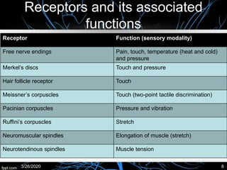
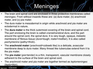
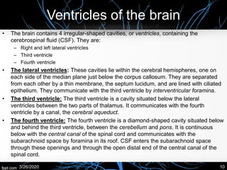
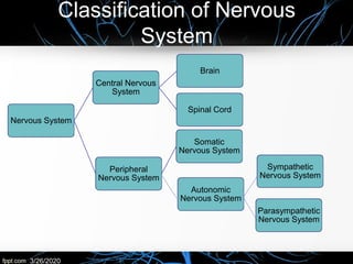
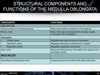
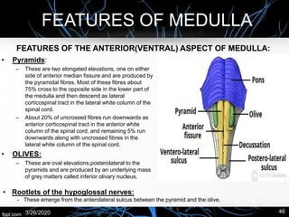
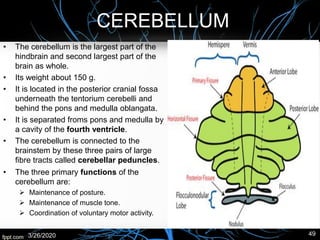
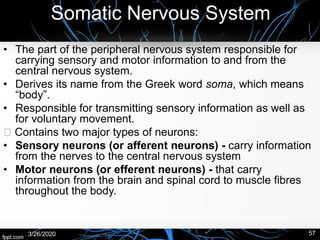
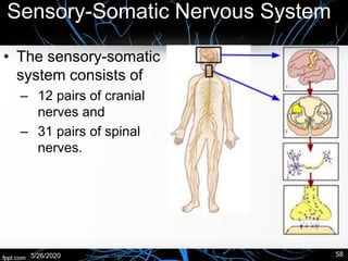
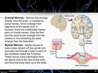
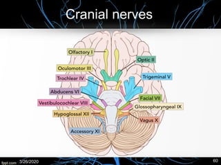
Introduction to nervous system, Divisions of Nervous System, Nervous System P...Shaista Jabeen
https://www.youtube.com/channel/UCrrAABI7QDRCJ1yMrQCip_w/videos
https://www.facebook.com/ShaistaJabeeen/
https://www.facebook.com/Human-Physiology-Lectures-100702741804409/
Introduction to nervous system, Divisions of Nervous System, Nervous System Physiology
Introduction to nervous system
Divisions of Nervous System
Nervous System Physiology
DIVISIONS OF NERVOUS SYSTEM
CENTRAL NERVOUS SYSTEM
PERIPHERAL NERVOUS SYSTEM
Short Notes
ppt pdf
This presentation based on a broad overview to the human central nervous system focusing over the parts of the system, different cell types present in the system, and special terminology used in the system.
Peripheral Nervous System, Audumbar MaliAudumbar Mali
Peripheral Nervous System,
Types of PNS,
Spinal nerves,
Types of neuron (3 basic types),
Plexus,
Cranial nerves,
Autonomic nervous system,
Structure of Neuron,
Human Anatomy and Physiology-I,
Syllabus As per PCI,
B. Pharm-I
special sense organs (anatomy and physiology) - a brief discussion Pallab Nath
brief discussion on special senses, Basic level class for technicians. topics discussed include eyes and vision, nose and sense of smell, tongue and sense of taste and ears and hearing
Nervous system consists of highly complex structure co-ordinates and controls the body along with the endocrine system.
Here we discussed about some important outlines concerned of psychobiology which is coming under unit 2 of syllabus of clinical speciality - mental health nursing.
The key points are,
- The anatomic review
- Brain & limbic system
- Nerve tissue-> Neurons & Neuroglia, Synapses, Synaptic cleft
- Neurotransmitters
- Autonomic nervous system, - sympathetic and parasympathetic nervous system.
Apart from these, its relation with different psychiatric disorders are also explained in brief.
The central nervous system (CNS) is made up of the brain and spinal cord. The brain controls most body functions, including awareness, movements, sensations, thoughts, speech and memory. The spinal cord is connected to the brain at the brain stem and is covered by the vertebrae of the spine.
Skeletal system. anatomy and physiology of skeletal system. appendicular skel...mamtabisht10
SKELETAL SYSTEM
bones, cartilage and ligaments are tightly joined to form a strong, flexible framework called skeletal system
anatomy and physiology of axial and appendicular skeletal system
Axial Skeleton: The axial skeleton includes the skull, spine, ribs and sternum.
Appendicular Skeleton:
The appendicular skeleton includes the appendages of the body, which are the shoulders, arms, hips, and legs.
a quick visual understanding of what actually nervous tissue is made up of at cellular level its functions nerve cell types chemical synapse detailed structure of neuron
Lecture notes and diagrams to help high school anatomy and physiology students learn the general functions of the nervous system and types of glial support nerve cells, types of neurons and anatomy of typical neurons.
THIS PRESENTATION IS UPLOADED TO HELP THE EDUCATOR OF MEDICAL, NURSING & ALLIE HEALTH SCIENCES TO TEACH THEIR STUDENTS ABOUT THE NERVOUS SYSTEM. IT WILL ALSO CREATE AWARENESS AMONG THE COMMON PEOPLE REGARDING NERVOUS SYSTEM.
This presentation is contain the nervous system. The anatomy and the physiology of the human nervous system . this presentation is the describe the central and peripheral system in the easy language . This presentation is also contain the cranial nerves and the spinal cord in detail and also all about the CNS AND THE PNS . This presentation is very useful for the student who study the in the BSc nursing and the GNM .
This lecture help the students such as medical ,nursing , and any health care provider to understand the basic information about anatomy of nervous system
Review of Nervous System, Unconsciousness, and CVA. The Nursing Core FunctionsAyinla Kazeem
This presentation was made at several sessions of Mandatory Continuing Professional Development Programme for Nigerian Nurses in Kwara State, and have undergone series of editing till date. While still working on the final editing to totally conform with global standard of practice, I deemed it necessary to share it in this forum.
cerebrovascular accidents - types, causes and its managementVarunMahajani
This PowerPoint presentation provides in-depth knowledge regarding cerebrovascular accidents types, stages of management, medical management, surgical management, nursing management, complications and their management
A 25 year old female patient was admitted to the female medicine ward with complaints of fever with chills since 1 and 1/2 months, bod ache, cough with expectoration since 10-15 days, weakness with giddiness.
14. a case study on diabetes mellitus type 1 with diabetic ketoacidosis cp in...Dr. Ajita Sadhukhan
A 26 year old male patient was admitted to the male medicine ward with complaints of nausea, vomiting, generalised weakness, anxiety, decreased appetite, headache since noon.
13. a case study on convulsions in a kco epilepsy with lactational amenorrhoeaDr. Ajita Sadhukhan
A 25 year old female patient was admitted to the female medicine ward with complaints of 2 and a half month amenorrhoea, epileptic fit convulsions at home, vertigo, generalised weakness and 1 episode of epileptic fit today evening.
A 46 year old female patient was admitted to the female medicine ward with complaints of breathlessness on walking, fever, right pedal oedema, giddiness on walking.
A 67 year old male patient was admitted to the male medicine ward with complaints of abdominal distension, bilateral lower limb oedema, pitting pedal oedema, distended and swelled scrotum and breathlessness since 15 days.
A 50 year old female patient was admitted to the female medicine ward with complaints of constipation (today), breathlessness, coughing, b/l pedal oedema, anasarca since 7 days.
A 28 year old male patient was admitted to the male medicine ward with complaints of fever since 1 week, bodyache, headache, slightly yellowish sclera and watery eyes.
A 35 year old female patient was admitted to the female medicine ward with complaints of bodyache with weakness, pain in knee joint since 2-3 months, difficulty in walking. she had a past history of TB lymphadenopathy.
A 45 year old female patient was admitted to the female medicine ward with complaints of severe joint pain in both extremities, difficulty in breathing, weakness, headache and eye pain, chest pain. She is a k/c/o hypertension since 1 year and hypoglycaemia since 1 month.
A 35-year old female patient was admitted to the female medicine ward with complaints of blackish discoloration of left toe, difficulty in walking since 5-6 months, joint pain since 15-20 years. she had a past history of malaria, convulsions and typhoid before 3-4 years.
A 70-year old male patient was admitted to the male medicine wards with complaints of cough with expectoration since 20 days, anorexia, pedal oedema, chest pain, haemoptasis since 10 days, low grade fever, weakness.
3. a case study on plasmodium falciparum with thrombocytopenia with viral hep...Dr. Ajita Sadhukhan
A 20-year old male patient was admitted to the male medicine ward with complaints of fever with chills since 1 week, headache, abdominal pain, nausea, vomiting, yellowish sclera, yellowish urine, anorexia, general weakness since 10 days.
2. a case study on hypertension with rheumatoid arthritis and erosive gastritisDr. Ajita Sadhukhan
A 50-year old female patient was admitted to the female medicine ward with complaints of anxiety and breathlessness since 7-8 days, decreased appetite and acidity. she was a known case of Rheumatoid Arthritis since 8 years . She was also a k/c/o hypertension since 10 years and had a past history of stroke.
A 45-year old male patient was admitted to the male medicine ward with symptoms of cough with expectoration, dyspnoea since 2 months and oedema of feet since 15-20 days.
Flu Vaccine Alert in Bangalore Karnatakaaddon Scans
As flu season approaches, health officials in Bangalore, Karnataka, are urging residents to get their flu vaccinations. The seasonal flu, while common, can lead to severe health complications, particularly for vulnerable populations such as young children, the elderly, and those with underlying health conditions.
Dr. Vidisha Kumari, a leading epidemiologist in Bangalore, emphasizes the importance of getting vaccinated. "The flu vaccine is our best defense against the influenza virus. It not only protects individuals but also helps prevent the spread of the virus in our communities," he says.
This year, the flu season is expected to coincide with a potential increase in other respiratory illnesses. The Karnataka Health Department has launched an awareness campaign highlighting the significance of flu vaccinations. They have set up multiple vaccination centers across Bangalore, making it convenient for residents to receive their shots.
To encourage widespread vaccination, the government is also collaborating with local schools, workplaces, and community centers to facilitate vaccination drives. Special attention is being given to ensuring that the vaccine is accessible to all, including marginalized communities who may have limited access to healthcare.
Residents are reminded that the flu vaccine is safe and effective. Common side effects are mild and may include soreness at the injection site, mild fever, or muscle aches. These side effects are generally short-lived and far less severe than the flu itself.
Healthcare providers are also stressing the importance of continuing COVID-19 precautions. Wearing masks, practicing good hand hygiene, and maintaining social distancing are still crucial, especially in crowded places.
Protect yourself and your loved ones by getting vaccinated. Together, we can help keep Bangalore healthy and safe this flu season. For more information on vaccination centers and schedules, residents can visit the Karnataka Health Department’s official website or follow their social media pages.
Stay informed, stay safe, and get your flu shot today!
Tom Selleck Health: A Comprehensive Look at the Iconic Actor’s Wellness Journeygreendigital
Tom Selleck, an enduring figure in Hollywood. has captivated audiences for decades with his rugged charm, iconic moustache. and memorable roles in television and film. From his breakout role as Thomas Magnum in Magnum P.I. to his current portrayal of Frank Reagan in Blue Bloods. Selleck's career has spanned over 50 years. But beyond his professional achievements. fans have often been curious about Tom Selleck Health. especially as he has aged in the public eye.
Follow us on: Pinterest
Introduction
Many have been interested in Tom Selleck health. not only because of his enduring presence on screen but also because of the challenges. and lifestyle choices he has faced and made over the years. This article delves into the various aspects of Tom Selleck health. exploring his fitness regimen, diet, mental health. and the challenges he has encountered as he ages. We'll look at how he maintains his well-being. the health issues he has faced, and his approach to ageing .
Early Life and Career
Childhood and Athletic Beginnings
Tom Selleck was born on January 29, 1945, in Detroit, Michigan, and grew up in Sherman Oaks, California. From an early age, he was involved in sports, particularly basketball. which played a significant role in his physical development. His athletic pursuits continued into college. where he attended the University of Southern California (USC) on a basketball scholarship. This early involvement in sports laid a strong foundation for his physical health and disciplined lifestyle.
Transition to Acting
Selleck's transition from an athlete to an actor came with its physical demands. His first significant role in "Magnum P.I." required him to perform various stunts and maintain a fit appearance. This role, which he played from 1980 to 1988. necessitated a rigorous fitness routine to meet the show's demands. setting the stage for his long-term commitment to health and wellness.
Fitness Regimen
Workout Routine
Tom Selleck health and fitness regimen has evolved. adapting to his changing roles and age. During his "Magnum, P.I." days. Selleck's workouts were intense and focused on building and maintaining muscle mass. His routine included weightlifting, cardiovascular exercises. and specific training for the stunts he performed on the show.
Selleck adjusted his fitness routine as he aged to suit his body's needs. Today, his workouts focus on maintaining flexibility, strength, and cardiovascular health. He incorporates low-impact exercises such as swimming, walking, and light weightlifting. This balanced approach helps him stay fit without putting undue strain on his joints and muscles.
Importance of Flexibility and Mobility
In recent years, Selleck has emphasized the importance of flexibility and mobility in his fitness regimen. Understanding the natural decline in muscle mass and joint flexibility with age. he includes stretching and yoga in his routine. These practices help prevent injuries, improve posture, and maintain mobilit
Couples presenting to the infertility clinic- Do they really have infertility...Sujoy Dasgupta
Dr Sujoy Dasgupta presented the study on "Couples presenting to the infertility clinic- Do they really have infertility? – The unexplored stories of non-consummation" in the 13th Congress of the Asia Pacific Initiative on Reproduction (ASPIRE 2024) at Manila on 24 May, 2024.
ARTIFICIAL INTELLIGENCE IN HEALTHCARE.pdfAnujkumaranit
Artificial intelligence (AI) refers to the simulation of human intelligence processes by machines, especially computer systems. It encompasses tasks such as learning, reasoning, problem-solving, perception, and language understanding. AI technologies are revolutionizing various fields, from healthcare to finance, by enabling machines to perform tasks that typically require human intelligence.
These simplified slides by Dr. Sidra Arshad present an overview of the non-respiratory functions of the respiratory tract.
Learning objectives:
1. Enlist the non-respiratory functions of the respiratory tract
2. Briefly explain how these functions are carried out
3. Discuss the significance of dead space
4. Differentiate between minute ventilation and alveolar ventilation
5. Describe the cough and sneeze reflexes
Study Resources:
1. Chapter 39, Guyton and Hall Textbook of Medical Physiology, 14th edition
2. Chapter 34, Ganong’s Review of Medical Physiology, 26th edition
3. Chapter 17, Human Physiology by Lauralee Sherwood, 9th edition
4. Non-respiratory functions of the lungs https://academic.oup.com/bjaed/article/13/3/98/278874
MANAGEMENT OF ATRIOVENTRICULAR CONDUCTION BLOCK.pdfJim Jacob Roy
Cardiac conduction defects can occur due to various causes.
Atrioventricular conduction blocks ( AV blocks ) are classified into 3 types.
This document describes the acute management of AV block.
Title: Sense of Smell
Presenter: Dr. Faiza, Assistant Professor of Physiology
Qualifications:
MBBS (Best Graduate, AIMC Lahore)
FCPS Physiology
ICMT, CHPE, DHPE (STMU)
MPH (GC University, Faisalabad)
MBA (Virtual University of Pakistan)
Learning Objectives:
Describe the primary categories of smells and the concept of odor blindness.
Explain the structure and location of the olfactory membrane and mucosa, including the types and roles of cells involved in olfaction.
Describe the pathway and mechanisms of olfactory signal transmission from the olfactory receptors to the brain.
Illustrate the biochemical cascade triggered by odorant binding to olfactory receptors, including the role of G-proteins and second messengers in generating an action potential.
Identify different types of olfactory disorders such as anosmia, hyposmia, hyperosmia, and dysosmia, including their potential causes.
Key Topics:
Olfactory Genes:
3% of the human genome accounts for olfactory genes.
400 genes for odorant receptors.
Olfactory Membrane:
Located in the superior part of the nasal cavity.
Medially: Folds downward along the superior septum.
Laterally: Folds over the superior turbinate and upper surface of the middle turbinate.
Total surface area: 5-10 square centimeters.
Olfactory Mucosa:
Olfactory Cells: Bipolar nerve cells derived from the CNS (100 million), with 4-25 olfactory cilia per cell.
Sustentacular Cells: Produce mucus and maintain ionic and molecular environment.
Basal Cells: Replace worn-out olfactory cells with an average lifespan of 1-2 months.
Bowman’s Gland: Secretes mucus.
Stimulation of Olfactory Cells:
Odorant dissolves in mucus and attaches to receptors on olfactory cilia.
Involves a cascade effect through G-proteins and second messengers, leading to depolarization and action potential generation in the olfactory nerve.
Quality of a Good Odorant:
Small (3-20 Carbon atoms), volatile, water-soluble, and lipid-soluble.
Facilitated by odorant-binding proteins in mucus.
Membrane Potential and Action Potential:
Resting membrane potential: -55mV.
Action potential frequency in the olfactory nerve increases with odorant strength.
Adaptation Towards the Sense of Smell:
Rapid adaptation within the first second, with further slow adaptation.
Psychological adaptation greater than receptor adaptation, involving feedback inhibition from the central nervous system.
Primary Sensations of Smell:
Camphoraceous, Musky, Floral, Pepperminty, Ethereal, Pungent, Putrid.
Odor Detection Threshold:
Examples: Hydrogen sulfide (0.0005 ppm), Methyl-mercaptan (0.002 ppm).
Some toxic substances are odorless at lethal concentrations.
Characteristics of Smell:
Odor blindness for single substances due to lack of appropriate receptor protein.
Behavioral and emotional influences of smell.
Transmission of Olfactory Signals:
From olfactory cells to glomeruli in the olfactory bulb, involving lateral inhibition.
Primitive, less old, and new olfactory systems with different path
micro teaching on communication m.sc nursing.pdfAnurag Sharma
Microteaching is a unique model of practice teaching. It is a viable instrument for the. desired change in the teaching behavior or the behavior potential which, in specified types of real. classroom situations, tends to facilitate the achievement of specified types of objectives.
Explore natural remedies for syphilis treatment in Singapore. Discover alternative therapies, herbal remedies, and lifestyle changes that may complement conventional treatments. Learn about holistic approaches to managing syphilis symptoms and supporting overall health.
Prix Galien International 2024 Forum ProgramLevi Shapiro
June 20, 2024, Prix Galien International and Jerusalem Ethics Forum in ROME. Detailed agenda including panels:
- ADVANCES IN CARDIOLOGY: A NEW PARADIGM IS COMING
- WOMEN’S HEALTH: FERTILITY PRESERVATION
- WHAT’S NEW IN THE TREATMENT OF INFECTIOUS,
ONCOLOGICAL AND INFLAMMATORY SKIN DISEASES?
- ARTIFICIAL INTELLIGENCE AND ETHICS
- GENE THERAPY
- BEYOND BORDERS: GLOBAL INITIATIVES FOR DEMOCRATIZING LIFE SCIENCE TECHNOLOGIES AND PROMOTING ACCESS TO HEALTHCARE
- ETHICAL CHALLENGES IN LIFE SCIENCES
- Prix Galien International Awards Ceremony
New Directions in Targeted Therapeutic Approaches for Older Adults With Mantl...i3 Health
i3 Health is pleased to make the speaker slides from this activity available for use as a non-accredited self-study or teaching resource.
This slide deck presented by Dr. Kami Maddocks, Professor-Clinical in the Division of Hematology and
Associate Division Director for Ambulatory Operations
The Ohio State University Comprehensive Cancer Center, will provide insight into new directions in targeted therapeutic approaches for older adults with mantle cell lymphoma.
STATEMENT OF NEED
Mantle cell lymphoma (MCL) is a rare, aggressive B-cell non-Hodgkin lymphoma (NHL) accounting for 5% to 7% of all lymphomas. Its prognosis ranges from indolent disease that does not require treatment for years to very aggressive disease, which is associated with poor survival (Silkenstedt et al, 2021). Typically, MCL is diagnosed at advanced stage and in older patients who cannot tolerate intensive therapy (NCCN, 2022). Although recent advances have slightly increased remission rates, recurrence and relapse remain very common, leading to a median overall survival between 3 and 6 years (LLS, 2021). Though there are several effective options, progress is still needed towards establishing an accepted frontline approach for MCL (Castellino et al, 2022). Treatment selection and management of MCL are complicated by the heterogeneity of prognosis, advanced age and comorbidities of patients, and lack of an established standard approach for treatment, making it vital that clinicians be familiar with the latest research and advances in this area. In this activity chaired by Michael Wang, MD, Professor in the Department of Lymphoma & Myeloma at MD Anderson Cancer Center, expert faculty will discuss prognostic factors informing treatment, the promising results of recent trials in new therapeutic approaches, and the implications of treatment resistance in therapeutic selection for MCL.
Target Audience
Hematology/oncology fellows, attending faculty, and other health care professionals involved in the treatment of patients with mantle cell lymphoma (MCL).
Learning Objectives
1.) Identify clinical and biological prognostic factors that can guide treatment decision making for older adults with MCL
2.) Evaluate emerging data on targeted therapeutic approaches for treatment-naive and relapsed/refractory MCL and their applicability to older adults
3.) Assess mechanisms of resistance to targeted therapies for MCL and their implications for treatment selection
New Directions in Targeted Therapeutic Approaches for Older Adults With Mantl...
Nervous system
1. Nervous System
• Ajita Sadhukhan
• Reeya Amin
• Ashwin Kujur
• Vishwa Gandhi
Pharm D 4th Year
3/26/2020 1
2. Introduction
Afferent Division
→ Receptors within Joints, Skin,
Skeletal muscle (somatic –
sensory fibres)
Efferent Division
→ Skeletal muscle fibres
(somatic – motor fibres)
Sensory receptors within
visceral organs (visceral
– sensory fibres)
Smooth muscle within
visceral organs, cardiac
muscle, glands (visceral –
autonomic motor fibres)
Central
Nervous
System
Functional Subdivisions of the Nervous System
3/26/2020 2
3. A Few Terminologies
Terms Definitions
Nerve Fibre Axon
Nerve Bundle of nerve fibres outside the CNS
Tract Bundle of nerve fibres inside the CNS
Ganglion Collection of nerve cell bodies outside the CNS
Nucleus Collection of nerve cell bodies inside the CNS
Sensory Neuron Neuron that transmits impulses from a sensory receptor to the
CNS
Motor Neuron Neuron that transmits impulses from the CNS to the effector
organ, e.g. muscle
Somatic motor nerve Nerve that stimulates contraction of skeletal muscles
Autonomic motor nerve Nerve that stimulates contraction/inhibition of smooth and
cardiac muscles
Nerve plexus Network of intercalated nerves
3/26/2020 3
5. Neuroglia
Cell Type Functions
Astrocytes • Help in the formation of blood-brain barrier, regulate ion, nutrient,
and dissolved gas concentrations
• Form scar tissue after injury to brain (gliosis)
Oligodendrocytes Form myelin sheath around the CNS axons
Microglia* Remove cellular debris, and pathogens in CNS by phagocytosis
Ependymal cells Line ventricles of the brain and central canal of the spinal cord.
Assist in production, circulation and monitoring of CSF.
Satellite cells Surround nerve cell bodies in peripheral ganglia and provide
nutrition to them
Schwann cells • Form myelin sheath around axons in PNS
• Helps in regeneration of nerve fibres after injury
*All the neuralgia are derived from ectoderm except microglia which are derived from
mesoderm.
3/26/2020 5
8. Receptors and its associated
functions
Receptor Function (sensory modality)
Free nerve endings Pain, touch, temperature (heat and cold)
and pressure
Merkel’s discs Touch and pressure
Hair follicle receptor Touch
Meissner’s corpuscles Touch (two-point tactile discrimination)
Pacinian corpuscles Pressure and vibration
Ruffini’s corpuscles Stretch
Neuromuscular spindles Elongation of muscle (stretch)
Neurotendinous spindles Muscle tension
3/26/2020 8
9. Meninges
• The brain and spinal cord are enclosed in three protective membranes called
meninges. From without inwards these are: (a) dura mater, (b) arachnoid
mater, and (c) pia mater.
• The dura mater is mesodermal in origin while arachnoid and pia mater are
ectodermal in nature.
• The dura mater is the thick outermost covering of the brain and spinal cord.
The part enclosing the brain is called cranial/cerebral dura, and the part
around the spinal cord, the spinal dura. It is very tough, opaque, inelastic
membrane of fibrous tissue (dura=tough; mater=mother). It is also called
pachymeninx (pathy=thick).
• The arachnoid mater (arachnoid=cobweb like) is a delicate, avascular
membrane deep to dura mater. Many thread like tuberculae extend from it to
the pia mater.
• The pia mater (pia=tender) is a thin, transparent, vascular membrane closely
adherent to the surface of the brain and spinal cord.
• The arachnoid mater and pia mater are together termed as leptomeninges
(lepto=thin).
3/26/2020 9
10. Ventricles of the brain
• The brain contains 4 irregular-shaped cavities, or ventricles, containing the
cerebrospinal fluid (CSF). They are:
– Right and left lateral ventricles
– Third ventricle
– Fourth ventricle
• The lateral ventricles: These cavities lie within the cerebral hemispheres, one on
each side of the median plane just below the corpus callosum. They are separated
from each other by a thin membrane, the septum lucidum, and are lined with ciliated
epithelium. They communicate with the third ventricle by interventricular foramina.
• The third ventricle: The third ventricle is a cavity situated below the lateral
ventricles between the two parts of thalamus. It communicates with the fourth
ventricle by a canal, the cerebral aqueduct.
• The fourth ventricle: The fourth ventricle is a diamond-shaped cavity situated below
and behind the third ventricle, between the cerebellum and pons. It is continuous
below with the central canal of the spinal cord and communicates with the
subarachnoid space by foramina in its roof. CSF enters the subarachnoid space
through these openings and through the open distal end of the central canal of the
spinal cord.
3/26/2020 10
11. Cerebrospinal fluid (CSF)
• CSF is somewhat similar to that of blood plasma and interstitial fluid. It is
present in the ventricular system within the CNS and in the subarachnoid
space surrounding the CNS. It bathes both the external and internal
surfaces of the brain and spinal cord and provides a protective cushion
between the CNS and the surrounding bones,
• In an adult, the total volume of CSF is about 150 ml, out of which only 30
ml is in ventricular system and remaining in the subarachnoid space.
• CSF is a clear, colourless and slightly alkaline fluid with a specific gravity
of 1005-1008. It contains urea, creatinine, inorganic salts, and traces of
protein and glucose similar to that of blood plasma. But it has a much
lower protein content than the plasma and the glucose content is about
half to that of blood, and the chloride content is slightly more in CSF.
• Normally CSF is almost cell-free having only 0-5 lymphocytes/cu. mm. In
bacterial meningitis, the fluid is cloudy, with raised protein content and
vastly increased number of cells.
3/26/2020 11
12. Special Properties of CSF
1. Due to presence of blood-CSF
barrier:
– Antibodies are not found in the CSF
hence infections of the CNS are often
fatal.
– Bile is not found in CSF, even in severe
jaundice.
– Most of the drugs cannot reach CSF.
2. There is no CSF-brain barrier,
hence if drugs are injected into
the subarachnoid space
(intrathecal injections), they
soon enter the extracellular
spaces around the neurons
and neuroglia.
3/26/2020 12
13. Functions of CSF
• It serves as a cushion between the CNS and the surrounding bones.
• CSF acts as a shock absorber (i.e., it prevents or diminishes the
transmission of jarring or shocking forces to the CNS).
• It supports the brain and the spinal cord, and maintains a uniform
pressure upon them. The brain simply floats in the CSF, and it has been
stated that a brain weighing 1,500 g in air, weighs no more than 50 g in
the cerebrospinal fluid.
• Change in the intracranial volume is sometimes compensated by the
controlled production and absorption of CSF. For example, if blood
volume of the brain increases then more CSF drains away. Conversely, if
brain’s blood volume reduces, more CSF is retained.
• It nourishes the CNS.
• CSF removes the metabolites (waste products) from the CNS.
• It serves as a pathway for pineal secretions to reach the pituitary gland.
3/26/2020 13
14. Nervous System
Central Nervous
System
Brain
Spinal Cord
Peripheral
Nervous System
Somatic
Nervous System
Autonomic
Nervous System
Parasympathetic
Nervous System
Sympathetic
Nervous System
Classification of Nervous
System
3/26/2020
17. Brain
• The brain is that part of the CNS which lies within the
cranial cavity.
• The functions of the brain are as follows:
– It receives information from, and controls the activities of trunk
and limbs mainly through its connections with the spinal cord.
– It receives the information from, and controls the activities of
head and neck structures through cranial nerves.
– It assimilates experiences, a requisite to higher mental
processes such as memory, learning and intelligence.
– It is also responsible for one’s personality, thoughts and
aspirations.
• Parts of the brain:
– The adult brain constitutes about one-fiftieth of the body
weight and weighs about 1400 g in males and 1200 g in
females.
– It consists of six major parts: The cerebrum, the diencephalon,
the midbrain, the medulla oblongata and the cerebellum.
– The midbrain, pons and medulla oblongata collectively form
the brain stem.
3/26/2020 17
19. Cerebrum
• The cerebrum is the largest part of the human brain that fills most of the cranial cavity. Its
large size is the result of progressive centralization (telencephalization) of the various higher
sensory and motor centres of the brain during evolution.
• The cerebrum is a heavily convoluted bilobed structure. The two lateral halves are called
cerebral hemispheres.
• A deep median cleft, the longitudinal cerebral fissure, incompletely separates the two
cerebral hemispheres. Both in front and behind, the cleft is complete, but in the central part the
cleft extends downwards up to the corpus callosum which is a large mass of white fibres joining
the two cerebral hemispheres across the medial plane.
• the longitudinal cerebral fissure is occupied by the following structures:
– Falx cerebri (a sickle-shaped fold of dura mater).
– Fold of arachnoid the follows the surfaces of falx cerebri.
– Pia mater covering the medial surface of falx cerebri.
– Anterior cerebral arteries and veins (which lie in the subarachnoid space between the arachnoid and pia)
• Each cerebral hemisphere consists of:
– An outer layer of grey matter called cerebral cortex,
– An inner mass of white matter,
– Large masses of grey matter embedded in the basal part of white matter called basal ganglia/basal
nuclei, and
– A cavity within it called lateral ventricle.
3/26/2020 19
20. External features of the cerebral
hemisphere
• Poles:
– Each cerebral hemisphere presents 3 poles – frontal, occipital and temporal. The
anterior end of the hemisphere is the frontal pole and the posterior end the
occipital pole. The temporal pole is below and in front at the junction of orbital
and tentorial surfaces.
• The frontal pole at thee anterior end of the hemisphere is more rounded than the occipital pole. It
lies opposite the medial part of the superciliary arch.
• The occipital pole at the posterior end of the hemisphere is more pointed than the frontal pole. It
lies at a short distance superolateral to the external occipital protuberance.
• The temporal pole between frontal and occipital poles points forwards. It fits into the anterior part
of the middle cranial fossa and is overhung by the lesser wing of the sphenoid.
• Surfaces:
– Each cerebral hemisphere has 3 surfaces – superolateral, medial, and inferior.
• The superolateral surface is the most convex and most extensive. It faces upwards and laterally
and conforms to the corresponding half of the cranial vault.
• The medial surface is flat and vertical. It presents a thick C-shaped cut surface of the corpus
callosum.
• The inferior surface is irregular to adopt the floors of anterior and middle cranial fossae. It is
divided into 2 parts by a deep horizontal groove or sulcus, the stem of lateral sulcus, viz. (a) a small
anterior part, the orbital surface, and (b)a large posterior part, the tentorial surface.
3/26/2020 20
21. • Borders:
– Each cerebral hemispheres presents with six borders, viz. superomedial, superciliary,
inferolateral, medial orbital, medial occipital and inferomedial.
• The superomedial border separates the superolateral surface from the medial surface.
• The superciliary border is at the junction of superolateral and orbital surfaces. Itlies just behind
the superciliary arch hence its name.
• The inferolateral border separates the superolateral surface from the tentnial surface. Posteriorly
this border exhibits a notch, the perioccipital notch about 3 cm in front of the occipital pole.
• The medial orbital border separates the medila surface from the orbital surface.
• The infeomedial/hippocampal border surrounds the cerebral peduncle. It is formed by the
medial aspect of uncus and parahippocampal gyrus.
• The medial occipital border separates the medial surface from the tentorial surface.
• Sulci and Gyri:
– The cerebral cortex (the surface layer of grey matter) is highly exclusive in man. To
accommodate it in the limited space available within the rigid cranial cavity, the surface of
cerebral hemisphere becomes folded, producing numerous convolutions separated by
fissures. These convolutions and fissures are termed gyri and sulci respectively. In
human brain the total surface area of cerebral hemisphere is about 2000 sq. cm but
approximately 2/3rd of this is hidden from surface view within the walls of the sulci.
– The gyri consist of a central core of white matter covered by a layer of grey matter.
3/26/2020 21
22. Lobes of the Cerebral Hemisphere
• The superolateral surface of the
cerebral hemisphere is arbitrarily
divided into 4 lobes – frontal, temporal,
lateral and occipital with the help of:
– Three main sulci central, lateral and parieto-
occipital, and
– Two imaginary lines
3/26/2020 22
23. Functions of the Cerebrum
• Three main varieties of activities associated with the cerebral cortex include:
– Mental activity involved in memory, intelligence, sense of responsibility, thinking,
reasoning, moral sense, and learning are attributed to the higher centres.
– Sensory reception including the perception of pain, temperature, touch, sight,
hearing, taste and smell.
– Initiation and control of sensory (voluntary) muscle contraction.
3/26/2020 23
24. Motor areas of the cerebrum
• The precentral (motor) area:
– This lies in the frontal lobe immediately anterior to the central sulcus. Cell bodies
initiate the contraction of the skeletal muscles.
• Nerve fibres initiated from the motor region passes downwards through the internal
capsule → medulla oblongata → crosses to the opposite side in the spinal cord.
• This means that the motor area in the right hemisphere of the cerebrum controls the
voluntary muscle movement on the left side of the body and vice-versa.
– The neuron with its cell body in the cerebrum is the upper motor neuron and the
other with its cell body in the spinal cord is the lower motor neuron.
– Damage to either of these neurons will result in paralysis.
– In the motor area of the cerebrum the body is represented upside down, i.e., the
cells nearest to the vertex (the point of intersection of lines) control the feet and
those in the lowest part control the head, neck, face and fingers.
– The sizes of the different areas of the cortex representing different areas of
the body are proportional to the complexity of movement of the body part, and
not its size.
3/26/2020 24
25. • The premotor area:
– It lies in the frontal lobe immediately anterior
to the motor area.
– Controls influence over the motor area,
ensuring an orderly series of movements. For
example, in tying a shoe lace or writing, many
muscles contract but the movements must be
coordinated and carried out in a particular
sequence.
– In the lower part of this area just above the
lateral sulcus there is a group of nerve cells
called the motor speech area which controls
the movements necessary for speech.
• The frontal lobe:
– This extends anteriorly from the premotor area
to include the remainder of the frontal lobe.
– It is a large area and it is highly developed in
humans than in other animals.
– It is thought that communication between this
and other regions in the cerebrum are
responsible for behaviour, character, and
emotional state of the individual.
3/26/2020 25
26. Sensory areas of the cerebrum
• The postcentral (sensory) area:
– This area is behind the sensory sulcus.
– Here sensations of pain, temperature, pressure, touch, knowledge of muscular
movement and positions of joints are perceived.
– The sensory area of the right hemisphere receives impulses from the left side of
the body and vice versa.
• The parietal area:
– This lies behind the postcentral (sensory) area and includes the greater part of
the parietal lobe of the cerebrum.
– Its functions are believed to be associated with obtaining and retaining
accurate knowledge of objects. It has been suggested that the objects can be
recognized by touch alone because of the knowledge from the past
experience (memory) retained in this area.
• The sensory speech area:
– This is situated in the lower part of the parietal lobe and extends into the
temporal lobe.
– It is here that the spoken word is perceived.
3/26/2020 26
27. • There is a dominant area in the left
hemisphere in the right handed people
and vice versa.
– The size of the areas representing
different parts of the body is proportional
to the extent of sensory interventions.
• The auditory (hearing) area:
– This lies immediately below the lateral
sulcus within the temporal lobe.
– The cells receive and interpret impulses
transmitted from the inner ear by the
cochlear (auditory) part of the
vestibulocochlear nerves (8th cranial
nerves).
• The olfactory (smell) area:
– This lies deep within the temporal lobe
where impulses from the nose via the
olfactory nerves (1st cranial nerves) are
received and interpreted.
3/26/2020 27
28. • The taste area:
– This is thought to lie just above the lateral sulcus in the deep layers of the
sensory area.
– This is the area where impulses from special nerve endings in taste buds in the
tongue and in the lining of the cheeks, palate and pharynx are perceived as
taste.
• The visual area:
– This lies behind the parieto-occipital sulcus and includes the greater part of the
occipital lobe.
– The optic nerves (2nd cranial nerves) pass from the eye to this area which
receives and interprets the impulses as visual impressions.
3/26/2020 28
29. Other areas of the cerebrum
• Deep within the cerebral
hemispheres there are
groups of cell bodies
called nuclei (previously
called ganglia) which act
as relay stations where
impulses are passed
from one neuron to the
next in a chain.
• Important masses of grey
matter include:
– Basal nuclei
– Thalamus
– hypothalamus
3/26/2020 29
30. • Basal nuclei:
– These are the areas of grey matter, lying deep within the cerebral
hemispheres having connections within the cerebral cortex and thalamus.
– The basal nuclei are thought to be involved in initiating muscle tone (normal
elasticity of muscle) in slow and coordinated activities.
– If control is inadequate or absent, movements are jerky, clumsy and
uncoordinated.
• Thalamus:
– The thalamus consists of two masses of nerve cells and fibres situated
within the corpus callosum (largest commissure of the brain connecting the
cerebral cortex of the two hemispheres), one on each side of the third ventricle.
– Sensory input from the skin, viscera and special sense organs is transmitted to
the thalamus before redistribution to the cerebrum.
• Hypothalamus:
– The hypothalamus is composed of a number of groups of nerve cells.
– It is situated below and in front of the thalamus, immediately above the pituitary
gland.
3/26/2020 30
31. – The hypothalamus is linked to the posterior lobe of the pituitary gland and nerve
fibres and to the anterior lobe by a complex system of blood vessels. Through
these connections, the hypothalamus controls the output of hormones from both
the lobes of the gland.
Other functions of the hypothalamus:
– Hypothalamus is associated with the control of:
• The ANS (autonomic nervous system) → involuntary functions
• Appetite and satiety (a state of being satisfactorily full and unable to take on more), thirst and water
balance
• Body temperature
• Emotional reactions, i.e., pleasure, fear and rage (a state of extreme anger)
• Biological clocks or circadian rhythms, i.e., sleeping and waking cycles, body temperature and
secretion of some hormones.
3/26/2020 31
34. • The brain stem consist of diffuse system of nerve cells and nerve fibres called
reticular formation.
• FUNCTIONS OF BRAIN STEM:
– It provides passage to various ascending descending tracts that connect the spinal cord to
the different part of the forebrain.
– It contains important autonomic reflex centres associated with the control of the
respiration, heart rate and blood pressure.
– It contains reticular activating system which control consciousness.
– It contains important nuclei of the last ten cranial nerves (i.e., 3rd to 12th)
– The midbrain is the upper and shortest part of the brain stem.
– It is about 2.5 cm long and 2.5 cm wide. It connects the hindbrain with the forebrain.
– The midbrain like the other parts of the brainstem consists of grey and white matter.
• ANTERIOR SURFACE:
– Anteriorly ,it presents two large bundles of fibres on each side of the midline, called crus
cerebri.
– The oculomotor nerve emerges from the medical aspect of the crus of the same side.
– The crus cerebri bounds the interpeduncular fossa on either side.
3/26/2020 34
36. STRUCTURAL COMPONENTS AND
FUNCTIONS OF MID BRAIN
COMPONENTS FUNCTIONS
GREY MATTER
Superior colliculi Reflex centres for visual reflexes.
Inferior colliculi Lower auditory centres,probably concerned with
reflexes involving auditory stimuli.
Red nuclei Involuntary control of muscle tone and posture.
Substantia nigra Regulate the activity of basal nuclei.
WHITE MATTER
Cerebral peduncles containing number of ascending
descending tracts
Provides passage to the fibres of motor and sensory
tracts.
3/26/2020 36
• POSTERIOR SIDE:
– Posteriorly, the midbrain presents four rounded swellings called colliculi-one superior and
inferior colliculi one on each side.
– Each colliculus is laterally related to a ridge called brachium.
• STRUCTURE OF MIDBRAIN:
– The midbrain comprises two lateral halves called-Cerebral peduncles: which is again
divided into two parts by a pigmented band of grey matter, substantia nigra.
1)Anterior part: Crus cerebri
2)Posterior part: Tegmentum
37. • Arterial supply: Basilar artery through its
posterior cerebral and superior cerebellar
arteries. Basilar artery also supplies
midbrain through direct branches.
3/26/2020 37
BLOOD SUPPLY OF THE
MIDBRAIN
39. PONS
• The pons is the middle part of
brainstem and situated between
the midbrain and medulla.
• On either side, the pons is
continuous as the middle
cerebellar peduncle, thus
forming a bridge between the
two cerebellar hemispheres,
hence its name ,pons.
• Pons is about 2.5 cm long and
forms the upper half of the floor
of the fourth ventricle.
• Structurally the pons consist of
grey and white matter
consisting number of nuclei and
fibre tracts respectively.
3/26/2020 39
40. STRUCTURAL COMPONENTS AND
FUNCTIONS OF THE PONS:
COMPONENTS FUNCTIONS
GREY MATTER
Pontine nuclei Relay stations of corticopontine fibres and give origin to
the pontocerebellar fibres.
Nuclei of 5th,6th,7t and 8th cranial nerves Give or receive nerve fibres of the cranial nerves.
Pontine respiratory centre Modifies the output of the respiratory centre in the
medulla.
WHITE MATTER
Ascending and descending tracts Subserves the motor and sensory functions.
Transverse pontocerebellar fibres Forms the distal segment of the recently evolved cortico
ponto cerebellar pathway.
3/26/2020 40
41. EXTERNAL FEATURES OF PONS:
• The pons has two surfaces: a ventral and a dorsal.
• And two borders : a superior and an inferior.
• VENTRAL SURFACE :
– It is also known as anterior surface.
– Ventral surface is convex in the both directions i.e. from before backwards and from side to
side.
– The trigeminal nerve is attached to this surface by two roots; a small motor and a large
sensory root.
• DORSAL SURFACE:
– It is also known as posterior surface.
– The dorsal surface of the pons is covered by the cerebellum and separated from it by the
cavity of fourth ventricle.
– The dorsal surface of pons is triangular in shape and forms upper part of fourth ventricles.
• SUPERIOR BORDER:
– The cerebral peduncles are attached to this border.
• INFERIOR BORDER:
– The upper end of the medulla is continuous with this border.
3/26/2020 41
42. INTERNAL STRUCTURE OF
PONS:
• A cross section at any level of pons shows two distinct regions:
➢ A large ventral or basilar part,
➢ A small dorsal or tegmental part.
The ventral or basilar part is continuous inferiorly with the pyramids of the medulla and
on each side with the middle cerebellar penducle.
The dorsal or tegmental part is direct upward continuation of the medulla excluding the
pyramids.
3/26/2020 42
BLOOD SUPPLY OF PONS:
• The pons is supplied by the following arteries:
➢ Numerous pontine branches from the basilar artery.
➢ Anterior inferior cerebellar artery.
43. MEDULLA OBLONGATA
• The medulla oblongata is the direct upward
continuation of the spinal cord,extending from the
foramen magnum to the lower border of the pons.
• The medulla is shaped like a truncated cone(bulb like)
hence its alternative name “bulb”.
• Medulla oblongata measures about 3 cm in lenghth, 2
cm in breadth and 1.25 cm in thickness.
• The medulla contains vital centres which are
essential for life. These are cardiac centre,
vasomotor centre and respiratory centre.
3/26/2020 43
45. STRUCTURAL COMPONENTS AND
FUNCTIONS OF THE MEDULLA OBLONGATA
COMPONENTS FUNCTIONS
GREY MATTER
Nucleus gracilis and nucleuscuneatus Relay propioceptive sensations to the thalamus
Olivary nuclei Relay information associated with voluntary muscle
movement to the cerebellum.
Vital centres
• Cardiac centre Regulate heart rate and force of contraction.
• Vasomotor centre Regulates distribution of blood flow in vessels.
• Respiratory centre Regulates respiratory movements.
other nuclei Relay ascending sensory information from spinal cord to
the higher centres.
WHITE MATTER
Ascending and descending tracts Connect the spinal cord with other parts of the brain.
3/26/2020 45
46. FEATURES OF MEDULLA
3/26/2020 46
FEATURES OF THE ANTERIOR(VENTRAL) ASPECT OF MEDULLA:
• Pyramids:
– These are two elongated elevations, one on either
side of anterior median fissure and are produced by
the pyramidal fibres. Most of these fibres about
75% cross to the opposite side in the lower part of
the medulla and then descend as lateral
corticospinal tract in the lateral white column of the
spinal cord.
– About 20% of uncrossed fibres run downwards as
anterior corticospinal tract in the anterior white
column of the spinal cord, and remaining 5% run
downwards along with uncrossed fibres in the
lateral white column of the spinal cord.
• OLIVES:
– These are oval elevations,posterolateral to the
pyramids and are produced by an underlying mass
of grey matters called inferior olivary nucleus.
• Rootlets of the hypoglossal nerves:
- These emerge from the anterolateral sulcus between the pyramid and the olive.
47. • The dorsal aspect the medulla is well demarcated into lower closed and upper open
parts.
• FEATURES OF CLOSED PART:
– The closed part on either side of the posterior median sulcus presents three longitudinal elevations.
• FEATURES OF THE OPEN PART:
– The open part of medulla forms the lower part of the forth ventricle, which present number of features
like medial sulcus, hypoglossal and vagal triangles.
3/26/2020 47
FEATURES ON THE POSTERIOR(DORSAL)
ASPECT OF MEDULLA:
INTERNAL STRUCTURE OF MEDULLA
• In the medulla the white matter surrounds the central mass of grey matter but in the
later the grey matter is broken up into the nuclei that are separated by nerve fibres.
• The internal structure of medulla is well appreciated by examining its transverse
section at the four levels.
➢ At the level of decussation of pyramids.
➢ At the level of decussation of medial lemnisci
➢ At the level of olives
➢ At the level just inferior to the pons.
48. BLOOD SUPPLY TO MEDULLA
• The medulla is
supplied by
following arteries.
➢ Two vertebral
arteries.
➢ Anterior and
posterior spinal
arteries.
➢ Anterior and
posterior inferior
cerebellar arteries.
➢ Basilar arteries.
3/26/2020 48
49. CEREBELLUM
• The cerebellum is the largest part of the
hindbrain and second largest part of the
brain as whole.
• Its weight about 150 g.
• It is located in the posterior cranial fossa
underneath the tentorium cerebelli and
behind the pons and medulla oblangata.
• It is separated froms pons and medulla by
a cavity of the fourth ventricle.
• The cerebellum is connected to the
brainstem by these three pairs of large
fibre tracts called cerebellar peduncles.
3/26/2020 49
• The three primary functions of the
cerebellum are:
➢ Maintenance of posture.
➢ Maintenance of muscle tone.
➢ Coordination of voluntary motor activity.
50. EXTERNAL FEATURES OF
CEREBELLUM:• The external features of the cerebellum comprise three parts, two surfaces, two notches,
and three well marked fissures.
• PARTS OF CEREBELLUM:
– The cerebellum consist of two large lateral hemisperical lobes, the cerebellar hemispheres which are united to
each other by a narrow median worm like portion calle vermis.
– The superior and inferior aspects of vermis are termed superior and inferior vermis respectively.
– The ridge like superior vermis is continuous on the either side with the superior surface of cerebellar hemisphere
imperreptively.
– The inferior vermis is more clearly demarcated from the hemispheres in the floor of vallecula cerebelli.
• SURFACES OF CEREBELLUM:
– The superior surface of the cerebellum is convex.
– The two cerebellar hemispheres are continuous with each other on this surface.
– The inferior surface presents a deep median notch called vallecula which separates the two cerebellar
hemispheres.
– The floor of the valeculla is formed by inferior vermis and is limited on each side by sulcus valleculae.
• NOTCHES OF CEREBELLUM:
– The anterior aspect of the cerebellum is marked by a wide shallow anterior cerebellar notch which accomodates
pons and medulla.
– The posterior cerebellar notch is deep and narrow, the lodges the flax cerebelli.
• FISSURES OF CEREBELLUM:
– The horizontal fissure is most conspicuous and runs along the lateral and posterior margins of the cerebellum.
It marks the junction between the superior and inferior surfaces of the cerebellum.
– The posterolateral fissure lies on the inferior surface of the cerebellum and separates the flocculomodular lobe
from the rest of the cerebellum.
– The V-shaped fissura prima on the superior surface cuts the superior vermis at the junction of its anterior two-
third and posterior one-third.
3/26/2020 50
51. • SUBDIVISIONS OF CEREBELLUM:
➢Anatomical subdivisons
➢Morphological subdivisons
• Anatomically cerebellum is divided in to three lobes:
➢ The anterior lobe.
➢ The posterior/middle lobe.
➢ The flocculonodular lobe.
• Based on functional criteria and phylogenetic the cerebellum is divided
into three parts:
➢ Vestibular cerebellum.
➢ Spinal cerebellum.
➢ Cerebral cerebellum.
3/26/2020 51
SUBDIVISONS OF CEREBELLUM:
52. SUBDIVISONS OF CEREBELLUM:
• Vestibular cerebellum: It mainly concerned with the maintenance of
equilibrium,posture and tone of trunk muscles.
• Spinal cerebellum: It mainly concerned with tone,posture and movements of limbs.
• Cerebral cerebellum: It mainly concerned with smooth performance of voluntary
movements.
3/26/2020 52
INTERNAL STRUCTURE OF CEREBELLUM
• The cerebellum is made up of a thin surface layer of grey matter, the cerebellar
cortex and a central core of white matter.embedded within the central core of white
matter are masses of grey matter called intracerebellar nuclei.
• GREY MATTER:
The grey matter of the cerebellum is represented by:
a)The cerebellar cortex and
b) The intracerebellar nuclei
• Cerebellar cortex consists of three distinct layers:
➢ An outer molecular layer.
➢ An intermediate purkinje cell layer.
➢ An inner granular layer.
53. – INTRACEREBELLAR
NUCLEI:
• The intracerebellar
nuclei are masses of
grey matter
embedded in the
white matter of the
cerebellum. on each
side of the midline
they are four in
number.
• WHITE MATTER:
– The white matter of
cerebellar is made up of
three types of fibres:
a)Instrinsic,
b)Afferent, and
c)Efferent.
3/26/2020 53
54. ARTERIAL SUPPLY OF THE
CEREBELLUM:
• The cerebellum is supplied by three pairs of
cerebellar arteries.
➢Superior cerebellar artery,
➢Anterior inferior cerebellar artery,
➢Posterior inferior cerebellar artery.
3/26/2020 54
55. What is Peripheral Nervous
System?
• The peripheral
nervous system
(PNS) is the division
of the nervous
system containing all
the nerves that lie
outside of the central
nervous system
(CNS).
• Primary role:
to connect the
CNS to the organs,
limbs and skin. These
nerves extend from the
central nervous system
to the outermost areas
of the body.
3/26/2020 55
57. Somatic Nervous System
• The part of the peripheral nervous system responsible for
carrying sensory and motor information to and from the
central nervous system.
• Derives its name from the Greek word soma, which means
“body”.
• Responsible for transmitting sensory information as well as
for voluntary movement.
Contains two major types of neurons:
• Sensory neurons (or afferent neurons) - carry information
from the nerves to the central nervous system
• Motor neurons (or efferent neurons) - that carry
information from the brain and spinal cord to muscle fibres
throughout the body.
3/26/2020 57
58. Sensory-Somatic Nervous System
• The sensory-somatic
system consists of
– 12 pairs of cranial
nerves and
– 31 pairs of spinal
nerves.
3/26/2020 58
59. • Cranial Nerves - Nerves that emerge
directly from the brain, in contrast to
spinal nerves, which emerge from
segments of the spinal cord. In
humans, there are traditionally twelve
pairs of cranial nerves. Only the first
and the second pair emerge from the
cerebrum; the remaining ten pairs
emerge from the brainstem.
• Spinal Nerves - Spinal nerves or
nerve roots, branch off the spinal cord
and pass out through a hole in each of
the vertebrae called the Foramen.
These nerves carry information from
the spinal cord to the rest of the body,
and from the body back up to the brain.
3/26/2020 59
61. • Sensory neurons running from stimulus
receptors that inform the CNS of the stimuli
• Motor neurons running from the CNS to the
muscles and glands - called effectors - that take
action.
It is responsible for monitoring conditions in the
internal environment and bringing about
appropriate changes in them. The contraction of
both smooth muscle and cardiac muscle is
controlled by motor neurons of the autonomic
system.
3/26/2020 61
63. Spinal nerves
• 31 nerves containing
the spinal-
cord and various
body region.
– 8 paired cervical
nerves
– 12 paired thoracic
nerves
– 5 paired lumber
nerves
– 5 paired sacral
nerves
– 1 pair of coccygeal
nerves
3/26/2020 63
64. Autonomic Nervous System
• Is the part of the
peripheral nervous
system responsible for
regulating involuntary
body functions, such as
blood flow, heartbeat,
digestion and breathing.
• Further divided into two
branches:
– Sympathetic system
regulates the flight-or-
fight responses.
– Parasympathetic
system helps maintain
normal body functions
and conserves physical
resources.
3/26/2020 64
66. Sympathetic Nervous System
• The sympathetic nervous system is located to the sympathetic chain, which connects
to skin, blood vessels and organs in the body cavity. The sympathetic chain is
located on both sides of the spine and consists of ganglia.
• stimulates heartbeat
• raises blood pressure
• dilates the pupils
• dilates the trachea and bronchi
• stimulates glycogenolysis — the conversion of liver glycogen into glucose
• shunts blood away from the skin and viscera to the skeletal muscles, brain, and heart
• inhibits peristalsis in the gastrointestinal (GI) tract inhibits contraction of the
bladder and rectum
• and, at least in rats and mice, increases the number of AMPA receptors in the
hippocampus and thus increases long-term potentiation (LTP).
3/26/2020 66
Sympathetic stimulation causes:
67. Parasympathetic Nervous System
• The Parasympathetic system is the branch of the Autonomic Nervous System (ANS)
responsible for the body’s ability to recuperate and return to a balanced state (known
as "homeostasis") after experiencing pain or stress.
3/26/2020 67
Parasympathetic stimulation causes:
• slowing down of the heartbeat
• lowering of blood pressure
• constriction of the pupils
• increased blood flow to the skin and viscera
• peristalsis of the GI tract
68. References
• Essentials of Medical Physiology by K.
Sembulingam, Prerna Sembulingam, 7th
edition, the Health Sciences Publisher.
• Clinical Neuroanatomy by Snell
• Clinical Neuroanatomy by Vikrant Singh
(Elsivier)
• Human Anatomy and Physiology by Ross and
WiIson
3/26/2020 68