Downloaded 114 times


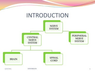
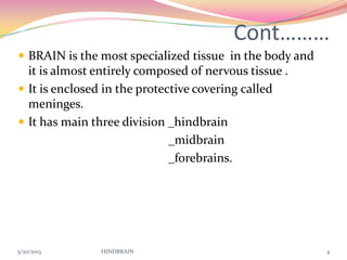
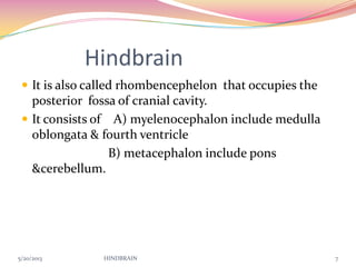
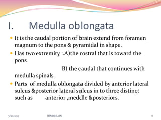


This document provides information about the hindbrain, which consists of the medulla oblongata, pons, and cerebellum. It describes the anatomy and functions of each part. The medulla oblongata extends from the foramen magnum to the pons and controls vital functions like breathing, blood pressure, and heart rate. The pons relays signals between the cerebrum and cerebellum and is involved in processes like sleep, respiration, and hearing. The cerebellum, located above the pons, aids in balance, muscle coordination, and motor skills.