Download as PDF, PPTX


![5353
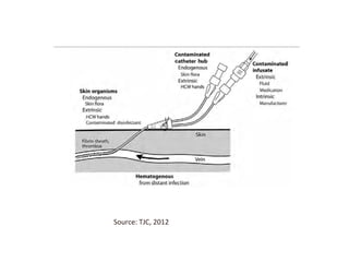
the right side of the neck carefully using a portable
ultrasound (US) machine (IMAGIC Agile, Kontron
Medical, WA, USA) with a linear, high frequency
transducer (7.5–12 MHz). Care was taken to apply
minimal pressure on the probe to prevent collapse of the
IJV. Imaging showed a single pulsatile vessel, which was
non compressible suggestive of the carotid artery with
right sided IJV has also been reported in a 12-year-old boy
during US evaluation prior to attempted cannulation.[4]
In
another report, IJV agenesis was discovered during neck
dissection. Patients who require removal of IJV due to
disease infiltration may have potentially life-threatening
complicationofcerebraledemaiftheotherIJVisaplastic.[5]
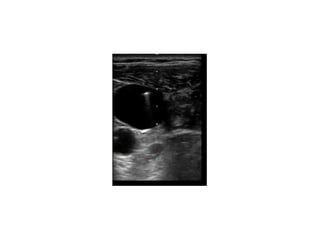
Figure 1: Ultrasound image of the right side of the neck showing absence
of internal jugular vein. CA: Carotid artery
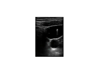
Figure 2: Ultrasound image of the left side of the neck showing normal
anatomy. CA: Carotid artery; IJV: Internal jugular vein](https://image.slidesharecdn.com/centralline-ncana-150930202125-lva1-app6892/85/Central-Line-in-Anesthesia-19-320.jpg)


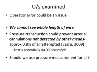
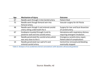


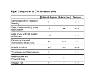


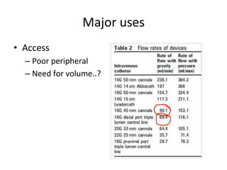
This document discusses central line placement, including: 1) It provides a brief history of central line development and discusses their increasing common use today. 2) Major uses of central lines include access for volume infusion, determination of cardiovascular function, and nutrition. 3) Potential complications include mechanical issues like arterial puncture or cannulation in 5-19% of cases, and infectious complications in 5-26% of cases.


