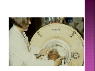
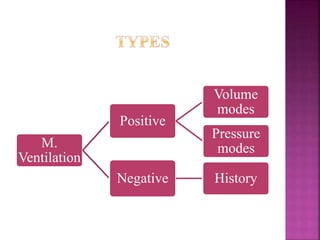
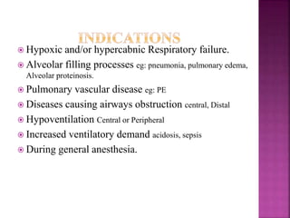
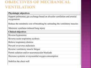
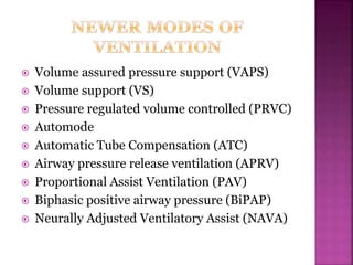
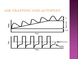
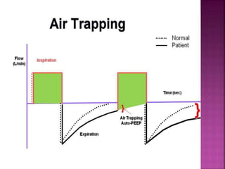
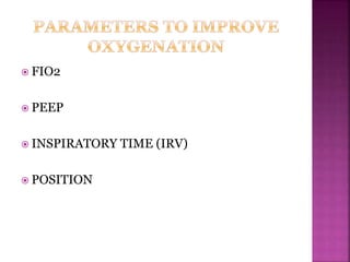
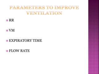
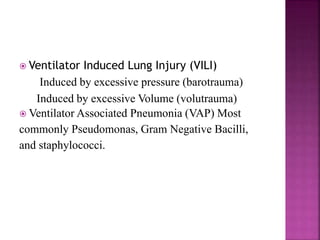
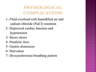
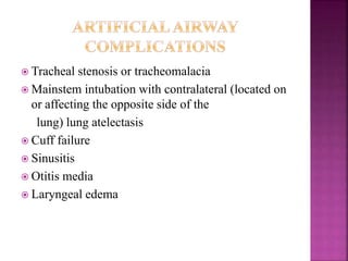
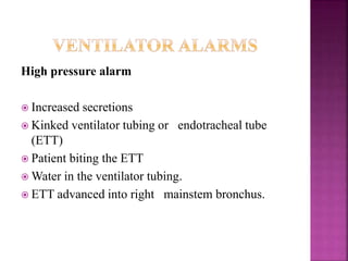
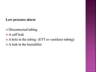
Dr. MAGDY KHAMES
INTENSIVIST
ZMH AL BATAYEH
1. Understand causes of respiratory failure
2. Have the knowledge to institute mechanical
ventilation safely
3. Understand the principles that guide
mechanical ventilation
4. Be able to apply these principles in clinical
practice
ICU with patients receiving mechanical ventilation via an iron lung.
Source: Reprinted by permission from the hospital archives, the Hospital
for Sick Children, Toronto.
The Drinker and Shaw tank-type ventilator of
1929 was one of the first negative-pressure
machines widely used for mechanical ventilation.
Better known as the iron lung, this metal cylinder
completely engulfed the patient up to the neck.
M.
Ventilation
Positive
Volume
modes
Pressure
modes
Negative History
 Hypoxic and/or hypercabnic Respiratory failure.
 Alveolar filling processes eg: pneumonia, pulmonary edema,
Alveolar proteinosis.
 Pulmonary vascular disease eg: PE
 Diseases causing airways obstruction central, Distal
 Hypoventilation Central or Peripheral
 Increased ventilatory demand acidosis, sepsis
 During general anesthesia.
Physiologic objectives
Support pulmonary gas exchange based on alveolar ventilation and arterial
oxygenation
Reduce the metabolic cost of breathing by unloading the ventilatory muscles
Minimize ventilator-induced lung injury
Clinical objectives
Reverse hypoxemia
Reverse acute respiratory acidosis
Relieve respiratory distress
Prevent or reverse atelectasis
Reverse ventilatory muscle fatigue
Permit sedation and/or neuromuscular blockade
Decrease systemic or myocardial oxygen consumption
Stabilize the chest wall
 Mode: is a combination of certain group of setting of MV
machine.
 Trigger: the way to initiate a ventilator-delivered breaths
(machine, patient)
 Target: a certain value you want to reach ( flow, pressure,
volume)
 Termination or cycle: the way the ventilator end the
delivered breath( time, volume, flow)
 Invasive: intubated patient.
 Noninvasive: via sealed face or nasal mask
 There are four distinct phases of ventilator breath
• cyclingInspiration to
expiration
• BaselineExpiration
• TriggerExpiration to
inspiration
• Targetinspiration
 Volume controlled: (volume targeted, machine
triggered, (volume, time, flow) cycled)
 Volume assist: ( volume targeted, patient triggered,
(volume, time, flow) cycled)
 Pressure controlled: ( pressure targeted , machine
triggered, (volume, time, flow) cycled)
 Pressure assist: ( pressure targeted , patient triggered,
(volume, time, flow) cycled)
 Pressure support:( pressure targeted , patient triggered,
flow cycled) used to augment patient breath.
Mode
Breath
strategy
(target)
Trigger Cycle
(breath
termination)
Types of breaths
Ventilator Patient Mandatory Assisted Spontaneous
CMV
Volume-
limited
Yes No Volume Yes No No
Pressure-
limited
Yes No Time Yes No No
AC
Volume-
limited
Yes Yes Volume Yes Yes No
Pressure-
limited
Yes Yes Time Yes Yes No
IMV
Volume-
limited
Yes Yes Volume Yes Yes* Yes*
Pressure-
limited (also
called
APRV)
Yes Yes Time Yes Yes* Yes*
PSV
Pressure-
limited
No Yes
Flow,
pressure, or
time
No Yes No
CPAP No Yes Flow No Yes No
Tube
compensatio
n
No Yes Flow No No Yes
Types of breaths:
Mandatory: Breaths are initiated by the ventilator and the ventilator performs the work of inspiration during those breaths
Assisted: Breaths are initiated by the patient, but the ventilator performs at least some of the work of inspiration for those patient
initiated breaths
Spontaneous: Breaths are initiated by the patient and the patient performs the entire work of inspiration for those patient initiated
breaths
 Volume assured pressure support (VAPS)
 Volume support (VS)
 Pressure regulated volume controlled (PRVC)
 Automode
 Automatic Tube Compensation (ATC)
 Airway pressure release ventilation (APRV)
 Proportional Assist Ventilation (PAV)
 Biphasic positive airway pressure (BiPAP)
 Neurally Adjusted Ventilatory Assist (NAVA)
Indications:
 Chronic obstructive pulmonary disease
 Cardiogenic pulmonary edema
 After discontinuation of mechanical ventilation
 Community-acquired pneumonia
 Asthma
 Immunocompromised state
 Postoperative respiratory distress
 Do-not-intubate status
 Neuromuscular respiratory failure
 Decompensated obstructive sleep apnea/
cor pulmonale
 Cystic fibrosis
 Acute respiratory distress syndrome
 Mild Pneumocystic carinii pneumonia
Absolute contraindications
 Coma
 Cardiac arrest
 Respiratory arrest
 Any condition requiring immediate intubation
Advantages and disadvantages
- Avoids complications associated with endotracheal
tube
- Lower rate of nosocomial infections and
pneumonias
- Shorter duration of mechanical ventilation
But
- High workload on personnel
- Patient selection and tolerance are critical
Criteria for patient selection
of NPPV(patient should be)
 Alert and cooperative (except COPD with CO2
coma)
 Haemodynamic stability
 No need for endotracheal intubation to
protect airways
remove excessive secretions
 No need for high PEEP
 No acute facial trauma, skull base fracture,
recent upper GI surgery
Criteria for discontinuing NPPV
 Inability to tolerate the mask
 Inability to improve gas exchange and dyspnia
 Need for endotracheal intubation
 Haemodynamic instability
 Signs of ischaemia on ECG
 Failure to improve mental status within 30 min
in CO2 coma or in hypoxaemic agitated
patients
 Verify the indication
 Determine invasive or noninvasive
 Select the ventilator mode
 Make ventilator setting
 Monitor your patient
MV basic setting:
Tidal volume(VT): 6 to10ml/IBW(kg) always consider it 70kg in average
adult.
Breathing Frequency(F): start with 15 b/m.
Positive end expiratory pressure(PEEP): 5 H2O is the physiological value.
Flow Rate: around 60L/minute.
I:E normal adult has 1:2.
FiO2: start by 100% the decrease according to SPO2.
MV setting in NPPV:
PS: begin with 10 H2O increase gradually to desired tidal volume and
tolerance of the patient
PEEP: begin with 5 H2O
FiO2: 100% then decrease as patient saturation permit target to be less than 50%
 FIO2
 PEEP
 INSPIRATORY TIME (IRV)
 POSITION
 RR
 VM
 EXPIRATORY TIME
 FLOW RATE
 Airway Complications.
 Mechanical complications.
 Physiological Complications.
 Artificial Airway Complications.
1- Aspiration
2- Decreased clearance of secretions
3- Nosocomial or ventilator-acquired
pneumonia
1- Hypoventilation with atelectasis with respiratory
acidosis or hypoxemia.
2- Hyperventilation with hypocapnia and respiratory
alkalosis
3- Barotrauma
a- Closed pneumothorax,
b- Tension pneumothorax,
c- Pneumomediastinum,
d- Subcutaneous emphysema.
4- Alarm “turned off”
5- Failure of alarms or ventilator
6- Inadequate nebulization or humidification
7- Overheated inspired air, resulting in hyperthermia
 Ventilator Induced Lung Injury (VILI)
Induced by excessive pressure (barotrauma)
Induced by excessive Volume (volutrauma)
 Ventilator Associated Pneumonia (VAP) Most
commonly Pseudomonas, Gram Negative Bacilli,
and staphylococci.
1- Fluid overload with humidified air and
sodium chloride (NaCl) retention
2- Depressed cardiac function and
hypotension
3- Stress ulcers
4- Paralytic ileus
5- Gastric distension
6- Starvation
7- Dyssynchronous breathing pattern
 Tracheal stenosis or tracheomalacia
 Mainstem intubation with contralateral (located on
or affecting the opposite side of the
lung) lung atelectasis
 Cuff failure
 Sinusitis
 Otitis media
 Laryngeal edema
High pressure alarm
 Increased secretions
 Kinked ventilator tubing or endotracheal tube
(ETT)
 Patient biting the ETT
 Water in the ventilator tubing.
 ETT advanced into right mainstem bronchus.
Low pressure alarm
 Disconnected tubing
 A cuff leak
 A hole in the tubing (ETT or ventilator tubing)
 A leak in the humidifier
Oxygen alarm
 The oxygen supply is insufficient or is not properly
connected.
High respiratory rate alarm
 Episodes of tachypnea.
 Anxiety.
 Pain.
 Hypoxia.
 Fever.
Apnea alarm
 During weaning, indicates that the patient has a
slow Respiratory rate and a period of apnea.
 Mechanical ventilation is an indispensable tool for
the ER physician
 Whether or not the patient requires ventilator
support is a crucial decision to make
 Proper understanding of ventilator function and
modes are vital to provide individualized therapy
to a wide range of patients
 Ventilator graphics can provide valuable
information regarding settings and pulmonary
characteristics
 Patient care during critical illness is vital – proper
co-ordination between machines, nurses and
doctors
 VILI and VAP are dreaded complications -
prevention is better than cure
1. Clinical Application of Mechanical Ventilation – David W Chang, 4th
Edition
2. Mechanical Ventilation – Vijay Deshpande, 2nd Edition
3. The ICU book – Paul L. Marino, 4th edition
4. Chatburn RL. Classification of Ventilator Modes. Respir Care 2007;
52(3)
5. www.ardsnet.org
6. www.frca.co.uk – Anaesthesia Tutorial of the Week
7. www.wikipedia.org
8. Ventilator Waveforms – Graphical representation of ventilatory data.
Puritan Bennett
9. Lindgren VA et al. Care for patients on mechanical ventilation. AJN
2005;105
10. Grossbach I et al. Overview of mechanical ventilatory support, and
managent of patient and ventilator related responses. Critical Care
Nurse 2011
11. Girard TD et al. Mechanical ventilation in ARDS – A state of the art
review. CHEST 2007; 131
THANK
YOU

Mechanical ventilation in emergency

  • 1.
  • 2.
    1. Understand causesof respiratory failure 2. Have the knowledge to institute mechanical ventilation safely 3. Understand the principles that guide mechanical ventilation 4. Be able to apply these principles in clinical practice
  • 3.
    ICU with patientsreceiving mechanical ventilation via an iron lung. Source: Reprinted by permission from the hospital archives, the Hospital for Sick Children, Toronto.
  • 4.
    The Drinker andShaw tank-type ventilator of 1929 was one of the first negative-pressure machines widely used for mechanical ventilation. Better known as the iron lung, this metal cylinder completely engulfed the patient up to the neck.
  • 6.
  • 7.
     Hypoxic and/orhypercabnic Respiratory failure.  Alveolar filling processes eg: pneumonia, pulmonary edema, Alveolar proteinosis.  Pulmonary vascular disease eg: PE  Diseases causing airways obstruction central, Distal  Hypoventilation Central or Peripheral  Increased ventilatory demand acidosis, sepsis  During general anesthesia.
  • 8.
    Physiologic objectives Support pulmonarygas exchange based on alveolar ventilation and arterial oxygenation Reduce the metabolic cost of breathing by unloading the ventilatory muscles Minimize ventilator-induced lung injury Clinical objectives Reverse hypoxemia Reverse acute respiratory acidosis Relieve respiratory distress Prevent or reverse atelectasis Reverse ventilatory muscle fatigue Permit sedation and/or neuromuscular blockade Decrease systemic or myocardial oxygen consumption Stabilize the chest wall
  • 9.
     Mode: isa combination of certain group of setting of MV machine.  Trigger: the way to initiate a ventilator-delivered breaths (machine, patient)  Target: a certain value you want to reach ( flow, pressure, volume)  Termination or cycle: the way the ventilator end the delivered breath( time, volume, flow)  Invasive: intubated patient.  Noninvasive: via sealed face or nasal mask
  • 11.
     There arefour distinct phases of ventilator breath • cyclingInspiration to expiration • BaselineExpiration • TriggerExpiration to inspiration • Targetinspiration
  • 12.
     Volume controlled:(volume targeted, machine triggered, (volume, time, flow) cycled)  Volume assist: ( volume targeted, patient triggered, (volume, time, flow) cycled)  Pressure controlled: ( pressure targeted , machine triggered, (volume, time, flow) cycled)  Pressure assist: ( pressure targeted , patient triggered, (volume, time, flow) cycled)  Pressure support:( pressure targeted , patient triggered, flow cycled) used to augment patient breath.
  • 13.
    Mode Breath strategy (target) Trigger Cycle (breath termination) Types ofbreaths Ventilator Patient Mandatory Assisted Spontaneous CMV Volume- limited Yes No Volume Yes No No Pressure- limited Yes No Time Yes No No AC Volume- limited Yes Yes Volume Yes Yes No Pressure- limited Yes Yes Time Yes Yes No IMV Volume- limited Yes Yes Volume Yes Yes* Yes* Pressure- limited (also called APRV) Yes Yes Time Yes Yes* Yes* PSV Pressure- limited No Yes Flow, pressure, or time No Yes No CPAP No Yes Flow No Yes No Tube compensatio n No Yes Flow No No Yes Types of breaths: Mandatory: Breaths are initiated by the ventilator and the ventilator performs the work of inspiration during those breaths Assisted: Breaths are initiated by the patient, but the ventilator performs at least some of the work of inspiration for those patient initiated breaths Spontaneous: Breaths are initiated by the patient and the patient performs the entire work of inspiration for those patient initiated breaths
  • 14.
     Volume assuredpressure support (VAPS)  Volume support (VS)  Pressure regulated volume controlled (PRVC)  Automode  Automatic Tube Compensation (ATC)  Airway pressure release ventilation (APRV)  Proportional Assist Ventilation (PAV)  Biphasic positive airway pressure (BiPAP)  Neurally Adjusted Ventilatory Assist (NAVA)
  • 16.
    Indications:  Chronic obstructivepulmonary disease  Cardiogenic pulmonary edema  After discontinuation of mechanical ventilation  Community-acquired pneumonia  Asthma  Immunocompromised state  Postoperative respiratory distress  Do-not-intubate status  Neuromuscular respiratory failure  Decompensated obstructive sleep apnea/ cor pulmonale  Cystic fibrosis  Acute respiratory distress syndrome  Mild Pneumocystic carinii pneumonia
  • 17.
    Absolute contraindications  Coma Cardiac arrest  Respiratory arrest  Any condition requiring immediate intubation
  • 18.
    Advantages and disadvantages -Avoids complications associated with endotracheal tube - Lower rate of nosocomial infections and pneumonias - Shorter duration of mechanical ventilation But - High workload on personnel - Patient selection and tolerance are critical
  • 19.
    Criteria for patientselection of NPPV(patient should be)  Alert and cooperative (except COPD with CO2 coma)  Haemodynamic stability  No need for endotracheal intubation to protect airways remove excessive secretions  No need for high PEEP  No acute facial trauma, skull base fracture, recent upper GI surgery
  • 20.
    Criteria for discontinuingNPPV  Inability to tolerate the mask  Inability to improve gas exchange and dyspnia  Need for endotracheal intubation  Haemodynamic instability  Signs of ischaemia on ECG  Failure to improve mental status within 30 min in CO2 coma or in hypoxaemic agitated patients
  • 21.
     Verify theindication  Determine invasive or noninvasive  Select the ventilator mode  Make ventilator setting  Monitor your patient
  • 22.
    MV basic setting: Tidalvolume(VT): 6 to10ml/IBW(kg) always consider it 70kg in average adult. Breathing Frequency(F): start with 15 b/m. Positive end expiratory pressure(PEEP): 5 H2O is the physiological value. Flow Rate: around 60L/minute. I:E normal adult has 1:2. FiO2: start by 100% the decrease according to SPO2.
  • 26.
    MV setting inNPPV: PS: begin with 10 H2O increase gradually to desired tidal volume and tolerance of the patient PEEP: begin with 5 H2O FiO2: 100% then decrease as patient saturation permit target to be less than 50%
  • 33.
     FIO2  PEEP INSPIRATORY TIME (IRV)  POSITION
  • 34.
     RR  VM EXPIRATORY TIME  FLOW RATE
  • 35.
     Airway Complications. Mechanical complications.  Physiological Complications.  Artificial Airway Complications.
  • 36.
    1- Aspiration 2- Decreasedclearance of secretions 3- Nosocomial or ventilator-acquired pneumonia
  • 37.
    1- Hypoventilation withatelectasis with respiratory acidosis or hypoxemia. 2- Hyperventilation with hypocapnia and respiratory alkalosis 3- Barotrauma a- Closed pneumothorax, b- Tension pneumothorax, c- Pneumomediastinum, d- Subcutaneous emphysema. 4- Alarm “turned off” 5- Failure of alarms or ventilator 6- Inadequate nebulization or humidification 7- Overheated inspired air, resulting in hyperthermia
  • 38.
     Ventilator InducedLung Injury (VILI) Induced by excessive pressure (barotrauma) Induced by excessive Volume (volutrauma)  Ventilator Associated Pneumonia (VAP) Most commonly Pseudomonas, Gram Negative Bacilli, and staphylococci.
  • 39.
    1- Fluid overloadwith humidified air and sodium chloride (NaCl) retention 2- Depressed cardiac function and hypotension 3- Stress ulcers 4- Paralytic ileus 5- Gastric distension 6- Starvation 7- Dyssynchronous breathing pattern
  • 40.
     Tracheal stenosisor tracheomalacia  Mainstem intubation with contralateral (located on or affecting the opposite side of the lung) lung atelectasis  Cuff failure  Sinusitis  Otitis media  Laryngeal edema
  • 41.
    High pressure alarm Increased secretions  Kinked ventilator tubing or endotracheal tube (ETT)  Patient biting the ETT  Water in the ventilator tubing.  ETT advanced into right mainstem bronchus.
  • 42.
    Low pressure alarm Disconnected tubing  A cuff leak  A hole in the tubing (ETT or ventilator tubing)  A leak in the humidifier
  • 43.
    Oxygen alarm  Theoxygen supply is insufficient or is not properly connected.
  • 44.
    High respiratory ratealarm  Episodes of tachypnea.  Anxiety.  Pain.  Hypoxia.  Fever.
  • 45.
    Apnea alarm  Duringweaning, indicates that the patient has a slow Respiratory rate and a period of apnea.
  • 46.
     Mechanical ventilationis an indispensable tool for the ER physician  Whether or not the patient requires ventilator support is a crucial decision to make  Proper understanding of ventilator function and modes are vital to provide individualized therapy to a wide range of patients  Ventilator graphics can provide valuable information regarding settings and pulmonary characteristics  Patient care during critical illness is vital – proper co-ordination between machines, nurses and doctors  VILI and VAP are dreaded complications - prevention is better than cure
  • 47.
    1. Clinical Applicationof Mechanical Ventilation – David W Chang, 4th Edition 2. Mechanical Ventilation – Vijay Deshpande, 2nd Edition 3. The ICU book – Paul L. Marino, 4th edition 4. Chatburn RL. Classification of Ventilator Modes. Respir Care 2007; 52(3) 5. www.ardsnet.org 6. www.frca.co.uk – Anaesthesia Tutorial of the Week 7. www.wikipedia.org 8. Ventilator Waveforms – Graphical representation of ventilatory data. Puritan Bennett 9. Lindgren VA et al. Care for patients on mechanical ventilation. AJN 2005;105 10. Grossbach I et al. Overview of mechanical ventilatory support, and managent of patient and ventilator related responses. Critical Care Nurse 2011 11. Girard TD et al. Mechanical ventilation in ARDS – A state of the art review. CHEST 2007; 131
  • 48.