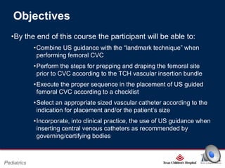
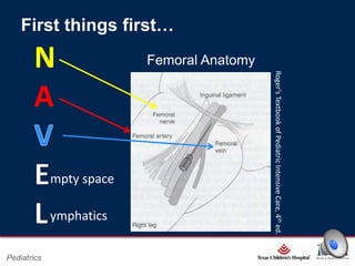
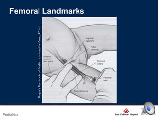
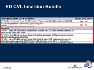
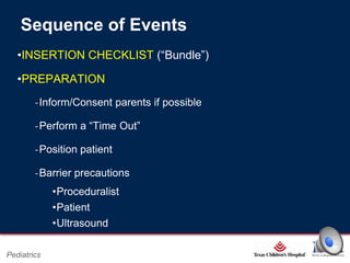
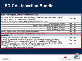
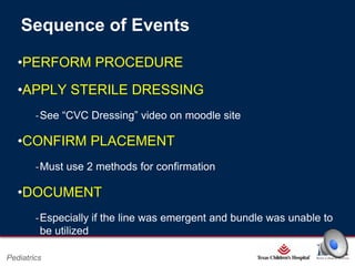
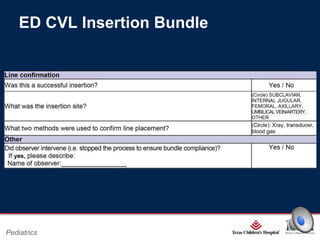
Central Venous
Catheterization:
“From the ICU to the EC”
A REFRESHER COURSE
Pediatrics
Objectives
•By the end of this course the participant will be able to:
•Combine US guidance with the “landmark technique” when
performing femoral CVC
•Perform the steps for prepping and draping the femoral site
prior to CVC according to the TCH vascular insertion bundle
•Execute the proper sequence in the placement of US guided
femoral CVC according to a checklist
•Select an appropriate sized vascular catheter according to the
indication for placement and/or the patient’s size
•Incorporate, into clinical practice, the use of US guidance when
inserting central venous catheters as recommended by
governing/certifying bodies

Page 1

Pediatrics

xxx00.#####.ppt 2/26/2014 6:39:21 PM
First things first…
Femoral Anatomy
Roger’s Textbook of Pediatric Intensive Care, 4th ed.

N
A
mpty space
ymphatics

Page 2

Pediatrics

xxx00.#####.ppt 2/26/2014 6:39:21 PM
Roger’s Textbook of Pediatric Intensive Care, 4th ed.

Femoral Landmarks

Page 3

Pediatrics

xxx00.#####.ppt 2/26/2014 6:39:21 PM
Indications
•Peripheral access unobtainable
•Medication/fluid administration
•Emergency resuscitation
•Monitoring of CVP and SvO2
•Parenteral nutrition
•Frequent blood sampling
•Hemodialysis/hemofiltration/apheresis
Page 4

Pediatrics

xxx00.#####.ppt 2/26/2014 6:39:21 PM
Contraindications
•Coagulopathy
•Skin infection at site of needle puncture
•Trauma
•Distorted anatomy
•Thrombosis

Page 5

Pediatrics

xxx00.#####.ppt 2/26/2014 6:39:21 PM
Complications Common to ALL Sites
•Infection

•Malposition

•Thrombosis

•Catheter fragment/
guidewire embolism

•Arterial puncture

•Cardiac dysrhythmias

•Arterial cannulation

‐IJ & Subclavian CVC
‐“high” femoral CVC

•Hemorrhage

•Air embolism

•Phlebitis

‐More so with IJ &
Subclavian CVC
Page 6

Pediatrics

xxx00.#####.ppt 2/26/2014 6:39:21 PM
FEMORAL CVC Complications
•Bladder puncture
•Retroperitoneal hemorrhage
•Malposition
‐ Ascending lumbar veins

May cause complications…
•Uncooperative patient
•Lack of experience/supervision

Page 7

Pediatrics

xxx00.#####.ppt 2/26/2014 6:39:21 PM
Choosing a Catheter
•Size?
•Length?
•Number of lumens?
•Patient
‐Weight/size
‐Size of vessel
‐Indications
Page 8

Pediatrics

xxx00.#####.ppt 2/26/2014 6:39:21 PM
Choosing a Catheter Size/Length
“Standard Catheter Lengths Available for Pediatric Use”,
according to booklet attached to catheter tray…

…NOT HELPFUL
Page 9

Pediatrics

xxx00.#####.ppt 2/26/2014 6:39:21 PM
ED CVL Insertion Bundle

Page 10

Pediatrics

xxx00.#####.ppt 2/26/2014 6:39:21 PM
Sequence of Events
•INSERTION CHECKLIST (“Bundle”)
•PREPARATION
‐Inform/Consent parents if possible

‐Perform a “Time Out”
‐Position patient
‐Barrier precautions
•Proceduralist
•Patient
•Ultrasound
Page 11

Pediatrics

xxx00.#####.ppt 2/26/2014 6:39:21 PM
ED CVL Insertion Bundle

Page 12

Pediatrics

xxx00.#####.ppt 2/26/2014 6:39:21 PM
Sequence of Events
•PERFORM PROCEDURE
•APPLY STERILE DRESSING
‐See “CVC Dressing” video on moodle site

•CONFIRM PLACEMENT
‐Must use 2 methods for confirmation

•DOCUMENT
‐Especially if the line was emergent and bundle was unable to
be utilized
Page 13

Pediatrics

xxx00.#####.ppt 2/26/2014 6:39:21 PM
ED CVL Insertion Bundle

Page 14

Pediatrics

xxx00.#####.ppt 2/26/2014 6:39:21 PM
REFERENCES
•Rey et al. Mechanical complications during central venous cannulations
in pediatric patients. Intensive Care Med 2009; 35: 1438–1443.
•Froehlich et al. Ultrasound-guided central venous catheter placement
decreases complications and decreases placement attempts compared
with the landmark technique in patients in a pediatric Intensive care unit.
Crit Care Med 2009; 37(3): 1090-1096
•Miller et al. Decreasing PICU Catheter-Associated Bloodstream
Infections: NACHRI's Quality Transformation Efforts. Pediatrics 2010;
125: 206-213
•Wheeler et al. A Hospital-wide Quality-Improvement Collaborative to
Reduce Catheter-Associated Bloodstream Infections. Pediatrics 2011;
126(4): e995-e1007
Page 15

Pediatrics

xxx00.#####.ppt 2/26/2014 6:39:21 PM
REFERENCES
•Kumar et al. Ultrasound guided vascular access: efficacy and safety.
Best Practice & Research Clinical Anaesthesiology 2009; 23: 299–311
•Srinivasan et al. Bedside ultrasound in pediatric critical care: A review.
Ped Crit Care Med 2011; 12(6): 667-674

•McGee et al. Preventing Complications of Central Venous
Catheterization. N Engl J Med 2003; 348(12): 1123-1133
•Moore et al. Point-of-Care Ultrasonography. N Engl J Med 2011; 364:
749-57.
•CDC for the Prevention of Intravascular Catheter-Related Infections,
2011. http://www.cdc.gov/hicpac/pdf/guidelines/bsi-guidelines-2011.pdf
Page 16

Pediatrics

xxx00.#####.ppt 2/26/2014 6:39:21 PM
REFERENCES
•American College of Emergency Physicians: Emergency ultrasound
guidelines. Ann Emerg Med. 2009; 53: 550-570
•Costello et al. Minimizing Complications Associated with Percutaneous
Central Venous Catheter Placement in Children: Recent Advances.
Pediatr Crit Care Med. 2013; 14: 273-283

Page 17

Pediatrics

xxx00.#####.ppt 2/26/2014 6:39:21 PM

2014 02 CVC in the EC_REFRESHER COURSE

  • 1.
    Central Venous Catheterization: “From theICU to the EC” A REFRESHER COURSE Pediatrics
  • 2.
    Objectives •By the endof this course the participant will be able to: •Combine US guidance with the “landmark technique” when performing femoral CVC •Perform the steps for prepping and draping the femoral site prior to CVC according to the TCH vascular insertion bundle •Execute the proper sequence in the placement of US guided femoral CVC according to a checklist •Select an appropriate sized vascular catheter according to the indication for placement and/or the patient’s size •Incorporate, into clinical practice, the use of US guidance when inserting central venous catheters as recommended by governing/certifying bodies Page 1 Pediatrics xxx00.#####.ppt 2/26/2014 6:39:21 PM
  • 3.
    First things first… FemoralAnatomy Roger’s Textbook of Pediatric Intensive Care, 4th ed. N A mpty space ymphatics Page 2 Pediatrics xxx00.#####.ppt 2/26/2014 6:39:21 PM
  • 4.
    Roger’s Textbook ofPediatric Intensive Care, 4th ed. Femoral Landmarks Page 3 Pediatrics xxx00.#####.ppt 2/26/2014 6:39:21 PM
  • 5.
    Indications •Peripheral access unobtainable •Medication/fluidadministration •Emergency resuscitation •Monitoring of CVP and SvO2 •Parenteral nutrition •Frequent blood sampling •Hemodialysis/hemofiltration/apheresis Page 4 Pediatrics xxx00.#####.ppt 2/26/2014 6:39:21 PM
  • 6.
    Contraindications •Coagulopathy •Skin infection atsite of needle puncture •Trauma •Distorted anatomy •Thrombosis Page 5 Pediatrics xxx00.#####.ppt 2/26/2014 6:39:21 PM
  • 7.
    Complications Common toALL Sites •Infection •Malposition •Thrombosis •Catheter fragment/ guidewire embolism •Arterial puncture •Cardiac dysrhythmias •Arterial cannulation ‐IJ & Subclavian CVC ‐“high” femoral CVC •Hemorrhage •Air embolism •Phlebitis ‐More so with IJ & Subclavian CVC Page 6 Pediatrics xxx00.#####.ppt 2/26/2014 6:39:21 PM
  • 8.
    FEMORAL CVC Complications •Bladderpuncture •Retroperitoneal hemorrhage •Malposition ‐ Ascending lumbar veins May cause complications… •Uncooperative patient •Lack of experience/supervision Page 7 Pediatrics xxx00.#####.ppt 2/26/2014 6:39:21 PM
  • 9.
    Choosing a Catheter •Size? •Length? •Numberof lumens? •Patient ‐Weight/size ‐Size of vessel ‐Indications Page 8 Pediatrics xxx00.#####.ppt 2/26/2014 6:39:21 PM
  • 10.
    Choosing a CatheterSize/Length “Standard Catheter Lengths Available for Pediatric Use”, according to booklet attached to catheter tray… …NOT HELPFUL Page 9 Pediatrics xxx00.#####.ppt 2/26/2014 6:39:21 PM
  • 11.
    ED CVL InsertionBundle Page 10 Pediatrics xxx00.#####.ppt 2/26/2014 6:39:21 PM
  • 12.
    Sequence of Events •INSERTIONCHECKLIST (“Bundle”) •PREPARATION ‐Inform/Consent parents if possible ‐Perform a “Time Out” ‐Position patient ‐Barrier precautions •Proceduralist •Patient •Ultrasound Page 11 Pediatrics xxx00.#####.ppt 2/26/2014 6:39:21 PM
  • 13.
    ED CVL InsertionBundle Page 12 Pediatrics xxx00.#####.ppt 2/26/2014 6:39:21 PM
  • 14.
    Sequence of Events •PERFORMPROCEDURE •APPLY STERILE DRESSING ‐See “CVC Dressing” video on moodle site •CONFIRM PLACEMENT ‐Must use 2 methods for confirmation •DOCUMENT ‐Especially if the line was emergent and bundle was unable to be utilized Page 13 Pediatrics xxx00.#####.ppt 2/26/2014 6:39:21 PM
  • 15.
    ED CVL InsertionBundle Page 14 Pediatrics xxx00.#####.ppt 2/26/2014 6:39:21 PM
  • 16.
    REFERENCES •Rey et al.Mechanical complications during central venous cannulations in pediatric patients. Intensive Care Med 2009; 35: 1438–1443. •Froehlich et al. Ultrasound-guided central venous catheter placement decreases complications and decreases placement attempts compared with the landmark technique in patients in a pediatric Intensive care unit. Crit Care Med 2009; 37(3): 1090-1096 •Miller et al. Decreasing PICU Catheter-Associated Bloodstream Infections: NACHRI's Quality Transformation Efforts. Pediatrics 2010; 125: 206-213 •Wheeler et al. A Hospital-wide Quality-Improvement Collaborative to Reduce Catheter-Associated Bloodstream Infections. Pediatrics 2011; 126(4): e995-e1007 Page 15 Pediatrics xxx00.#####.ppt 2/26/2014 6:39:21 PM
  • 17.
    REFERENCES •Kumar et al.Ultrasound guided vascular access: efficacy and safety. Best Practice & Research Clinical Anaesthesiology 2009; 23: 299–311 •Srinivasan et al. Bedside ultrasound in pediatric critical care: A review. Ped Crit Care Med 2011; 12(6): 667-674 •McGee et al. Preventing Complications of Central Venous Catheterization. N Engl J Med 2003; 348(12): 1123-1133 •Moore et al. Point-of-Care Ultrasonography. N Engl J Med 2011; 364: 749-57. •CDC for the Prevention of Intravascular Catheter-Related Infections, 2011. http://www.cdc.gov/hicpac/pdf/guidelines/bsi-guidelines-2011.pdf Page 16 Pediatrics xxx00.#####.ppt 2/26/2014 6:39:21 PM
  • 18.
    REFERENCES •American College ofEmergency Physicians: Emergency ultrasound guidelines. Ann Emerg Med. 2009; 53: 550-570 •Costello et al. Minimizing Complications Associated with Percutaneous Central Venous Catheter Placement in Children: Recent Advances. Pediatr Crit Care Med. 2013; 14: 273-283 Page 17 Pediatrics xxx00.#####.ppt 2/26/2014 6:39:21 PM

Editor's Notes

  • #3 Laterally to medially NAVEL is a common mnemonic used to recall the anatomy of the femoral/inguinal regionFemoral vein lies in femoral sheath, medial to femoral artery, immediately below inguinal ligamentFV formed by joining of deep/superficial veins of the leg, above inguinal ligament becomes external iliac and joins with internal iliac to become common iliac, both common iliacs jon to become IVC
  • #4 There are a few of ways to identify the landmarks used to perform femoral CVC:FA is at midpoint btwn ASIS and SP, 2cm below inguinal ligament and 1 cm or 1FB medial to FA pulsationThumb over Pubic tubercle and index finger over ASIS, vessels lie within the webbed space(Use diagram)Anatomic variation (up to 20%)
  • #5 General Ind/Contraind for CVL placement, none specific to femoral siteThese lists are not necessarily exhaustive but just capture the more common ones
  • #6 RelativeFew, if any, absolute contraindicatons (i.e. refusal of consent)Risks vs. Benefits
  • #8 Fem CVC bladder puncture (DECOMPRESS BLADDER), retroperitoneal hemorrhageUncoop pt poses risk to themselves and proceduralistSedation/analgesia for patient comfort, facilitate placement and reduce complications related to patient movementINEXPERIENCE or lack of supervisionLast 2 bullets arent necessarily complications but can LEAD to complications
  • #9 Not one size fits all, may consider measuring
  • #11 Position-hip abduction/external rotationConfirmation-transduction, blood gas, radiographic
  • #12 CHG reduces the risk of catheter colonizationOther entering room must wear cap/mask
  • #13 Position-hip abduction/external rotationConfirmation-transduction, blood gas, radiographic