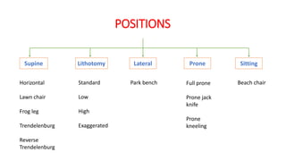
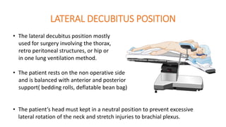
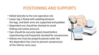
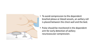
PRESENTER:
DR. ARYA DASMAHAPATRA
1ST YEAR POST GRADUATE TRAINEE,
DEPARTMENT OF ANAESTHESIOLOGY,
MEDICAL COLLEGE KOLKATA
MODERATOR:
DR. DEBABANHI BARUA
ASSISTANT PROFESSOR,
DEPARTMENT OF ANAESTHESIOLOGY,
MEDICAL COLLEGE KOLKATA
DIFFERENT PATIENT
POSITIONING IN
ANAESTHESIA
PREAMBLE
• Patient positioning is a substantial responsibility that requires the
coordination of the entire team in operation theatres. A balance between
optimal surgical positioning and patient well-being is sometimes required.
• It is prudent to adapt optimal positioning as it can pre-empt adverse
outcomes.
• Patient positioning and postural limitations should be assessed during the
pre-anaesthetic check-up(PAC).
GOALS OF PROPER POSITIONING
• To promote proper physiological alignment.
• To maintain patient’s airway and avoid constriction on the chest cavity.
• To minimize interference with circulation.
• To gain optimum exposure to operative and anaesthetist site.
• To protect skeletal and neuromuscular structures.
• To provide comfort, stability, safety and dignity to the patient.
GENERAL CONSIDERATIONS
• The patient’s position during anaesthesia care should be neutral-one that
would be well tolerated if the patient were awake and unsedated.
• Weight-bearing surfaces and joints should be well padded and curvatures
including the lumber spine, should be supported.
• The head should be in midline without substantial flexion and extension.
Eyes should be closed without external pressure.
• Tilting of table before draping, using safety straps and prevention of falls
from the table, are fundamental.
ASSESSMENT
We should assess the following prior to positioning of the patient:
• Duration of the surgery.
• Surgeon’s preference of position.
• Required position for procedure.
• Anaesthesia to be administered.
• Patient’s risk factors (age, weight, skin condition, pre-existing conditions,
etc.)
• Patient’s privacy and medical needs.
• Basics of anatomy and physiology.
TABLE ACCESSORIES AND ATTACHMENTS
Elevated
arm rest
Lateral arm
support
Stirrups
Detachable
foot rest
Metal socket
Hydraulic wheeled base
stand
Sliding bars
Breakable
head rest
Arm board
Manual lever
Pillows, Head rings, Sandbags, Roll supports, Soft pads, Mattress
PHYSIOLOGICAL CHANGES RELATED TO
CHANGES IN BODY POSITION
• Most changes are related to gravitational effects on cardiovascular system
and respiratory system.
• Alterations in position redistribute blood within the venous, arterial and
pulmonary vasculature.
• Pulmonary mechanics and pulmonary perfusion also vary with different
body positions.
CARDIOVASCULAR CONCERNS
Supine position from
erect posture
Pooled blood from
lower extremities
towards heart
Preload, stroke volume,
cardiac output
increases
Arterial blood pressure
increases
Activates afferent
baroreceptors by IXth
and Xth nerve
Reduces sympathetic
outflow
Increases
parasympathetic
impulses to the SA
node and myocardium.
Compensatory
decrease in HR & CO
 GA, MR, PPV, neuraxial blockade
all interfere with the venous return
to the heart, arterial tone and auto
regulatory mechanism.
PULMONARY CONCERNS
Supine position from
erect posture
Cephalad displacement
of the diaphragm &
abdominal contents
Reduction in
FRC, TV, TLC
• Anesthetized persons who are spontaneously breathing have a reduced
TV, FRC and an increased closing volume.
• Positive pressure ventilation with muscle relaxation may ameliorate
ventilation-perfusion mismatches under GA by maintaining adequate
minute ventilation.
• Gravity affects the preferential perfusion of the dependent portions of
lung.
POSITIONS
Supine Lithotomy Lateral Prone Sitting
Horizontal
Lawn chair
Frog leg
Trendelenburg
Reverse
Trendelenburg
Standard
Low
High
Exaggerated
Park bench Full prone
Prone jack
knife
Prone
kneeling
Beach chair
SUPINE
POSITION
POSITIONING, PHYSIOLOGY, MERITS,
DEMERITS, VARIATIONS AND COMPLICATIONS
POSITIONING
• Most commonly used position for surgeries.
• The head, neck and spine all retain neutrality.
• Arms can be abducted, adducted
but should be placed in as neutral
a position as possible.
• When adducted arms are securely
placed next to the body. Abducted arms
are kept on arm boards beside the table.
• Attention should be given to pad bony prominences.
ARM POSITIONING
When arms are adducted, they are usually
held alongside the body with a “drawsheet”
that passes under the body.
Abduction should be limited to less than 90o to
minimize brachial plexus injury.
LAWN CHAIR POSITION
• Patient’s hips at the break of the table.
• Reduces stress on back, hips and knees
as they are in flexed position.
• Sometimes, this position is better
tolerated than full supine position.
• Better venous drainage in lower limb as they
are slightly over the level of heart.
• Abdominal wall tension is reduced, as xiphoid
to pubic distance is reduced.
FROG LEG POSITION
• Allows access to perineum, medial thighs,
genitalia and rectum.
• Positioned supine, hips and knees are flexed with
hips externally rotated and soles facing each other.
• Care must be taken to minimize stress
and postoperative pain in the hips and to
prevent dislocation by appropriately supporting
the knees.
TRENDELENBURG POSITION
• Frequently used to improve exposure during abdominal and laparoscopic
surgeries.
• Position produces haemodynamic
and respiratory changes.
• This position is often preferred
during central line placement to
prevent air embolism and to offset
hypotension by temporarily increasing
venous return.
• The cephalad movement of diaphragm
decreases FRC, thus, decreasing pulmonary
compliance.
• This position increases CVP, ICP, IOP and can have significant respiratory
consequences.
• Prolonged head down can lead to swelling of face, conjunctiva, larynx and
tongue with an increased potential for post-operative upper airway
obstruction.
• Care must be taken to prevent patients in steep head down positions from
slipping cephalad on the surgical table. Shoulder braces are not recommended.
 In spontaneously ventilating patients, the work of breathing
increases.
 In mechanically ventilated patients, airway pressures must be
higher to ensure adequate ventilation.
REVERSE TRENDELENBURG POSITION
• Head up tilt is often employed to facilitate
upper abdominal surgeries.
• The position of head above the heart
reduces cerebral perfusion pressure and
may also cause systemic hypotension
as venous return decreases.
• Invasive arterial pressure transducer, if used, should
be zeroed at the level of the Circle of Willis.
SUPINE CONCERNS
• Greatest concerns are circulation and pressure points.
• Most common nerve damages:
1. Brachial plexus: arm abduction >90 degrees.
2. Radial and ulnar: compression against the OR
bed, metal attachments.
3. Peroneal and tibial: crossing of feet and
planter flexion of ankles and feet.
 Vulnerable bony prominences: Due to rubbing and
sustained pressure(occiput, spine, scapula,
olecranon, sacrum, calcaneus)
SUPINE POSITION PRESSURE POINTS
COMPLICATIONS
• Pressure alopecia
Caused by ischaemic hair follicles, is related to prolonged immobilization of
the head with its full weight falling on a limited area, usually the occiput.
• Backache
May occur because the normal lordotic curvature is often lost during GA with
muscle relaxation or a neuraxial block.
• Peripheral nerve injury
Brachial plexus injury, ulnar neuropathy etc.
LITHOTOMY
POSITION
POSITIONING, PHYSIOLOGY, MERITS,
DEMERITS, VARIATIONS AND COMPLICATIONS
POSITIONING
• The classic lithotomy position is frequently
used during gynaecologic, rectal and urologic
surgeries.
• The hips are flexed 80 to 100 degrees
from the trunk, and the legs are abducted
30 to 45 degrees from the midline.
• The knees are flexed until the lower legs are parallel
to the torso and supports or stirrups hold the legs.
Foot section of the table is lowered or detached.
The lower extremities should be padded to
prevent compression against the stirrups.
Initiation of the lithotomy position requires
coordinated positioning of the lower extremities
by two assistants to avoid torsion of the lumber
spine. Both the legs should be raised together,
simultaneously flexing the hips and knees.
After surgery, the patient must also be
returned to the same position in a
coordinated manner.
ARM POSITIONING
• The recommended position of the arms is
on armrests far from the table hinge point.
• If the arms are on the surgical table
alongside the patient, then the hands
and fingers may lie near the open edge
of the lower section of the table.
• Crush injury of fingers may occur when the foot of the table is raised.
HIGH LITHOTOMY POSITION
• Frequently used for procedures that
requires a vaginal or perineal approach.
• The patient is in the supine position
with legs raised and abducted by stirrups.
• Once the feet are positioned in stirrups,
the foot-board is removed and the bottom
section of the bed is lowered.
LOW LITHOTOMY POSITION
• All the positioning techniques used in high lithotomy apply.
• Placed in supine position with the legs raised
and abducted in crutch-like or full lower leg
support stirrups.
• The angle between the patient’s thigh and trunk
is not as acute as for the high lithotomy position.
• Used in vaginal procedures, perineal access,
transurethral instrumentation.
EXAGGERATED LITHOTOMY POSITION
• Transperineal access to the retropubic area.
• The patient’s pelvis be flexed ventrally on the spine.
• The thighs almost forcibly flexed on the trunk.
• The lower legs aimed skyward so they are
out of the way.
• The long axis of the symphysis pubis almost parallel
to the floor.
STIRRUPS
KNEE CRADLE ALLEN STIRRUP CANDY CANE
LITHOTOMY CONCERNS
• When the legs are elevated, venous return increases, causing
a transient increase in cardiac output, central venous, intracranial
pressure in otherwise healthy patients.
• The lithotomy position causes the abdominal viscera to displace
the diaphragm cephalad, reducing lung compliance and tidal volume.
• The normal lordotic curvature of the lumber spine is lost, potentially
aggravating any previous lower back pain.
POTENTIAL
NERVE
INJURIES
COMPARTMENT SYNDROME
• Rare complication caused by inadequate tissues perfusion that is
associated with the lithotomy position.
• Local arterial pressure decreases 0.78 mm Hg for each cm the
leg is raised above the right atrium.
• Decompression fasciotomy –tissue pressure >30 mm Hg.
• Irreversible muscle damage –pressure >50 mm Hg.
• Long surgery time is a distinguishing characteristic associated with this.
LATERAL
POSITION
POSITIONING, PHYSIOLOGY, MERITS,
DEMERITS, VARIATIONS AND COMPLICATIONS
LATERAL DECUBITUS POSITION
• The lateral decubitus position mostly
used for surgery involving the thorax,
retro peritoneal structures, or hip or
in one lung ventilation method.
• The patient rests on the non operative side
and is balanced with anterior and posterior
support( bedding rolls, deflatable bean bag)
• The patient’s head must kept in a neutral position to prevent excessive
lateral rotation of the neck and stretch injuries to brachial plexus.
POSITIONING AND SUPPORTS
• Rolled laterally to the non-operative site
• Lower leg is flexed with padding between
the legs, and both arms are supported and padded.
• Dependent ear should be checked to avoid
folding and undue pressure.
• Eyes should be securely taped closed before
repositioning and frequently checked for compression.
• Kidney rest must be properly placed under the
the dependent iliac crest to prevent compression
of the inferior vena cava.
• To avoid compression to the dependent
brachial plexus or blood vessels, an axillary roll
is placed between the chest wall and the bed.
• Pulse should be monitored in the dependent
arm for early detection of axillary
neurovascular compression.
PHYSIOLOGICAL CHANGES
Patient in lateral
decubitus position
Lateral weight of
mediastinum and
cephalad abdominal
pressure
Overventilation of
nondependent lung
Potentially affecting
gas exchange and
ventilation
Ventilation-perfusion
matching worsens
Increased perfusion
in dependent lung
due to gravity
FLEXED LATERAL DECUBITUS POSITION
• Patient may be flexed in lateral position
• To spread the ribs during thoracotomies
• To improve exposure of the retroperitoneum
for renal surgeries.
• The point of flexion should lie under the iliac
crest, rather than under the flank or lower ribs
to optimize ventilation of the dependent lung.
PRONE
POSITION
POSITIONING, PHYSIOLOGY, MERITS,
DEMERITS, VARIATIONS AND COMPLICATIONS
POSITIONING
• The prone or ventral decubitus position is primarily used for surgical
access to the posterior fossa of
the skull, the posterior spine,
the buttocks, perirectal area and
the lower extremities.
• Patient’s legs should be padded
and flexed slightly at knee and hip.
• Arms may be tucked sidewise neutrally or on
the armboard next to the head.
• The head supported facedown or turned to
the side.
• When GA is planned, the patient is first intubated on the stretcher.
The tube is well secured to prevent dislodgement. Wire-reinforced tube is
considered.
• With the coordination of the entire operating room staff, the patient
is turned prone onto the OR table, keeping the neck in line with the
spine during the move.
• Extra padding under the elbow is needed to prevent compromise
of the ulnar nerve.
CAREFUL PRONE POSITIONING
POSITIONING AIDS AND SUPPORTS
MAYFIELD(PIN) HEAD HOLDER
• It support the head without any direct
pressure on the face.
• These pins allow access to the airway and
firmly hold the head in one position that
can be finely adjusted for optimal neuro-
surgical exposure.
• Rigid fixation is provided for cervical spine and posterior intracranial
surgery.
• Extreme head positions may increase the risk of cervical cord injury.
HORSESHOE HEAD REST
• It supports only the forehead and malar regions.
Allows excellent access to the airway and eyes.
The face is seen from the below.
• Head height is adjusted to position the
neck in a natural position.
• This head rest is rigid and therefore
potentially dangerous if the head
moves.
 MIRROR SYSTEM
• Bony structures of head and face are
well supported.
• Monitoring of the eyes and airway is
facilitated with a plastic mirror.
• The eyes should be taped closed.
PRONE POSITION WITH WILSON FRAME
• Arms are abducted less than 90 degrees.
• The chest and abdomen are supported
away to the bed to minimize abdominal
pressure to preserve pulmonary compliance.
• Soft head pillows has cutouts for eyes and nose.
• Breasts and genitalia should be free from torsion.
• To promote low abdominal and thoracic pressures, multiple
commercial rolls and bolsters are available.
PRESSURE POINTS
PRONE CONCERNS
• Because the abdominal wall is easily displaced, external pressure
on the abdomen may elevate intra abdominal pressure.
• External pressure on the abdomen may push the diaphragm cephalad,
decreasing FRC, pulmonary compliance and increasing peak airway
pressure.
• Careful attention must be paid to the ability of the abdomen to hang
free and to move with respiration.
• Abdominal pressure also may impede venous return through compression
of the inferior vena cava.
COMPLICATIONS
• AIRWAY
• Accidental extubation
• Obstruction of ET tube
• Facial, airway oedema
• NECK INJURY
• Excessive lateral torsion or hyper flexion leads
to post operative pain, cervical nerve root or
vascular compression
• Accentuation of of pre-existing trauma.
• VISUAL LOSS
JACK KNIFE POSITION
• Used for anal surgeries, pilonidal sinus excision.
• Places patient prone with head & feet at a lower
level.
• The hips are over the centre break of the OR
bed between the body and leg sections.
• Chest rolls are placed to raise the chest.
KNEE CHEST POSITION
• Exaggerated jack knife position
• Used for sigmoidoscopy, lumbar
laminectomy
• Severe hypotension can happen due to
pooling of blood in lower limb.
SITTING
POSITION
POSITIONING, PHYSIOLOGY, MERITS,
DEMERITS, VARIATIONS AND COMPLICATIONS
POSITIONING
• The sitting position is preferred in approaching
the posterior cervical spine and the posterior fossa.
• The head may be fixed in pins or taped in with
adequate support.
• Arms must be supported to prevent shoulder
traction and stretching of the brachial plexus.
• The knees are slightly flexed for balance and to reduce stretching
• The legs are kept as high as possible to promote venous return.
• Position produces excellent surgical exposure ,decreased blood in the
operating field, superior access to the airway, reduces facial swelling.
BEACH CHAIR POSITION
• Used for shoulder surgeries, including
arthroscopic procedures.
• The arms must be supported to prevent
stretching of the brachial plexus without
pressure on the ulnar area of the elbow.
• Associated with neurologic injury, cervical
neurapraxia and hypotensive bradycardia
(when used epinephrine containing
interscalene block)
SITTING CONCERNS
• Because of the pooling of blood into the lower body, patients under GA are
prone to hypotensive episodes.
• Excessive cervical flexion can impede arterial and venous blood
flow, causing hypo perfusion or venous congestion of the brain.
• Because of the elevation of the surgical field above the heart, and
inability of the dura venous sinuses to collapse because of their
bony attachment, the risk of venous air embolism is a constant
concern.
COMPLICATIONS
• Venous air embolism
• Hypotension
• Airway obstruction
• Macroglossia
• Pneumocephalus
• Quadriplegia
PERIPHERAL
NERVE
INJURY
FACTS
• Peripheral nerve injury remains second most occurred
perioperative complication after death at the top.
The fraction is ever rising.
• Although most patients with nerve injuries recover,
in the 5280 closed claims from 1990 to 2007, 23% of the
injuries were permanent; 15% occurred after regional
anaesthesia, 5% occurred after GA, and a few cases
occurred after monitored anaesthesia care.
• Ulnar neuropathy is the most common postoperative
nerve injury, followed by injury to the brachial plexus,
lumbo sacral nerve roots and spinal cord.
NERVE INJURY CLAIMS IN ASA CLOSED CLAIMS
DATABASE
ULNAR
32%
BRACHIAL
20%
SPINAL
9%
LUMBOSACRAL
15%
SCIATIC
6%
MEDIAN
5%
RADIAL
4%
FEMORAL
3%
OTHERS
6%
ULNAR
13%
BRACHIAL
17%
SPINAL
23%
LUMBOSACRAL
17%
SCIATIC
6%
MEDIAN
5%
RADIAL
4%
FEMORAL
4%
OTHERS
11%
1990-20101970-1989
ULNAR NERVE INJURY
• Most common nerve injury.
• Often injured when compressed between the posterior aspect of medial
epicondyle of elbow and arm board or bed. Most likely with elbow flexed or
forearm pronated.
• Symptoms include inability to abduct or oppose the fifth finger, diminished
sensation in the fourth and fifth fingers and eventual atrophy of the intrinsic
muscle of the hands creating a claw like hand.
• The cause of ulnar nerve palsy is multifactorial and not always preventable.
Very thin or obese patients were at increased risk.
• ASA closed claims project also demonstrated that perioperative ulnar
neuropathy occurred predominantly in older males.
RECOMMENDATION FOR PREVENTION OF
ULNAR NERVE INJURY
• Avoid excessive pressure on the post condylar
groove of the humerus. Flexion may cause injury to ulnar nerve.
• Prolonged pressure on spiral groove should be avoided.
• Extension of elbow may stretch median nerve.
• Keep the hand and forearm either supinated or
in a neutral position.
• Abduction of the arms should be between 30 and
90 degrees on a surgical armrest.
BRACHIAL PLEXUS INJURY
• Second most common type of nerve injury.
• Injury occurs often when the plexus is stretched or compressed
between the clavicle and first rib because of the proximity and
mobility of both the clavicle and the humerus.
• It is associated with use of shoulder braces in patients undergoing
surgery in the trendelenburg position.
• Patient often complains of sensory deficit in the distribution of ulnar
nerve which is associated with intraoperative arm abduction> 90 degrees.
RECOMMENDATION FOR PREVENTION OF
BRACHIAL PLEXUS INJURY
• Avoid excessive lateral rotation of the head, either in
in the supine or prone position.
• Limit abduction of the arm <90 degrees in the supine
position.
• When utilizing a steep head down position , avoid the
use of shoulder braces and beanbags.
• When possible use non sliding mattresses.
EVALUATION AND TREATMENT OF
PERIOPERATIVE NERVE INJURY
• With proper diagnosis and management, most nerve injuries resolve.
• Nerve conduction studies permit the assessment of both motor and sensory
nerve.
• For motor neuropathy, an electromyogram (EMG) can be performed.
• Most sensory neuropathies are generally transient and require only reassurance
and follow-ups.
• Most motor neuropathies include demyelination of peripheral fibres of a nerve
trunk (neurapraxia) generally take 4 to 6 weeks for recovery.
• Injury to the axon within an intact nerve sheath (axonotmesis) or complete
nerve disruption (neurotmesis) can cause severe pain and disability.
• Interim physical therapy is recommended to prevent contracture and muscle
atrophy.
PERIOPERATIVE EYE INJURY AND VISUAL LOSS
• Corneal abrasion continues to be the most common type of perioperative
eye injury and is associated with direct trauma to the cornea from face
masks, surgical drapes, or other foreign objects.
• Ischaemic optic neuropathy and to a lesser extent, central retinal artery
occlusion from direct retinal pressure are the conditions most cited as
potential causes.
• Patients undergone spine surgery, are the mostly affected.
• During reversal, patients often try to rub their eyes with pulse oximeter
probe, armboards, and IV lines inadvertently endangering their eyes.
RISK FACTORS
• Long duration of surgery specially in the prone position.
• Prolonged hypotension.
• Large blood loss.
• Anaemia or haemodilution.
• Large crystalloid use.
• IOP increases in the dependent eye in the lateral decubitus.
RECOMMENDATION FOR PREVENTION OF
PERIOPERATIVE EYE INJURY
• Early and careful taping of the eyelids after the induction of anaesthesia.
• An awareness of dangling objects when leaning over patients.
• Avoid direct pressure on the eye to prevent CRAO.
• Position the high-risk patient to ensure that the head is level with or
higher than the heart.
• Frequent eye checks should be documented when patients are in prone.
• Post operative evaluation of vision should be done.
“Anaesthesia, that’s one technique: If it hurts, invent a different
pain.”

DIFFERENT PATIENT POSITIONING IN ANAESTHESIA

  • 1.
    PRESENTER: DR. ARYA DASMAHAPATRA 1STYEAR POST GRADUATE TRAINEE, DEPARTMENT OF ANAESTHESIOLOGY, MEDICAL COLLEGE KOLKATA MODERATOR: DR. DEBABANHI BARUA ASSISTANT PROFESSOR, DEPARTMENT OF ANAESTHESIOLOGY, MEDICAL COLLEGE KOLKATA DIFFERENT PATIENT POSITIONING IN ANAESTHESIA
  • 2.
    PREAMBLE • Patient positioningis a substantial responsibility that requires the coordination of the entire team in operation theatres. A balance between optimal surgical positioning and patient well-being is sometimes required. • It is prudent to adapt optimal positioning as it can pre-empt adverse outcomes. • Patient positioning and postural limitations should be assessed during the pre-anaesthetic check-up(PAC).
  • 3.
    GOALS OF PROPERPOSITIONING • To promote proper physiological alignment. • To maintain patient’s airway and avoid constriction on the chest cavity. • To minimize interference with circulation. • To gain optimum exposure to operative and anaesthetist site. • To protect skeletal and neuromuscular structures. • To provide comfort, stability, safety and dignity to the patient.
  • 4.
    GENERAL CONSIDERATIONS • Thepatient’s position during anaesthesia care should be neutral-one that would be well tolerated if the patient were awake and unsedated. • Weight-bearing surfaces and joints should be well padded and curvatures including the lumber spine, should be supported. • The head should be in midline without substantial flexion and extension. Eyes should be closed without external pressure. • Tilting of table before draping, using safety straps and prevention of falls from the table, are fundamental.
  • 5.
    ASSESSMENT We should assessthe following prior to positioning of the patient: • Duration of the surgery. • Surgeon’s preference of position. • Required position for procedure. • Anaesthesia to be administered. • Patient’s risk factors (age, weight, skin condition, pre-existing conditions, etc.) • Patient’s privacy and medical needs. • Basics of anatomy and physiology.
  • 6.
    TABLE ACCESSORIES ANDATTACHMENTS Elevated arm rest Lateral arm support Stirrups Detachable foot rest Metal socket Hydraulic wheeled base stand Sliding bars Breakable head rest Arm board Manual lever Pillows, Head rings, Sandbags, Roll supports, Soft pads, Mattress
  • 7.
    PHYSIOLOGICAL CHANGES RELATEDTO CHANGES IN BODY POSITION • Most changes are related to gravitational effects on cardiovascular system and respiratory system. • Alterations in position redistribute blood within the venous, arterial and pulmonary vasculature. • Pulmonary mechanics and pulmonary perfusion also vary with different body positions.
  • 8.
    CARDIOVASCULAR CONCERNS Supine positionfrom erect posture Pooled blood from lower extremities towards heart Preload, stroke volume, cardiac output increases Arterial blood pressure increases Activates afferent baroreceptors by IXth and Xth nerve Reduces sympathetic outflow Increases parasympathetic impulses to the SA node and myocardium. Compensatory decrease in HR & CO  GA, MR, PPV, neuraxial blockade all interfere with the venous return to the heart, arterial tone and auto regulatory mechanism.
  • 9.
    PULMONARY CONCERNS Supine positionfrom erect posture Cephalad displacement of the diaphragm & abdominal contents Reduction in FRC, TV, TLC • Anesthetized persons who are spontaneously breathing have a reduced TV, FRC and an increased closing volume. • Positive pressure ventilation with muscle relaxation may ameliorate ventilation-perfusion mismatches under GA by maintaining adequate minute ventilation. • Gravity affects the preferential perfusion of the dependent portions of lung.
  • 10.
    POSITIONS Supine Lithotomy LateralProne Sitting Horizontal Lawn chair Frog leg Trendelenburg Reverse Trendelenburg Standard Low High Exaggerated Park bench Full prone Prone jack knife Prone kneeling Beach chair
  • 11.
  • 12.
    POSITIONING • Most commonlyused position for surgeries. • The head, neck and spine all retain neutrality. • Arms can be abducted, adducted but should be placed in as neutral a position as possible. • When adducted arms are securely placed next to the body. Abducted arms are kept on arm boards beside the table. • Attention should be given to pad bony prominences.
  • 13.
    ARM POSITIONING When armsare adducted, they are usually held alongside the body with a “drawsheet” that passes under the body. Abduction should be limited to less than 90o to minimize brachial plexus injury.
  • 14.
    LAWN CHAIR POSITION •Patient’s hips at the break of the table. • Reduces stress on back, hips and knees as they are in flexed position. • Sometimes, this position is better tolerated than full supine position. • Better venous drainage in lower limb as they are slightly over the level of heart. • Abdominal wall tension is reduced, as xiphoid to pubic distance is reduced.
  • 15.
    FROG LEG POSITION •Allows access to perineum, medial thighs, genitalia and rectum. • Positioned supine, hips and knees are flexed with hips externally rotated and soles facing each other. • Care must be taken to minimize stress and postoperative pain in the hips and to prevent dislocation by appropriately supporting the knees.
  • 16.
    TRENDELENBURG POSITION • Frequentlyused to improve exposure during abdominal and laparoscopic surgeries. • Position produces haemodynamic and respiratory changes. • This position is often preferred during central line placement to prevent air embolism and to offset hypotension by temporarily increasing venous return. • The cephalad movement of diaphragm decreases FRC, thus, decreasing pulmonary compliance.
  • 17.
    • This positionincreases CVP, ICP, IOP and can have significant respiratory consequences. • Prolonged head down can lead to swelling of face, conjunctiva, larynx and tongue with an increased potential for post-operative upper airway obstruction. • Care must be taken to prevent patients in steep head down positions from slipping cephalad on the surgical table. Shoulder braces are not recommended.  In spontaneously ventilating patients, the work of breathing increases.  In mechanically ventilated patients, airway pressures must be higher to ensure adequate ventilation.
  • 18.
    REVERSE TRENDELENBURG POSITION •Head up tilt is often employed to facilitate upper abdominal surgeries. • The position of head above the heart reduces cerebral perfusion pressure and may also cause systemic hypotension as venous return decreases. • Invasive arterial pressure transducer, if used, should be zeroed at the level of the Circle of Willis.
  • 19.
    SUPINE CONCERNS • Greatestconcerns are circulation and pressure points. • Most common nerve damages: 1. Brachial plexus: arm abduction >90 degrees. 2. Radial and ulnar: compression against the OR bed, metal attachments. 3. Peroneal and tibial: crossing of feet and planter flexion of ankles and feet.  Vulnerable bony prominences: Due to rubbing and sustained pressure(occiput, spine, scapula, olecranon, sacrum, calcaneus)
  • 20.
  • 21.
    COMPLICATIONS • Pressure alopecia Causedby ischaemic hair follicles, is related to prolonged immobilization of the head with its full weight falling on a limited area, usually the occiput. • Backache May occur because the normal lordotic curvature is often lost during GA with muscle relaxation or a neuraxial block. • Peripheral nerve injury Brachial plexus injury, ulnar neuropathy etc.
  • 22.
  • 23.
    POSITIONING • The classiclithotomy position is frequently used during gynaecologic, rectal and urologic surgeries. • The hips are flexed 80 to 100 degrees from the trunk, and the legs are abducted 30 to 45 degrees from the midline. • The knees are flexed until the lower legs are parallel to the torso and supports or stirrups hold the legs. Foot section of the table is lowered or detached.
  • 24.
    The lower extremitiesshould be padded to prevent compression against the stirrups. Initiation of the lithotomy position requires coordinated positioning of the lower extremities by two assistants to avoid torsion of the lumber spine. Both the legs should be raised together, simultaneously flexing the hips and knees. After surgery, the patient must also be returned to the same position in a coordinated manner.
  • 25.
    ARM POSITIONING • Therecommended position of the arms is on armrests far from the table hinge point. • If the arms are on the surgical table alongside the patient, then the hands and fingers may lie near the open edge of the lower section of the table. • Crush injury of fingers may occur when the foot of the table is raised.
  • 26.
    HIGH LITHOTOMY POSITION •Frequently used for procedures that requires a vaginal or perineal approach. • The patient is in the supine position with legs raised and abducted by stirrups. • Once the feet are positioned in stirrups, the foot-board is removed and the bottom section of the bed is lowered.
  • 27.
    LOW LITHOTOMY POSITION •All the positioning techniques used in high lithotomy apply. • Placed in supine position with the legs raised and abducted in crutch-like or full lower leg support stirrups. • The angle between the patient’s thigh and trunk is not as acute as for the high lithotomy position. • Used in vaginal procedures, perineal access, transurethral instrumentation.
  • 28.
    EXAGGERATED LITHOTOMY POSITION •Transperineal access to the retropubic area. • The patient’s pelvis be flexed ventrally on the spine. • The thighs almost forcibly flexed on the trunk. • The lower legs aimed skyward so they are out of the way. • The long axis of the symphysis pubis almost parallel to the floor.
  • 29.
    STIRRUPS KNEE CRADLE ALLENSTIRRUP CANDY CANE
  • 30.
    LITHOTOMY CONCERNS • Whenthe legs are elevated, venous return increases, causing a transient increase in cardiac output, central venous, intracranial pressure in otherwise healthy patients. • The lithotomy position causes the abdominal viscera to displace the diaphragm cephalad, reducing lung compliance and tidal volume. • The normal lordotic curvature of the lumber spine is lost, potentially aggravating any previous lower back pain.
  • 31.
  • 32.
    COMPARTMENT SYNDROME • Rarecomplication caused by inadequate tissues perfusion that is associated with the lithotomy position. • Local arterial pressure decreases 0.78 mm Hg for each cm the leg is raised above the right atrium. • Decompression fasciotomy –tissue pressure >30 mm Hg. • Irreversible muscle damage –pressure >50 mm Hg. • Long surgery time is a distinguishing characteristic associated with this.
  • 33.
  • 34.
    LATERAL DECUBITUS POSITION •The lateral decubitus position mostly used for surgery involving the thorax, retro peritoneal structures, or hip or in one lung ventilation method. • The patient rests on the non operative side and is balanced with anterior and posterior support( bedding rolls, deflatable bean bag) • The patient’s head must kept in a neutral position to prevent excessive lateral rotation of the neck and stretch injuries to brachial plexus.
  • 35.
    POSITIONING AND SUPPORTS •Rolled laterally to the non-operative site • Lower leg is flexed with padding between the legs, and both arms are supported and padded. • Dependent ear should be checked to avoid folding and undue pressure. • Eyes should be securely taped closed before repositioning and frequently checked for compression. • Kidney rest must be properly placed under the the dependent iliac crest to prevent compression of the inferior vena cava.
  • 36.
    • To avoidcompression to the dependent brachial plexus or blood vessels, an axillary roll is placed between the chest wall and the bed. • Pulse should be monitored in the dependent arm for early detection of axillary neurovascular compression.
  • 37.
    PHYSIOLOGICAL CHANGES Patient inlateral decubitus position Lateral weight of mediastinum and cephalad abdominal pressure Overventilation of nondependent lung Potentially affecting gas exchange and ventilation Ventilation-perfusion matching worsens Increased perfusion in dependent lung due to gravity
  • 38.
    FLEXED LATERAL DECUBITUSPOSITION • Patient may be flexed in lateral position • To spread the ribs during thoracotomies • To improve exposure of the retroperitoneum for renal surgeries. • The point of flexion should lie under the iliac crest, rather than under the flank or lower ribs to optimize ventilation of the dependent lung.
  • 39.
  • 40.
    POSITIONING • The proneor ventral decubitus position is primarily used for surgical access to the posterior fossa of the skull, the posterior spine, the buttocks, perirectal area and the lower extremities. • Patient’s legs should be padded and flexed slightly at knee and hip. • Arms may be tucked sidewise neutrally or on the armboard next to the head. • The head supported facedown or turned to the side.
  • 41.
    • When GAis planned, the patient is first intubated on the stretcher. The tube is well secured to prevent dislodgement. Wire-reinforced tube is considered. • With the coordination of the entire operating room staff, the patient is turned prone onto the OR table, keeping the neck in line with the spine during the move. • Extra padding under the elbow is needed to prevent compromise of the ulnar nerve.
  • 42.
  • 43.
    POSITIONING AIDS ANDSUPPORTS MAYFIELD(PIN) HEAD HOLDER • It support the head without any direct pressure on the face. • These pins allow access to the airway and firmly hold the head in one position that can be finely adjusted for optimal neuro- surgical exposure. • Rigid fixation is provided for cervical spine and posterior intracranial surgery. • Extreme head positions may increase the risk of cervical cord injury.
  • 44.
    HORSESHOE HEAD REST •It supports only the forehead and malar regions. Allows excellent access to the airway and eyes. The face is seen from the below. • Head height is adjusted to position the neck in a natural position. • This head rest is rigid and therefore potentially dangerous if the head moves.
  • 45.
     MIRROR SYSTEM •Bony structures of head and face are well supported. • Monitoring of the eyes and airway is facilitated with a plastic mirror. • The eyes should be taped closed.
  • 46.
    PRONE POSITION WITHWILSON FRAME • Arms are abducted less than 90 degrees. • The chest and abdomen are supported away to the bed to minimize abdominal pressure to preserve pulmonary compliance. • Soft head pillows has cutouts for eyes and nose. • Breasts and genitalia should be free from torsion. • To promote low abdominal and thoracic pressures, multiple commercial rolls and bolsters are available.
  • 47.
  • 48.
    PRONE CONCERNS • Becausethe abdominal wall is easily displaced, external pressure on the abdomen may elevate intra abdominal pressure. • External pressure on the abdomen may push the diaphragm cephalad, decreasing FRC, pulmonary compliance and increasing peak airway pressure. • Careful attention must be paid to the ability of the abdomen to hang free and to move with respiration. • Abdominal pressure also may impede venous return through compression of the inferior vena cava.
  • 49.
    COMPLICATIONS • AIRWAY • Accidentalextubation • Obstruction of ET tube • Facial, airway oedema • NECK INJURY • Excessive lateral torsion or hyper flexion leads to post operative pain, cervical nerve root or vascular compression • Accentuation of of pre-existing trauma. • VISUAL LOSS
  • 50.
    JACK KNIFE POSITION •Used for anal surgeries, pilonidal sinus excision. • Places patient prone with head & feet at a lower level. • The hips are over the centre break of the OR bed between the body and leg sections. • Chest rolls are placed to raise the chest.
  • 51.
    KNEE CHEST POSITION •Exaggerated jack knife position • Used for sigmoidoscopy, lumbar laminectomy • Severe hypotension can happen due to pooling of blood in lower limb.
  • 52.
  • 53.
    POSITIONING • The sittingposition is preferred in approaching the posterior cervical spine and the posterior fossa. • The head may be fixed in pins or taped in with adequate support. • Arms must be supported to prevent shoulder traction and stretching of the brachial plexus. • The knees are slightly flexed for balance and to reduce stretching • The legs are kept as high as possible to promote venous return. • Position produces excellent surgical exposure ,decreased blood in the operating field, superior access to the airway, reduces facial swelling.
  • 54.
    BEACH CHAIR POSITION •Used for shoulder surgeries, including arthroscopic procedures. • The arms must be supported to prevent stretching of the brachial plexus without pressure on the ulnar area of the elbow. • Associated with neurologic injury, cervical neurapraxia and hypotensive bradycardia (when used epinephrine containing interscalene block)
  • 55.
    SITTING CONCERNS • Becauseof the pooling of blood into the lower body, patients under GA are prone to hypotensive episodes. • Excessive cervical flexion can impede arterial and venous blood flow, causing hypo perfusion or venous congestion of the brain. • Because of the elevation of the surgical field above the heart, and inability of the dura venous sinuses to collapse because of their bony attachment, the risk of venous air embolism is a constant concern.
  • 56.
    COMPLICATIONS • Venous airembolism • Hypotension • Airway obstruction • Macroglossia • Pneumocephalus • Quadriplegia
  • 57.
  • 58.
    FACTS • Peripheral nerveinjury remains second most occurred perioperative complication after death at the top. The fraction is ever rising. • Although most patients with nerve injuries recover, in the 5280 closed claims from 1990 to 2007, 23% of the injuries were permanent; 15% occurred after regional anaesthesia, 5% occurred after GA, and a few cases occurred after monitored anaesthesia care. • Ulnar neuropathy is the most common postoperative nerve injury, followed by injury to the brachial plexus, lumbo sacral nerve roots and spinal cord.
  • 59.
    NERVE INJURY CLAIMSIN ASA CLOSED CLAIMS DATABASE ULNAR 32% BRACHIAL 20% SPINAL 9% LUMBOSACRAL 15% SCIATIC 6% MEDIAN 5% RADIAL 4% FEMORAL 3% OTHERS 6% ULNAR 13% BRACHIAL 17% SPINAL 23% LUMBOSACRAL 17% SCIATIC 6% MEDIAN 5% RADIAL 4% FEMORAL 4% OTHERS 11% 1990-20101970-1989
  • 61.
    ULNAR NERVE INJURY •Most common nerve injury. • Often injured when compressed between the posterior aspect of medial epicondyle of elbow and arm board or bed. Most likely with elbow flexed or forearm pronated. • Symptoms include inability to abduct or oppose the fifth finger, diminished sensation in the fourth and fifth fingers and eventual atrophy of the intrinsic muscle of the hands creating a claw like hand. • The cause of ulnar nerve palsy is multifactorial and not always preventable. Very thin or obese patients were at increased risk. • ASA closed claims project also demonstrated that perioperative ulnar neuropathy occurred predominantly in older males.
  • 62.
    RECOMMENDATION FOR PREVENTIONOF ULNAR NERVE INJURY • Avoid excessive pressure on the post condylar groove of the humerus. Flexion may cause injury to ulnar nerve. • Prolonged pressure on spiral groove should be avoided. • Extension of elbow may stretch median nerve. • Keep the hand and forearm either supinated or in a neutral position. • Abduction of the arms should be between 30 and 90 degrees on a surgical armrest.
  • 63.
    BRACHIAL PLEXUS INJURY •Second most common type of nerve injury. • Injury occurs often when the plexus is stretched or compressed between the clavicle and first rib because of the proximity and mobility of both the clavicle and the humerus. • It is associated with use of shoulder braces in patients undergoing surgery in the trendelenburg position. • Patient often complains of sensory deficit in the distribution of ulnar nerve which is associated with intraoperative arm abduction> 90 degrees.
  • 64.
    RECOMMENDATION FOR PREVENTIONOF BRACHIAL PLEXUS INJURY • Avoid excessive lateral rotation of the head, either in in the supine or prone position. • Limit abduction of the arm <90 degrees in the supine position. • When utilizing a steep head down position , avoid the use of shoulder braces and beanbags. • When possible use non sliding mattresses.
  • 65.
    EVALUATION AND TREATMENTOF PERIOPERATIVE NERVE INJURY • With proper diagnosis and management, most nerve injuries resolve. • Nerve conduction studies permit the assessment of both motor and sensory nerve. • For motor neuropathy, an electromyogram (EMG) can be performed. • Most sensory neuropathies are generally transient and require only reassurance and follow-ups. • Most motor neuropathies include demyelination of peripheral fibres of a nerve trunk (neurapraxia) generally take 4 to 6 weeks for recovery. • Injury to the axon within an intact nerve sheath (axonotmesis) or complete nerve disruption (neurotmesis) can cause severe pain and disability. • Interim physical therapy is recommended to prevent contracture and muscle atrophy.
  • 66.
    PERIOPERATIVE EYE INJURYAND VISUAL LOSS • Corneal abrasion continues to be the most common type of perioperative eye injury and is associated with direct trauma to the cornea from face masks, surgical drapes, or other foreign objects. • Ischaemic optic neuropathy and to a lesser extent, central retinal artery occlusion from direct retinal pressure are the conditions most cited as potential causes. • Patients undergone spine surgery, are the mostly affected. • During reversal, patients often try to rub their eyes with pulse oximeter probe, armboards, and IV lines inadvertently endangering their eyes.
  • 67.
    RISK FACTORS • Longduration of surgery specially in the prone position. • Prolonged hypotension. • Large blood loss. • Anaemia or haemodilution. • Large crystalloid use. • IOP increases in the dependent eye in the lateral decubitus.
  • 68.
    RECOMMENDATION FOR PREVENTIONOF PERIOPERATIVE EYE INJURY • Early and careful taping of the eyelids after the induction of anaesthesia. • An awareness of dangling objects when leaning over patients. • Avoid direct pressure on the eye to prevent CRAO. • Position the high-risk patient to ensure that the head is level with or higher than the heart. • Frequent eye checks should be documented when patients are in prone. • Post operative evaluation of vision should be done.
  • 69.
    “Anaesthesia, that’s onetechnique: If it hurts, invent a different pain.”