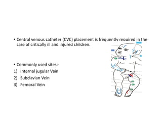
CVC Insertion and infection
Prevention
Dr. Prakash Thakur
• Central venous catheter (CVC) placement is frequently required in the
care of critically ill and injured children.
• Commonly used sites:-
1) Internal jugular Vein
2) Subclavian Vein
3) Femoral Vein
Indications
1) Reliable venous access for medication administration
2) Monitoring of central venous pressure
3) Central venous oxygen saturation
4) Parenteral Nutrition
5) Frequent blood sampling
6) Haemodialysis & CRRT
7) Apheresis
Naik VM, Mantha SS, Rayani BK. Vascular access in children. Indian J Anaesth 2019;63:737-45.
Naik VM, Mantha SS, Rayani BK. Vascular access in children. Indian J Anaesth
2019;63:737-45.
Insertion techniques for all sites
• 1) Strict hand scrubbing prior to placement.
• 2) Skin antisepsis with chlorhexidine.
• 3) Full barrier precautions (operator wearing hair covering, mask,
sterile gown and gloves, and use of a large sterile-field drape) with
attention to visibility of the tracheal tube/ventilator tubing and
peripheral vascular access site where medications are being
administered.
• 4) Sedation and analgesia plus local anaesthesia should be routinely
used for Pediatric CVC placement, both for patient comfort and to
facilitate placement and reduce complications related to patient
movement.
• 5) If possible, an additional provider should be monitoring the
administration of sedation and analgesia while the operator is
concentrating on the vascular access procedure itself.
6) CVCs are placed using the wire-guided (Seldinger) technique, in
which a needle or catheter-over-needle unit is introduced into the
desired vein, blood is aspirated, and a guidewire is placed through the
needle or catheter.
7) Advancing the guidewire through the veins into the chambers of the
heart, particularly into the ventricle, may cause cardiac arrhythmias.
8) Next step is dilation step is frequently required and is performed by
passing the dilator over the guidewire after the needle has been
removed.
9) Care must be taken to insert the dilator only to the estimated depth
of the vessel, as the stiffness of the dilator may penetrate the posterior
wall of the vessel.
Catheters should first be flushed and then filled with saline or diluted
heparin flush solution prior to insertion, and then occluded to reduce
the chance of air embolism.
In hypovolemic patients, volume resuscitation through a peripheral or
intraosseous site prior to attempted CVC placement may increase
vein size and facilitate successful cannulation.
• In children with severe hypoxemia or cyanotic congenital heart
disease, recognition of inadvertent arterial puncture or placement
can be difficult owing to poorly saturated arterial blood.
• To check that the catheter is not inserted into an adjacent artery.
1) Analysing a blood sample from the line for blood gas results-
unfortunately blood gas results may lie in between definitively
arterial and venous values.
2) Pressure transducer in line-look for venous or arterial wave form-
Time taking process.
3) A sterile, saline-filled, extension tubing set may be attached to the
needle or short catheter and the distal end of the IV tubing should be
opened, and the tubing should be raised to ˜10 cm above the body
surface (e.g., above the level of the presumed central venous pressure
of the patient).
Arterial placement should result in pulsatile blood that pushes saline
from the tubing at this level.
while venous placement frequently results in oscillations of the fluid
column with respiration.
• In equivocal cases, a sterile pressure transducer set can be attached
to the tubing to verify pressures and differentiate arterial and venous
waveforms.
• Most authorities recommend placement at or just above the junction
of the superior vena cava and right atrium for upper body catheters
, to minimize risk of atrial perforation or ventricular arrhythmia.
• The catheter should be secured with suture or a suture less catheter
securement device, as per the hospital policy and procedure.
• Confirmation and documentation of catheter tip location with
ultrasound and/or x-ray is recommended.
Complication
• Based on balancing the benefits and risks:
1) Bleeding - most common immediate Complication –Frequent in Subclavian
Vein Canulation - frequently avoided in very young and coagulopathic
patients because of inability to effectively compress the subclavian vessels
2) Deep venous thrombosis is found with all catheters sites and is
associated with diabetic ketoacidosis, as well as oncologic conditions.
3) Air or clot embolus
4) Vessel puncture or injury, nerve or lymphatic injury
5 ) Pneumothorax-Subclavian and Internal jugular approaches.
Retroperitoneal haemorrhage - Femoral approaches.
6) Wire induced arrhythmia
7) Catheter displacement.
8) Catheter-related bloodstream infection (CRBSI), a common complication
of CVC, can be substantially reduced by using a “bundle” of practices during
insertion and ongoing care of CVCs . Less with subclavian Line Insertion, Not
proven in children
9) The risk of catheter-induced erosion increases with stiffer catheters, when
the catheter tip rubs against a vessel bifurcation or the thin right atrium, or
remains in place for a long time.
• Cannulation complications can be reduced using bedside ultrasound.
Jonge RC, Polderman KH, Gemke RJ. Central venous catheter use in the pediatric patient: mechanical and infectious
complications. Pediatr Crit Care Med. 2005;6:329–39
Practice Guidelines for Central Venous Access 2020, Anesthesiology 2020; 132:8–43.
Internal Jugular Catheterization
• Anatomy Of Internal Juglar vein
• Internal jugular catheterization can be achieved via multiple
approaches. When available, ultrasound guidance is preferable.
• Right-sided approaches are preferred owing to potential injury to the
thoracic duct on the left side.
• The carotid artery should be palpated, as it lies medial to the internal
jugular vein within the carotid sheath.
1) Positioned supine and in a slight (15-30 degree) Trendelenburg
position, with a roll under the shoulders and with the head turned
away from the puncture site.
2). Drape the child with visible Ipsilateral Nipple.
3 Basic Approach
• Anterior approach, the needle is introduced
along the anterior margin of the SCM muscle,
halfway between the mastoid process and
sternum and directed toward the ipsilateral
nipple . MOST Preferred
• Middle approach, the needle enters the apex
of a triangle formed by the clavicle and the
heads of the SCM. The skin should be
punctured with the needle at a 30-60-degree
angle while the needle is directed toward the
ipsilateral nipple.
• Posterior approach, the needle should be
introduced along the posterior border of the
SCM cephalad to its bifurcation into the
sternal and clavicular heads .
Naik VM, Mantha SS, Rayani BK. Vascular access in children.
Indian J Anaesth 2019;63:737-45.
• 4) In all approaches, the needle should be advanced during exhalation
to minimize the chance of pneumothorax.
• 5) The syringe should be aspirated as the needle is advanced. When
the vein is entered and free flow of venous blood is established.
• 6) The needle should be stabilized and the syringe removed while the
hub of the needle is covered to prevent air entrainment.
• 7) The guidewire should then be introduced and advanced a distance
that approximates the distance to the junction of the superior vena
cava and right atrium.
• During guidewire introduction it is helpful to have an assistant
watching the patient's electrocardiogram (ECG) and announcing the
provocation of dysrhythmias by the guidewire.
• 8) Nonarterial placement confirmation, securing, and chest x-ray
should be obtained as mentioned before.
Subclavian Venous Catheterization
Surface Anatomy
• 1) Positioning of the patient in a head-down position (Trendelenburg)
of ˜30 degrees (increases upper body venous pressures causing
distention of the central veins and minimises air embolism traveling
to the brain)
• Most common positioning technique is to extend the patient’s neck,
turn the patients head away from the site of cannulation, and place
a rolled towel beneath the patient’s shoulder blades, along the axis
of the thoracic spine.
2) In the smaller intubated patient, sedation, analgesia, and temporary
neuromuscular blockade will facilitate proper patient positioning and
reduce complications related to patient movement.
3) In intubated patients, care should be taken to avoid kinking,
disconnecting, or dislodging the tracheal tube.
4) Bilateral breath sounds should be verified after proper patient
positioning.
5.The infraclavicular approach is most commonly used .
Alternative approaches include the supraclavicular approach using
ultrasound guidance.
6) The junction of the medial 2/3 third and lateral 1/3 should be
located, and a small (25 gauge) needle should be used to infiltrate local
anaesthesia .
7) The needle should be introduced just under the clavicle at the
junction of the medial 2/3 third and lateral 1/3 and slowly advanced
while negative pressure is applied with an attached syringe .
8) The needle should be inserted parallel with the frontal plane and
directed medially and slightly cephalad, under the clavicle toward the
sternal notch.
9) When patients are mechanically ventilated, the needle is advanced
while someone holds the ventilator in an expiratory hold position to
minimize the risk of pneumothorax.
10) When free flow of venous blood is obtained, the needle should be
stabilized and the syringe removed while a fingertip is placed over the
needle hub to prevent air entrainment.
11) The guidewire should be introduced during inspiration in a patient
on positive-pressure ventilation or during exhalation in a
spontaneously breathing patient (to avoid air embolus).
12) The Seldinger technique as described earlier should then be
followed. Once the CVC is placed, nonarterial cannulation should be
determined, the catheter should be secured with suture or with a
sutureless securement device, and a chest X-ray should be obtained to
verify catheter location prior to using the catheter and to rule out
complications, such as pneumothorax or hemothorax.
Femoral Venous Catheterization
Surface Anatomy
Femoral venous cannulation
1) The lower extremity should be positioned with slight external
rotation at the hip and flexion at the knee (frog-leg appearance).
2) A rolled towel under the buttock may facilitate successful venous
access, particularly in smaller children.
3) The femoral artery should be located by palpation and/or ultrasound
or, in the pulseless patient, assumed to be at the midpoint between the
pubic symphysis and anterior superior iliac spine.
4) Local anaesthesia to the area over the intended puncture site should
be considered and can reduce the need for sedation and analgesia.
• The needle should be inserted 1-2 cm below the inguinal ligament,
just medial to the femoral artery, and slowly advanced while negative
pressure is applied to a syringe attached to the introducer needle.
• The needle should be directed at a 15-60-degree angle toward the
umbilicus, depending on the size of the child, with a flatter approach
used in infants than in older children.
• Once the free flow of venous blood is observed, the syringe should be
removed while the needle is carefully stabilized and the guidewire
introduced gently.
• The guidewire should pass easily with minimal resistance; force
should not be applied to overcome a great deal of resistance. Once
the guidewire is in place, the Seldinger technique should be
employed.
• Checking for venous and not arterial placement should be done as
described earlier.
• Some experts recommend confirmation with ultrasound or a lateral
abdominal x-ray when femoral venous catheters are placed to
document that the catheter has not been placed in the lumbar
venous plexus .
• The catheter should be secured with suture or sutureless catheter
securement device.
Catheter Related Blood Stream Infection
• It is estimated that 15 million central venous catheter (CVC) days
occur in ICUs each year .
• CRBSI increase morbidity and healthcare costs.
• CRBSI is a clinical definition that requires more extensive testing to
pinpoint the central line as the source of the infection.
CDC Guidelines for the Prevention of Intravascular Catheter-Related Infections, 2011
• CRBSI is either a bacteraemia or a fungemia documented with at least
one peripherally obtained blood culture obtained from a vein and not
a catheter.
• The growth of an organism in the bloodstream must be documented
by
(a) a positive semiquantitative or quantitative culture of a catheter
segment with an organism identical in species and antibiogram as
isolated from a peripheral blood culture.
• (b) Simultaneously drawn peripheral and central line quantitative
blood cultures with greater than a 5:1 ratio in catheter blood versus
peripheral blood colony counts.
(c) A differential in timing of culture positivity of >2 hours between the
catheter and peripheral blood culture, where the catheter culture is
positive first.
• A catheter -associated bloodstream infection (CABSI) has less
rigorous criteria and requires the presence of central line being in place
during the 48 hours prior to the drawing of the positive culture and
compelling evidence that the infection is related to the line. This
definition is helpful for surveillance but can overestimate the true
incidence of bloodstream infections
• Coagulase-negative staphylococci are the most common cause of
pediatric nosocomial BSIs, accounting for 20% to nearly 50% of
isolates.
• Gram-negative bacteria account for 25% of PICU nosocomial BSIs. S.
aureus and Candida species are also responsible.
• Klebsiella species are the third most common isolates at 7.3% behind
coagulase-negative staphylococci and S. aureus in the pediatric
oncology population
Diagnosis of CRBSI
• Most symptoms such as fever, chills, and rigors have poor specificity
and as a result CVCs should not be removed for fever alone. Site
inflammation with or without purulence and positive bloodstream
cultures have better specificity but low sensitivity.
• Surveillance cultures are not recommended, and cultures should only
be obtained when a CRBSI is suspected.
• Cultures positive for common nosocomial BSIs such as coagulase-
negative staphylococci, S. aureus, and Candida species without an
identified source of infection and in the presence of a CVC should
raise suspicion for the possibility of a CRBSI
• Use of clinical judgment is important, and if the patient has fever with
only mild to moderate symptoms, the catheter should not be
routinely removed.
• Two blood cultures should be obtained when a catheter related
infection is suspected and at least one of them, if not both, should be
drawn percutaneously. When paired central and peripheral cultures
are sent.
• It is recommended that they either be quantitative cultures or be
qualitative cultures with continuously monitored differential time to
positivity to aid in the diagnosis of a CRBSI
• A differential time to positivity at least 2 hours earlier for the central-
line culture compared with the peripheral culture is indicative of a
central venous line infection, with a sensitivity of 91% and a
specificity of 94%.
• After removing the central line, It is not recommended to send
catheter tip for cultures.
Diagnosis
Treatment
Prevention of Catheter-Related Infections
(1) Migration of skin bacteria
down external surface of
catheter-Most Common
(2) Hematogenous spread from
distal sites
(3) Contaminated infusate
(4) Colonization of catheter
hub
(5) Contamination of
transducer or IV tubing.
• Prevention strategies begin with education and training of the critical
care staff on the placement and maintenance of central lines and give
them feedback to the staff about rates of infection to raise
awareness.
• Mandatory education of physicians and nurses as well as the
establishment of a unit based infection-control nurse position can
reduce the rate of CRBSI.
• A daily discussion of the need for central access to aid in timely line
removal.
1) Hand Washing: Strong emphasis should be placed on thorough hand
washing prior to insertion & Aseptic technique should be maintained
throughout insertion .
2) Skin Preparation: Skin preparation should be performed with a 2%
aqueous chlorhexidine gluconate solution.
• Chlorhexidine has been proven to lower catheter-related infections
compared with both 10% povidone-iodine and 70% alcohol.
• Chlorhexidine preparations should not be used on infants younger than 2
months of age due to possible absorption through the immature skin.
Paternoster M, Niola M, Graziano V. Avoiding chlorhexidine burns in preterm infants. J ObstetGynecol Neonatal Nurs
2017;46:267-71.
3)Maximal Barrier Precautions: After adequate skin preparation, full
barrier precautions, which include a large sterile or full-body drape,
long-sleeved sterile gown, cap, mask, and sterile gloves, should be
taken.
Use of full barrier precautions reduces the risk of infection compared
with standard precautions consisting of a small drape and sterile
gloves.
4.Catheter Site Dressing: Once the line is secured, a dressing should be
applied. The use of transparent, semipermeable polyurethane
dressings further secures the device and allows for visual inspection of
the catheter and insertion site..
Regardless of the type of dressing utilized, it should be replaced when
damp, soiled, loosened, or visual inspection of the site is needed.
• Examination of the use of chlorhexidine-impregnated sponge
dressings at the site of catheter insertion revealed, through a meta-
analysis, a reduction in bacterial colonization and showed a trend
toward the prevention of catheter-related infections .
• A randomized control trial in critically ill adults showed a significant
reduction in catheter related infections with the use of a
chlorhexidine-impregnated sponge dressing even when background
rates were low.
• The chlorhexidine-impregnated sponge is effective only in preventing
extraluminal surface infections and not the intraluminal infections,
which are more common in long-term indwelling catheters
• Thank You
CRBSI Bundle
• Insertion bundle
• Maximal sterile barrier precautions(surgical mask, sterile gloves, cap,
sterile gown, and large sterile drape)
• Skin cleaning with alcohol-based chlorhexidine(rather than iodine)12
• Avoidance of the femoral veinfor central venous access in adult patients;
use of subclavian rather than jugular veins13
• Dedicated staff for central line insertion, and competency
training/assessment
• Standardized insertion packs
• Availability of insertion guidelines (including indications for central line use)
and use of checklists with trained observers
• Use of ultrasound guidance for insertion of internal jugular lines
• Maintenance bundle
• Daily review of central line necessity
• Prompt removal of unnecessary lines
• Disinfection prior to manipulation of the line
• Daily chlorhexidine washes (in ICU, patients > 2 months)
• Disinfect catheter hubs, ports, connectors, etc before using the catheter
• Change dressings and disinfect site with alcohol-based chlorhexidine every
5 – 7 days (change earlier if soiled)
• Replace administration sets within 96 hours (immediately if used for blood
products or lipids)
• Ensure appropriate nurse-to-patient ratio in ICU (1:2 or 1:1)

Cvc

  • 1.
    CVC Insertion andinfection Prevention Dr. Prakash Thakur
  • 2.
    • Central venouscatheter (CVC) placement is frequently required in the care of critically ill and injured children. • Commonly used sites:- 1) Internal jugular Vein 2) Subclavian Vein 3) Femoral Vein
  • 3.
    Indications 1) Reliable venousaccess for medication administration 2) Monitoring of central venous pressure 3) Central venous oxygen saturation 4) Parenteral Nutrition 5) Frequent blood sampling 6) Haemodialysis & CRRT 7) Apheresis
  • 4.
    Naik VM, ManthaSS, Rayani BK. Vascular access in children. Indian J Anaesth 2019;63:737-45.
  • 5.
    Naik VM, ManthaSS, Rayani BK. Vascular access in children. Indian J Anaesth 2019;63:737-45.
  • 7.
    Insertion techniques forall sites • 1) Strict hand scrubbing prior to placement. • 2) Skin antisepsis with chlorhexidine. • 3) Full barrier precautions (operator wearing hair covering, mask, sterile gown and gloves, and use of a large sterile-field drape) with attention to visibility of the tracheal tube/ventilator tubing and peripheral vascular access site where medications are being administered.
  • 8.
    • 4) Sedationand analgesia plus local anaesthesia should be routinely used for Pediatric CVC placement, both for patient comfort and to facilitate placement and reduce complications related to patient movement. • 5) If possible, an additional provider should be monitoring the administration of sedation and analgesia while the operator is concentrating on the vascular access procedure itself.
  • 9.
    6) CVCs areplaced using the wire-guided (Seldinger) technique, in which a needle or catheter-over-needle unit is introduced into the desired vein, blood is aspirated, and a guidewire is placed through the needle or catheter. 7) Advancing the guidewire through the veins into the chambers of the heart, particularly into the ventricle, may cause cardiac arrhythmias. 8) Next step is dilation step is frequently required and is performed by passing the dilator over the guidewire after the needle has been removed.
  • 10.
    9) Care mustbe taken to insert the dilator only to the estimated depth of the vessel, as the stiffness of the dilator may penetrate the posterior wall of the vessel. Catheters should first be flushed and then filled with saline or diluted heparin flush solution prior to insertion, and then occluded to reduce the chance of air embolism. In hypovolemic patients, volume resuscitation through a peripheral or intraosseous site prior to attempted CVC placement may increase vein size and facilitate successful cannulation.
  • 11.
    • In childrenwith severe hypoxemia or cyanotic congenital heart disease, recognition of inadvertent arterial puncture or placement can be difficult owing to poorly saturated arterial blood. • To check that the catheter is not inserted into an adjacent artery. 1) Analysing a blood sample from the line for blood gas results- unfortunately blood gas results may lie in between definitively arterial and venous values. 2) Pressure transducer in line-look for venous or arterial wave form- Time taking process.
  • 12.
    3) A sterile,saline-filled, extension tubing set may be attached to the needle or short catheter and the distal end of the IV tubing should be opened, and the tubing should be raised to ˜10 cm above the body surface (e.g., above the level of the presumed central venous pressure of the patient). Arterial placement should result in pulsatile blood that pushes saline from the tubing at this level. while venous placement frequently results in oscillations of the fluid column with respiration.
  • 13.
    • In equivocalcases, a sterile pressure transducer set can be attached to the tubing to verify pressures and differentiate arterial and venous waveforms. • Most authorities recommend placement at or just above the junction of the superior vena cava and right atrium for upper body catheters , to minimize risk of atrial perforation or ventricular arrhythmia.
  • 14.
    • The cathetershould be secured with suture or a suture less catheter securement device, as per the hospital policy and procedure. • Confirmation and documentation of catheter tip location with ultrasound and/or x-ray is recommended.
  • 15.
    Complication • Based onbalancing the benefits and risks: 1) Bleeding - most common immediate Complication –Frequent in Subclavian Vein Canulation - frequently avoided in very young and coagulopathic patients because of inability to effectively compress the subclavian vessels 2) Deep venous thrombosis is found with all catheters sites and is associated with diabetic ketoacidosis, as well as oncologic conditions. 3) Air or clot embolus 4) Vessel puncture or injury, nerve or lymphatic injury
  • 16.
    5 ) Pneumothorax-Subclavianand Internal jugular approaches. Retroperitoneal haemorrhage - Femoral approaches. 6) Wire induced arrhythmia 7) Catheter displacement.
  • 17.
    8) Catheter-related bloodstreaminfection (CRBSI), a common complication of CVC, can be substantially reduced by using a “bundle” of practices during insertion and ongoing care of CVCs . Less with subclavian Line Insertion, Not proven in children 9) The risk of catheter-induced erosion increases with stiffer catheters, when the catheter tip rubs against a vessel bifurcation or the thin right atrium, or remains in place for a long time. • Cannulation complications can be reduced using bedside ultrasound. Jonge RC, Polderman KH, Gemke RJ. Central venous catheter use in the pediatric patient: mechanical and infectious complications. Pediatr Crit Care Med. 2005;6:329–39
  • 18.
    Practice Guidelines forCentral Venous Access 2020, Anesthesiology 2020; 132:8–43.
  • 19.
    Practice Guidelines forCentral Venous Access 2020, Anesthesiology 2020; 132:8–43.
  • 20.
    Internal Jugular Catheterization •Anatomy Of Internal Juglar vein
  • 21.
    • Internal jugularcatheterization can be achieved via multiple approaches. When available, ultrasound guidance is preferable. • Right-sided approaches are preferred owing to potential injury to the thoracic duct on the left side. • The carotid artery should be palpated, as it lies medial to the internal jugular vein within the carotid sheath.
  • 22.
    1) Positioned supineand in a slight (15-30 degree) Trendelenburg position, with a roll under the shoulders and with the head turned away from the puncture site. 2). Drape the child with visible Ipsilateral Nipple.
  • 23.
    3 Basic Approach •Anterior approach, the needle is introduced along the anterior margin of the SCM muscle, halfway between the mastoid process and sternum and directed toward the ipsilateral nipple . MOST Preferred • Middle approach, the needle enters the apex of a triangle formed by the clavicle and the heads of the SCM. The skin should be punctured with the needle at a 30-60-degree angle while the needle is directed toward the ipsilateral nipple. • Posterior approach, the needle should be introduced along the posterior border of the SCM cephalad to its bifurcation into the sternal and clavicular heads . Naik VM, Mantha SS, Rayani BK. Vascular access in children. Indian J Anaesth 2019;63:737-45.
  • 24.
    • 4) Inall approaches, the needle should be advanced during exhalation to minimize the chance of pneumothorax. • 5) The syringe should be aspirated as the needle is advanced. When the vein is entered and free flow of venous blood is established. • 6) The needle should be stabilized and the syringe removed while the hub of the needle is covered to prevent air entrainment.
  • 25.
    • 7) Theguidewire should then be introduced and advanced a distance that approximates the distance to the junction of the superior vena cava and right atrium. • During guidewire introduction it is helpful to have an assistant watching the patient's electrocardiogram (ECG) and announcing the provocation of dysrhythmias by the guidewire. • 8) Nonarterial placement confirmation, securing, and chest x-ray should be obtained as mentioned before.
  • 27.
  • 28.
    • 1) Positioningof the patient in a head-down position (Trendelenburg) of ˜30 degrees (increases upper body venous pressures causing distention of the central veins and minimises air embolism traveling to the brain) • Most common positioning technique is to extend the patient’s neck, turn the patients head away from the site of cannulation, and place a rolled towel beneath the patient’s shoulder blades, along the axis of the thoracic spine.
  • 29.
    2) In thesmaller intubated patient, sedation, analgesia, and temporary neuromuscular blockade will facilitate proper patient positioning and reduce complications related to patient movement. 3) In intubated patients, care should be taken to avoid kinking, disconnecting, or dislodging the tracheal tube. 4) Bilateral breath sounds should be verified after proper patient positioning.
  • 30.
    5.The infraclavicular approachis most commonly used . Alternative approaches include the supraclavicular approach using ultrasound guidance. 6) The junction of the medial 2/3 third and lateral 1/3 should be located, and a small (25 gauge) needle should be used to infiltrate local anaesthesia . 7) The needle should be introduced just under the clavicle at the junction of the medial 2/3 third and lateral 1/3 and slowly advanced while negative pressure is applied with an attached syringe .
  • 31.
    8) The needleshould be inserted parallel with the frontal plane and directed medially and slightly cephalad, under the clavicle toward the sternal notch. 9) When patients are mechanically ventilated, the needle is advanced while someone holds the ventilator in an expiratory hold position to minimize the risk of pneumothorax. 10) When free flow of venous blood is obtained, the needle should be stabilized and the syringe removed while a fingertip is placed over the needle hub to prevent air entrainment.
  • 32.
    11) The guidewireshould be introduced during inspiration in a patient on positive-pressure ventilation or during exhalation in a spontaneously breathing patient (to avoid air embolus). 12) The Seldinger technique as described earlier should then be followed. Once the CVC is placed, nonarterial cannulation should be determined, the catheter should be secured with suture or with a sutureless securement device, and a chest X-ray should be obtained to verify catheter location prior to using the catheter and to rule out complications, such as pneumothorax or hemothorax.
  • 34.
  • 35.
    Femoral venous cannulation 1)The lower extremity should be positioned with slight external rotation at the hip and flexion at the knee (frog-leg appearance). 2) A rolled towel under the buttock may facilitate successful venous access, particularly in smaller children.
  • 36.
    3) The femoralartery should be located by palpation and/or ultrasound or, in the pulseless patient, assumed to be at the midpoint between the pubic symphysis and anterior superior iliac spine. 4) Local anaesthesia to the area over the intended puncture site should be considered and can reduce the need for sedation and analgesia.
  • 37.
    • The needleshould be inserted 1-2 cm below the inguinal ligament, just medial to the femoral artery, and slowly advanced while negative pressure is applied to a syringe attached to the introducer needle. • The needle should be directed at a 15-60-degree angle toward the umbilicus, depending on the size of the child, with a flatter approach used in infants than in older children.
  • 38.
    • Once thefree flow of venous blood is observed, the syringe should be removed while the needle is carefully stabilized and the guidewire introduced gently. • The guidewire should pass easily with minimal resistance; force should not be applied to overcome a great deal of resistance. Once the guidewire is in place, the Seldinger technique should be employed.
  • 39.
    • Checking forvenous and not arterial placement should be done as described earlier. • Some experts recommend confirmation with ultrasound or a lateral abdominal x-ray when femoral venous catheters are placed to document that the catheter has not been placed in the lumbar venous plexus . • The catheter should be secured with suture or sutureless catheter securement device.
  • 40.
    Catheter Related BloodStream Infection • It is estimated that 15 million central venous catheter (CVC) days occur in ICUs each year . • CRBSI increase morbidity and healthcare costs. • CRBSI is a clinical definition that requires more extensive testing to pinpoint the central line as the source of the infection. CDC Guidelines for the Prevention of Intravascular Catheter-Related Infections, 2011
  • 41.
    • CRBSI iseither a bacteraemia or a fungemia documented with at least one peripherally obtained blood culture obtained from a vein and not a catheter. • The growth of an organism in the bloodstream must be documented by (a) a positive semiquantitative or quantitative culture of a catheter segment with an organism identical in species and antibiogram as isolated from a peripheral blood culture.
  • 42.
    • (b) Simultaneouslydrawn peripheral and central line quantitative blood cultures with greater than a 5:1 ratio in catheter blood versus peripheral blood colony counts. (c) A differential in timing of culture positivity of >2 hours between the catheter and peripheral blood culture, where the catheter culture is positive first.
  • 43.
    • A catheter-associated bloodstream infection (CABSI) has less rigorous criteria and requires the presence of central line being in place during the 48 hours prior to the drawing of the positive culture and compelling evidence that the infection is related to the line. This definition is helpful for surveillance but can overestimate the true incidence of bloodstream infections
  • 44.
    • Coagulase-negative staphylococciare the most common cause of pediatric nosocomial BSIs, accounting for 20% to nearly 50% of isolates. • Gram-negative bacteria account for 25% of PICU nosocomial BSIs. S. aureus and Candida species are also responsible. • Klebsiella species are the third most common isolates at 7.3% behind coagulase-negative staphylococci and S. aureus in the pediatric oncology population
  • 45.
    Diagnosis of CRBSI •Most symptoms such as fever, chills, and rigors have poor specificity and as a result CVCs should not be removed for fever alone. Site inflammation with or without purulence and positive bloodstream cultures have better specificity but low sensitivity. • Surveillance cultures are not recommended, and cultures should only be obtained when a CRBSI is suspected.
  • 46.
    • Cultures positivefor common nosocomial BSIs such as coagulase- negative staphylococci, S. aureus, and Candida species without an identified source of infection and in the presence of a CVC should raise suspicion for the possibility of a CRBSI • Use of clinical judgment is important, and if the patient has fever with only mild to moderate symptoms, the catheter should not be routinely removed.
  • 47.
    • Two bloodcultures should be obtained when a catheter related infection is suspected and at least one of them, if not both, should be drawn percutaneously. When paired central and peripheral cultures are sent. • It is recommended that they either be quantitative cultures or be qualitative cultures with continuously monitored differential time to positivity to aid in the diagnosis of a CRBSI
  • 48.
    • A differentialtime to positivity at least 2 hours earlier for the central- line culture compared with the peripheral culture is indicative of a central venous line infection, with a sensitivity of 91% and a specificity of 94%. • After removing the central line, It is not recommended to send catheter tip for cultures.
  • 49.
  • 50.
  • 51.
    Prevention of Catheter-RelatedInfections (1) Migration of skin bacteria down external surface of catheter-Most Common (2) Hematogenous spread from distal sites (3) Contaminated infusate (4) Colonization of catheter hub (5) Contamination of transducer or IV tubing.
  • 52.
    • Prevention strategiesbegin with education and training of the critical care staff on the placement and maintenance of central lines and give them feedback to the staff about rates of infection to raise awareness. • Mandatory education of physicians and nurses as well as the establishment of a unit based infection-control nurse position can reduce the rate of CRBSI. • A daily discussion of the need for central access to aid in timely line removal.
  • 53.
    1) Hand Washing:Strong emphasis should be placed on thorough hand washing prior to insertion & Aseptic technique should be maintained throughout insertion . 2) Skin Preparation: Skin preparation should be performed with a 2% aqueous chlorhexidine gluconate solution. • Chlorhexidine has been proven to lower catheter-related infections compared with both 10% povidone-iodine and 70% alcohol. • Chlorhexidine preparations should not be used on infants younger than 2 months of age due to possible absorption through the immature skin. Paternoster M, Niola M, Graziano V. Avoiding chlorhexidine burns in preterm infants. J ObstetGynecol Neonatal Nurs 2017;46:267-71.
  • 54.
    3)Maximal Barrier Precautions:After adequate skin preparation, full barrier precautions, which include a large sterile or full-body drape, long-sleeved sterile gown, cap, mask, and sterile gloves, should be taken. Use of full barrier precautions reduces the risk of infection compared with standard precautions consisting of a small drape and sterile gloves.
  • 55.
    4.Catheter Site Dressing:Once the line is secured, a dressing should be applied. The use of transparent, semipermeable polyurethane dressings further secures the device and allows for visual inspection of the catheter and insertion site.. Regardless of the type of dressing utilized, it should be replaced when damp, soiled, loosened, or visual inspection of the site is needed.
  • 56.
    • Examination ofthe use of chlorhexidine-impregnated sponge dressings at the site of catheter insertion revealed, through a meta- analysis, a reduction in bacterial colonization and showed a trend toward the prevention of catheter-related infections .
  • 57.
    • A randomizedcontrol trial in critically ill adults showed a significant reduction in catheter related infections with the use of a chlorhexidine-impregnated sponge dressing even when background rates were low. • The chlorhexidine-impregnated sponge is effective only in preventing extraluminal surface infections and not the intraluminal infections, which are more common in long-term indwelling catheters
  • 58.
  • 59.
    CRBSI Bundle • Insertionbundle • Maximal sterile barrier precautions(surgical mask, sterile gloves, cap, sterile gown, and large sterile drape) • Skin cleaning with alcohol-based chlorhexidine(rather than iodine)12 • Avoidance of the femoral veinfor central venous access in adult patients; use of subclavian rather than jugular veins13 • Dedicated staff for central line insertion, and competency training/assessment • Standardized insertion packs • Availability of insertion guidelines (including indications for central line use) and use of checklists with trained observers • Use of ultrasound guidance for insertion of internal jugular lines
  • 60.
    • Maintenance bundle •Daily review of central line necessity • Prompt removal of unnecessary lines • Disinfection prior to manipulation of the line • Daily chlorhexidine washes (in ICU, patients > 2 months) • Disinfect catheter hubs, ports, connectors, etc before using the catheter • Change dressings and disinfect site with alcohol-based chlorhexidine every 5 – 7 days (change earlier if soiled) • Replace administration sets within 96 hours (immediately if used for blood products or lipids) • Ensure appropriate nurse-to-patient ratio in ICU (1:2 or 1:1)