This document discusses carbohydrate metabolism pathways including glycolysis, the citric acid cycle, gluconeogenesis, and glycogen metabolism. It provides detailed information on glycolysis, including its definition, sites in the body, steps, energy production, oxidation of NADH, importance and functions. It also discusses glycogen metabolism including glycogenesis and glycogenolysis. The document concludes with sections on disorders of carbohydrate metabolism including pentosuria and galactosemia.
This PPT contains content of Gluconeogenesis, Steps involved in Gluconeogenesis, (Gluconeogenesis from Pyruvate, Gluconeogenesis from lactate, Gluconeogenesis from amino acids, Gluconeogenesis from glycerol, Gluconeogenesis from Propionate), Regulation and significance of Gluconeogenesis
This PPT contains content of Gluconeogenesis, Steps involved in Gluconeogenesis, (Gluconeogenesis from Pyruvate, Gluconeogenesis from lactate, Gluconeogenesis from amino acids, Gluconeogenesis from glycerol, Gluconeogenesis from Propionate), Regulation and significance of Gluconeogenesis
Glucose transporters are a wide group of membrane proteins that facilitate the transport of glucose across the plasma membrane, a process known as facilitated diffusion. Because glucose is a vital source of energy for all life, these transporters are present in all phyla.
the all the content in this profile is completed by the teachers, students as well as other health care peoples.
thank you, all the respected peoples, for giving the information to complete this presentation.
this information is free to use by anyone.
Carbohydrates are the sugars, starches and fibers found in fruits, grains, vegetables and milk products. Though often maligned in trendy diets, carbohydrates — one of the basic food groups — are important to a healthy diet.
Supplying a huge array of metabolic intermediates for biosynthetic reactions. Normally carbohydrate metabolism supplies more than half of the energy requirements of the body. In fact the brain largely depends upon carbohydrate
Carbohydrate metabolism comprises glycolysis, HMP shunt, Gluconeogenesis, Glycogenolysis, TCA cycle, with Glucose-6-phosphate dehydrogenase deficiency disorder.
intro of glycolysis there cycle and step - function-significance-defination-glucogenesis cycle-significance of gluconeogenesis-function of gluconeogenesis-conclusion
Similar to Carbohydrate metabolism and its disorders.pdf (20)
Herbal Drug Technology
Herbs as Raw Materials: Definition of herb, herbal medicine, herbal medicinal product and herbal drug preparation, source of herbs, selection, identification and authentication of herbal materials, processing of herbal raw material.
Herbal Excipients : Herbal Excipients – Significance of substances of natural origin as excipients, – colorants, sweeteners, binders, diluents, viscosity builders, dis-integrants, flavors & perfumes.
Herbal Formulations: Stages involved in herbal formulations, Orthodox formulations and methods of delivery of herbal extracts, Novel formulations of herbal extracts.
Introduction to proteomics, techniques to study proteomics such as protein electrophoresis, chromatography and mass spectrometry and protein database analysis, case studies derived from scientific literature including comparisons between healthy and diseased tissues, new approaches to analyse metabolic pathways, comprehensive analysis of protein-protein interactions in different cell types.
Metabolomics-Introduction, metabolism, intermediary metabolism, metabolic pathways, metabolites, metabolome, metabolic turnover, techniques used in metabolomics, metabolite profiling methods, data analysis, metabolomic resources, role of metabolomics in system biology.
Introduction to proteomics, techniques to study proteomics such as protein electrophoresis, chromatography and mass spectrometry and protein database analysis, case studies derived from scientific literature including comparisons between healthy and diseased tissues, new approaches to analyse metabolic pathways, comprehensive analysis of protein-protein interactions in different cell types.
The analysis of global gene expression and transcription factor regulation, global approaches to alternative splicing and its regulation, long noncoding RNAs, gene expression models of signalling pathways, from gene expression to disease phenotypes, introduction to isoform sequencing, systematic and integrative analysis of gene expression to identify feature genes underlying human diseases.
Genome projects
Definition of genome, history of genome projects, whole genome sequencing, Maxam Gilbert sequencing, sanger sequencing, explanation on the first sequenced organisms (Bacteriophage, bacteria, archaeon, virus, bakers yeast, nematode.
Model organism-Arabidopsis thaliana, Mus musculus, Oryza sativa, Pan troglodyte etc.
Human genome project, milestones and significance.
Epigenetics studies stably heritable traits that cannot be explained by changes in DNA sequence.
Covalent modifications in chromatin
DNA- DNA methylation (CpG); hydroxymethylation
Histone - lysine acetylation, lysine and arginine methylation, serine and threonine phosphorylation, and lysine ubiquitination and sumoylation
Epigenetic mechanisms:
Modified histones as post translational modification
DNA methylation – 5mC the 5th base, methyl transferases; genetic imprinting.
Epigenomics: complete set of epigenetic modifications on the genetic material of a cell.
Specific epigenetic regulation
RNA interference
X inactivation (Lyonization)
Genomic imprinting
Epigenetics in development and diseases.
Comparative genomics: Genomic features are compared, evolutionary relationship
The major principle of comparative genomics is that common features of two organisms will often be encoded within the DNA that is evolutionarily conserved between them. orthologous sequences,
Started as soon as the whole genomes of two organisms became available (that is, the genomes of the bacteria Haemophilus influenzae and Mycoplasma genitalium) in 1995, comparative genomics is now a standard component of the analysis of every new genome sequence. comparative genomics studies of small model organisms (for example the model Caenorhabditis elegans and closely related Caenorhabditis briggsae) are of great importance to advance our understanding of general mechanisms of evolution
Computational tools for analyzing sequences and complete genomes. Application of comparative genomics in agriculture and medicine.
Mapping and sequencing genomes: Genetic and physical mapping, Sequencing genomes different strategies, High-throughput sequencing, next-generation sequencing technologies, comparative genomics, population genomics, epigenetics, Human genome project, pharmacogenomics, genomic medicine, applications of genomics to improve public health.
Disorders of liver and kidney, Nitrogen metabolism.pdfshinycthomas
Disorders of liver and kidney – Jaundice, fatty liver, normal and abnormal functions of liver and kidney. Inulin and urea clearance.
Abnormalities of nitrogen metabolism
Lipid metabolism and its disorders.pdfshinycthomas
Disorders of Lipids – Plasma lipoproteins, cholesterol, triglycerides and phospholipids in health and disease, hyperlipidemia, hyperlipoproteinemia, Gaucher’s disease, Tay-Sach’s and Niemann-Pick disease, ketone bodies.
a) Definition, classification, structure, stereochemistry and reactions of amino acids;
b) Classification of proteins on the basis of solubility and shape, structure, and biological functions. Primary structure - determination of amino acid sequences of proteins, the peptide bond, Ramachandran plot.
c) Secondary structure - weak interactions involved - alpha helix and beta sheet and beta turns structure, Pauling and Corey model for fibrous proteins, Collagen triple helix, and super secondary structures - helix-loop-helix.
d) Tertiary structure - alpha and beta domains. Quaternary structure - structure of haemoglobin, Solid state synthesis of peptides, Protein-Protein interactions, Concept of chaperones.
Nucleic acid-DNA and RNA
Gene-part of DNA
Functions of DNA
RNA-Functions, different types of RNA-Ribosomal RNAs (rRNAs), Messenger RNAs (mRNAs), Transfer RNAs (tRNAs)-Other RNA-Small nuclear RNA (snRNA), Micro RNA (miRNA), Small interfering RNA (siRNA), Heterogenous RNA (hnRNA).
Nucleic acid-nucleotides-nucleoside
Components of nucleotide-a nitrogenous (nitrogen-containing) base (pyrimidine and purine), (2) a pentose, and (3) a phosphate
Structure of pentose sugar, and 5 major bases (cytosine, thymine, uracil-pyrimidine bases and adenine, guanine-purine bases).
Deoxyribonucleotides and Ribo nucleotides-Molecular structure of deoxyadenosine monophosphate (dAMP), deoxyguanosine monophosphate (dGMP), deoxythymidine monophosphate (dTMP), deoxycytidine monophosphate (dCMP) and Adenosine monophosphate (AMP), Guanosine monophosphate (GMP), Cytosine monophosphate (CMP) and Uridine monophosphate (UMP), Watson crick base pairing, Hoogsteen base pairing,
Helical structure-Heterocylic N -Glycosides, Syn and Anti Conformers, detailed structure of single strand and double stranded DNA.
DNA Nucleotides and Tautomeric Form-Tautomers of Adenine, Cytosine, Guanine, and Thymine
Template strand, non coding strand and coding strand
Hydrogen bonds, phosphodiester linkage, hydrolysis of DNA and RNA.
Different forms of DNA-A, B, and Z forms.
Palindrome sequence, Linear DNA, Cruciform DNA, H DNA (Triplex DNA), Denaturation of DNA, Hyperchromicity, Tm, Renaturation of DNA, Tertiary structure of DNA, Difference of DNA and RNA, RNA structural elements, primary. secondary and tertiary structure of RNA. Detailed structure and functions of tRNA, mRNA, rRNA, miRNA, siRNA, hn RNA, snRNA.
Nucleic acid hybridization, C0t analysis, Buoyant density of DNA, Isopycnic centrifugation.
Lipids-Introduction, properties and functions.
Classification-Simple lipids, complex lipids and derived lipids.
Lipids contain fatty acid and alcohol.
Saturated and Unsaturated fatty acids. Nomenclature of fatty acids, Cis-trans isomerism, essential fatty acids
Simple lipids-Fats, waxes
Compound lipids-Structure, function with examples of Phospholipids, Glycolipids, sulpholipids and lipoproteins.
Derived lipids: Structure, types, and functions of steroids, terpenes and carotenoids.
Lipoproteins-classified into chylomicrons, very low-density lipoproteins (VLDL), low density lipoproteins (LDL) and high-density lipoproteins (HDL) and their function.
Eicosanoids-prostanoids, leukotrienes (LTs), and lipoxins (LXs).
Functions of Eicosanoids
Lipids, micelles and liposomes.
Vitamins-Introduction, Water soluble and fat soluble vitamins.
Water soluble vitamins-B complex vitamins: thiamin (vitamin B1), riboflavin (vitamin B2), niacin (vitamin B3), vitamin B6 (pyridoxine), folate (folic acid), vitamin B12, biotin and pantothenic acid-their source, structure, properties, metabolism, physiological significance, deficiency disease and human requirements.
Fat soluble vitamins: Fat soluble vitamins, Vitamin A, D, E and K and their their source, structure, properties, metabolism, physiological significance, deficiency disease and human requirements.
Vitamin A-Carotene in plants-α-carotenes, β-carotenes and γ-carotenes, 3 forms of vitamin A-Retinol, Retinal, Retinoic acid.
Vitamin D3-cholecalciferol,
Vitamin E -Tocopherol, Vitamin K-Phylloquinone or Anti hemorrhagic Vitamin or Coagulation Vitamin
Professional air quality monitoring systems provide immediate, on-site data for analysis, compliance, and decision-making.
Monitor common gases, weather parameters, particulates.
Nutraceutical market, scope and growth: Herbal drug technologyLokesh Patil
As consumer awareness of health and wellness rises, the nutraceutical market—which includes goods like functional meals, drinks, and dietary supplements that provide health advantages beyond basic nutrition—is growing significantly. As healthcare expenses rise, the population ages, and people want natural and preventative health solutions more and more, this industry is increasing quickly. Further driving market expansion are product formulation innovations and the use of cutting-edge technology for customized nutrition. With its worldwide reach, the nutraceutical industry is expected to keep growing and provide significant chances for research and investment in a number of categories, including vitamins, minerals, probiotics, and herbal supplements.
Richard's entangled aventures in wonderlandRichard Gill
Since the loophole-free Bell experiments of 2020 and the Nobel prizes in physics of 2022, critics of Bell's work have retreated to the fortress of super-determinism. Now, super-determinism is a derogatory word - it just means "determinism". Palmer, Hance and Hossenfelder argue that quantum mechanics and determinism are not incompatible, using a sophisticated mathematical construction based on a subtle thinning of allowed states and measurements in quantum mechanics, such that what is left appears to make Bell's argument fail, without altering the empirical predictions of quantum mechanics. I think however that it is a smoke screen, and the slogan "lost in math" comes to my mind. I will discuss some other recent disproofs of Bell's theorem using the language of causality based on causal graphs. Causal thinking is also central to law and justice. I will mention surprising connections to my work on serial killer nurse cases, in particular the Dutch case of Lucia de Berk and the current UK case of Lucy Letby.
Richard's aventures in two entangled wonderlandsRichard Gill
Since the loophole-free Bell experiments of 2020 and the Nobel prizes in physics of 2022, critics of Bell's work have retreated to the fortress of super-determinism. Now, super-determinism is a derogatory word - it just means "determinism". Palmer, Hance and Hossenfelder argue that quantum mechanics and determinism are not incompatible, using a sophisticated mathematical construction based on a subtle thinning of allowed states and measurements in quantum mechanics, such that what is left appears to make Bell's argument fail, without altering the empirical predictions of quantum mechanics. I think however that it is a smoke screen, and the slogan "lost in math" comes to my mind. I will discuss some other recent disproofs of Bell's theorem using the language of causality based on causal graphs. Causal thinking is also central to law and justice. I will mention surprising connections to my work on serial killer nurse cases, in particular the Dutch case of Lucia de Berk and the current UK case of Lucy Letby.
A brief information about the SCOP protein database used in bioinformatics.
The Structural Classification of Proteins (SCOP) database is a comprehensive and authoritative resource for the structural and evolutionary relationships of proteins. It provides a detailed and curated classification of protein structures, grouping them into families, superfamilies, and folds based on their structural and sequence similarities.
Predicting property prices with machine learning algorithms.pdf
Carbohydrate metabolism and its disorders.pdf
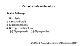
1. 1. Glycolysis
2. Citric acid cycle
3. Gluconeogenesis
4. Glycogen metabolism
(a) Glycogenesis (b) Glycogenolysis
Carbohydrate metabolism
Major Pathways
Dr. Shiny C Thomas, Department of Biosciences, ADBU
2. I. Glycolysis (Embden Meyerhof Pathway):
A. Definition:
1. Glycolysis means oxidation of glucose to give pyruvate (in the
presence of oxygen) or lactate (in the absence of oxygen).
B. Site:
cytoplasm of all tissue cells, but it is of physiological importance in:
1. Tissues with no mitochondria: mature RBCs, cornea and lens.
2. Tissues with few mitochondria: Testis, leucocytes, medulla of the
kidney, retina, skin and gastrointestinal tract.
3. Tissues undergo frequent oxygen lack: skeletal muscles especially
during exercise.
3. C. Steps:
Stages of glycolysis
1. Stage one (the energy requiring stage):
a) One molecule of glucose is converted into two molecules of
glycerosldhyde-3-phosphate.
b) These steps requires 2 molecules of ATP (energy loss)
2. Stage two (the energy producing stage(:
a) The 2 molecules of glyceroaldehyde-3-phosphate are converted
into
pyruvate (aerobic glycolysis) or lactate (anaerobic glycolysis(.
b) These steps produce ATP molecules (energy production).
6. Energy production of glycolysis:
Net energy
ATP utilized
ATP produced
2 ATP
2ATP
From glucose to
glucose -6-p.
From fructose -6-p to
fructose 1,6 p.
4 ATP
(Substrate level
phosphorylation)
2ATP from 1,3 DPG.
2ATP from
phosphoenol
pyruvate
In absence of oxygen
(anaerobic
glycolysis)
6 ATP
Or
8 ATP
2ATP
-From glucose to
glucose -6-p.
From fructose -6-p to
fructose 1,6 p.
4 ATP
(substrate level
phosphorylation)
2ATP from 1,3 BPG.
2ATP from
phosphoenol
pyruvate.
In presence of
oxygen (aerobic
glycolysis)
+ 4ATP or 6ATP
(from oxidation of 2
NADH + H in
mitochondria).
7. E. oxidation of extramitochondrial NADH+H+:
1. cytoplasmic NADH+H+ cannot penetrate mitochondrial membrane,
however, it can be used to produce energy (4 or 6 ATP) by respiratory
chain phosphorylation in the mitochondria.
2. This can be done by using special carriers for hydrogen of NADH+H+
These carriers are either dihydroxyacetone phosphate (Glycerophosphate
shuttle) or oxaloacetate (aspartate malate shuttle).
a) Glycerophosphate shuttle:
1) It is important in certain muscle and nerve cells.
2) The final energy produced is 4 ATP.
3) Mechanism:
- The coenzyme of cytoplasmic glycerol-3- phosphate dehydrogenase
is NAD+.
- The coenzyme of mitochondrial glycerol-3-phosphate dehydrogenase is
FAD.
- Oxidation of FADH, in respiratory chain gives 2 ATP. As glycolysis
gives 2 cytoplasmic NADH + H+ → 2 mitochondrial FADH, 2 x 2
ATP → = 4 ATP.
b) Malate – aspartate shuttle:
1) It is important in other tissues particularly liver and heart.
2) The final energy produced is 6 ATP.
8. Differences between aerobic and
anaerobic glycolysis:
Anaerobic
Aerobic
Lactate
Pyruvate
1. End product
2 ATP
6 or 8 ATP
2 .energy
Through Lactate
formation
Through respiration
chain in mitochondria
3. Regeneration of
NAD+
Not available as lactate
is cytoplasmic substrate
Available and 2 Pyruvate
can oxidize to give 30
ATP
4. Availability to TCA in
mitochondria
9. Importance of lactate production in anerobic glycolysis:
1. In absence of oxygen, lactate is the end product of glycolysis:
Glucose → Pyruvate → Lactate
2. In absence of oxygen, NADH + H+ is not oxidized by the
respiratory chain.
3. The conversion of pyruvate to lactate is the mechanism for
regeneration of NAD+.
4. This helps continuity of glycolysis, as the generated NAD+
will be
used once more for oxidation of another glucose molecule.
10. Substrate level phosphorylation:
This means phosphorylation of ADP to ATP at the reaction itself .in
glycolysis there are 2 examples:
- 1.3 Bisphosphoglycerate + ADP 3 Phosphoglycerate + ATP
- Phospho-enol pyruvate + ADP Enolpyruvate + ATP
I. Special features of glycolysis in RBCs:
1. Mature RBCs contain no mitochondria, thus:
a) They depend only upon glycolysis for energy production (=2 ATP).
b) Lactate is always the end product.
2. Glucose uptake by RBCs is independent on insulin hormone.
3. Reduction of met-hemoglobin: Glycolysis produces NADH+H+, which
used for reduction of met-hemoglobin in red cells.
11. Biological importance (functions) of glycolysis:
1. Energy production:
a) anaerobic glycolysis gives 2 ATP.
b) aerobic glycolysis gives 8 ATP.
2. Oxygenation of tissues:
Through formation of 2,3 bisphosphoglycerate, which decreases the
affinity of Hemoglobin to O2.
3. Provides important intermediates:
a) Dihydroxyacetone phosphate: can give glycerol-3phosphate, which is
used for synthesis of triacylglycerols and phospholipids (lipogenesis).
b) 3 Phosphoglycerate: which can be used for synthesis of amino acid
serine.
c) Pyruvate: which can be used in synthesis of amino acid alanine.
4. Aerobic glycolysis provides the mitochondria with pyruvate, which gives
acetyl CoA Krebs' cycle.
12. Reversibility of glycolysis (Gluconeogenesis):
1. Reversible reaction means that the same enzyme can catalyzes the
reaction in both directions.
2. all reactions of glycolysis -except 3- are reversible.
3. The 3 irreversible reactions (those catalyzed by kinase enzymes) can be
reversed by using other enzymes.
Glucose-6-p → Glucose
F1, 6 Bisphosphate → Fructose-6-p
Pyruvate → Phosphoenol pyruvate
4. During fasting, glycolysis is reversed for synthesis of glucose from non-
carbohydrate sources e.g. lactate. This mechanism is called:
gluconeogenesis.
13. As pyruvate enters the mitochondrion, a multienzyme complex modifies
pyruvate to acetyl CoA which enters the Krebs cycle in the matrix.
A carboxyl group is removed as CO2.
A pair of electrons is transferred from the remaining two-carbon
fragment to NAD+ to form NADH.
19. Glycogenesis:
• Glycogenesis is the formation of glycogen from glucose. Glycogen is
synthesized depending on the demand for glucose and ATP (energy).
• If both are present in relatively high amounts, then the excess of insulin
promotes the glucose conversion into glycogen for storage in liver and
muscle cells.
• In the synthesis of glycogen, one ATP is required per glucose
incorporated into the polymeric branched structure of glycogen.
• Actually, glucose-6-phosphate is the cross-roads compound. Glucose-6-
phosphate is synthesized directly from glucose or as the end product of
gluconeogenesis.
20. Glycogenolysis
• In glycogenolysis, glycogen stored in the liver and muscles, is converted
first to glucose-1- phosphate and then into glucose-6-phosphate.
• Two hormones which control glycogenolysis are a peptide, glucagon
from the pancreas and epinephrine from the adrenal glands.
• Glucagon is released from the pancreas in response to low blood
glucose and epinephrine is released in response to a threat or stress.
• Both hormones act upon enzymes to stimulate glycogen phosphorylase
to begin glycogenolysis and inhibit glycogen synthetase (to stop
glycogenesis).
21. • Glycogen is a highly branched polymeric structure containing glucose as the
basic monomer.
• First individual glucose molecules are hydrolyzed from the chain, followed by
the addition of a phosphate group at C-1.
• In the next step the phosphate is moved to the C-6 position to give glucose 6-
phosphate, a cross road compound.
• Glucose-6-phosphate is the first step of the glycolysis pathway if glycogen is
the carbohydrate source and further energy is needed.
• If energy is not immediately needed, the glucose-6-phosphate is converted to
glucose for distribution in the blood to various cells such as brain cells.
22. Disorders of Carbohydrate metabolism
Pentosuria
• It is characterized by the constant excretion of small amounts of L-xylulose.
• Other forms of pentosuria from which this condition has to be
distinguished are alimentary pentosuria, in which arabinose or xylose is
found in the urine after ingestion of a large amount of fruit or fruit product,
and possibly also ribosuria, which has recently been claimed to accompany
some cases of muscular dystrophy.
Pentosuria (MIM 260800), first described in 1892 (6), is characterized by high
urinary excretion (1–4 gm/d) of the pentose sugar L-xylulose. In 1970, the
critical enzyme deficiency was identified as L-xylulose reductase
23. • The phenotype results from a defect in the glucuronic
acid oxidation pathway.
• In this pathway, the carboxyl carbon atom of D-
glucuronic acid is removed in a series of reactions, giving
rise to the pentose L-xylulose, which is then converted
to xylitol, and hence to D- xylulose, which may be
phosphorylated to participate in reactions of the
pentose phosphate pathway, leading to its conversion to
hexose phosphate
24. • In pentosuria, failure to convert L-xylulose to xylitol
leads to accumulation of L-xylulose.
• Pentosuria is completely benign, but in the first half of
the 20th century attracted attention because it was
confused with diabetes.
• For as long as standard testing for urine sugars did not
differentiate between glucose (the six-carbon sugar of
diabetes mellitus) and pentose (the five-carbon sugar
excreted in pentosuria), persons with pentosuria were
often inappropriately treated with insulin, leading to
hypoglycemic reactions.
25. • Once a specific test for glycosuria was developed,
individuals with pentosuria no longer came to clinical
attention.
• Pentosuria is found almost exclusively among persons of
Ashkenazi Jewish ancestry.
• The confusion of pentosuria and glycosuria in this
population in the early 20th century motivated the
intensive study of pentosuria by Margaret Lasker (1884–
1976), a clinical biochemist working at Montefiore
Hospital in New York.
• Margaret Lasker developed an accurate method for testing
for L-xylulose in urine, a major contribution to resolving
diagnostic errors.
26. • Based on extensive pedigree and survival analyses of
persons with pentosuria and their families, she
concluded that the trait was inherited as an autosomal
recessive and had no impact on mortality.
• The gene DCXR, encoding L-xylulose reductase, was
cloned in 2002 and its tetrameric structure was
elucidated in 2004, but mutations responsible for
pentosuria were not identified.
27. Pentosuria: Deficiency of Xylitol Dehydrogenase
• The glucuronic acid oxidation pathway presumably is
not essential for human carbohydrate metabolism, since
individuals in whom the pathway is blocked suffer no ill
effects.
• A metabolic variation, called idiopathic pentosuria,
results from reduced activity of NADP linked L-xylulose
reductase, the enzyme that catalyzes the reduction of
xylulose to xylitol.
Hence affected individuals excrete large amounts of
pentose into the urine especially following intake of
glucuronic acid.
28. Galactosemia
What is Galactosemia?
• It is an inborn error in carbohydrates due to a deficiency
in one of the enzymes (galactose-1-phosphate uridyl
transferase) that is involved in the breakdown of simple
sugar galactose. This enzyme is called galactose-1-
phosphate uridyl transferase
• Galactose is primarily a part of a larger sugar called
lactose, which is found in all dairy products and many
baby formulas.
• The signs and symptoms of galactosemia result from an
inability to use galactose to produce energy.
29. What causes the disease?
Mutations in the GALT gene are a potential cause of the galactose-1-phosphate
uridyl transferase enzyme deficiency.
What are the clinical features of the disease?
• The affected babies are usually normal at birth, however, at their first few
weeks of life, after drinking milk that contains lactose, they start presenting
symptoms such as vomiting, diarrhea, weight loss, failure to gain weight,
poor feeding, jaundice, lethargy, hypoglycemia, liver damage, cataract,
bleeding, and E. coli sepsis.
• Even with early treatment, however, children with galactosemia are at an
increased risk for developmental delays, speech problems (verbal dyspraxia),
abnormalities of motor function, and osteoporosis.
• In females, premature ovarian failure is possible.
30. How is the diagnosis confirmed?
• The diagnosis of Galactosemia is established by measuring the amount
of galactose, galactose-1-phosphate, and enzymatic activity in the
blood sample and confirmed by DNA molecular testing of the GALT
gene.
What is the treatment of the disease?
• A galactose-restricted diet is effective in preventing many of the
complications of galactosemia, including the liver and kidney problems.
• It may also reduce the risk for developmental delays. A Biochemical
genetics specialist and a Metabolic Genetics dietitian should coordinate
the treatment.
31. Causes of galactosemia:
• Galactosemia is an inherited autosomal-recessive disorder of galactose
metabolism.
• People with galactosemia cannot tolerate any form of milk (human or
otherwise).
• The sugar lactose (a disaccharide present in milk) is made up of equal
parts of glucose and galactose; thus a deficiency of the enzymes involved
in galactose metabolism can lead to severe clinical consequences.
• Ingestion of milk produces toxic levels of galactose and its metabolite
galactose-1-phosphate (gal-1-P) in the infant.
• The classical and most severe form is caused by a deficiency of the
enzyme galactose-1-phosphate uridyl transferase (GALT).
32. • Two other enzyme deficiencies also cause galactosemia, one is epimerase
and the other is galactokinase.
• In cases with a deficiency of one of these enzymes the initial newborn
screen will show elevated galactose level with normal GALT enzyme activity.
• This incongruent result would suggest the possibility of one of the other
enzyme deficiencies. Children with galactosemia due to deficiency of these
other enzymes may not become as severely ill as the infants with classical
galactosemia.
• However, they may be mentally retarded or have cataracts if not treated.
The incidence of these disorders is significantly lower than GALT related
galactosemia.
33. Galactosemia: Inability to Transform Galactose into Glucose
• Reactions of galactose are of particular interest because in humans they
are subject to genetic defects resulting in the hereditary disorder
galactosemia.
• When a defect is present, individuals are unable to metabolize the
galactose derived from lactose (milk sugar) to glucose metabolites, often
with resultant cataract formation, growth failure, mental retardation, or
eventual death from liver damage.
34. • The genetic disturbance is expressed as a cellular deficiency of either
galactokinase, causing a relatively mild disorder characterized by early
cataract formation, or of galactose 1phosphate uridylyltransferase,
resulting in severe disease.
• Galactose is reduced to galactitol in a reaction similar to the reduction of
glucose to sorbitol.
• Galactitol is the initiator of cataract formation in the lens and may play a
role in the central nervous system damage.
• Accumulation of galactose 1phosphate is responsible for liver failure; the
toxic effects of galactose metabolites disappear when galactose is removed
from the diet.
35. Glycogen Storage Diseases
• There are a number of well characterized glycogen storage diseases, all
due to inherited defects of one or more of the enzymes involved in the
synthesis and degradation of glycogen.
• The liver is usually the tissue most affected, but heart and muscle
glycogen metabolism can also be defective.
36. Von Gierke's Disease
• The most common glycogen storage disease, referred to as type I or von
Gierke's disease, is caused by a deficiency of liver, intestinal mucosa, and
kidney glucose 6 phosphatase.
• Thus diagnosis by small bowel biopsy is possible.
• Patients with this disease can be further sub classified into those lacking
the glucose 6phosphatase enzyme per se (type Ia) and those lacking the
glucose 6phosphatase translocase (type Ib).
37. • A genetic abnormality in glucose 6phosphate hydrolysis occurs in only about
1 person in 200,000 and is transmitted as an autosomal recessive trait.
• Clinical manifestations include fasting hypoglycemia, lactic acidemia
hyperlipidemia, and hyperuricemia with gouty arthritis.
• The fasting hypoglycemia is readily explained as a
consequence of the glucose 6 phosphatase deficiency, the enzyme required to
obtain glucose from liver glycogen and gluconeogenesis.
38. • The liver of these patients does release some glucose by the action of the
glycogen debranching enzyme.
• The lactic acidemia occurs because the liver cannot use lactate effectively
for glucose synthesis.
• In addition, the liver inappropriately produces lactic acid in response to
glucagon.
• This hormone should trigger glucose release without lactate production;
however, the opposite occurs because of the lack of glucose
6phosphatase.
39. • Hyperuricemia results from increased purine degradation in the liver;
• Hyperlipidemia results because of increased availability of lactic acid for
lipogenesis and lipid mobilization from the adipose tissue caused by high
glucagon levels in response to hypoglycemia.
• The manifestations of von Gierke's disease can greatly be diminished by
providing carbohydrate throughout the day to prevent hypoglycemia.
• During sleep this can be done by infusion of carbohydrate into the gut by
a nasogastric tube.
40. Pompe's Disease
• Type II glycogen storage disease or Pompe's disease is caused by the
absence of a 1,4 glucosidase (or acid maltase), an enzyme normally
found in lysosomes.
• The absence of this enzyme leads to the accumulation of glycogen in
virtually every tissue.
• This is somewhat surprising, but lysosomes take up glycogen granules
and become defective with respect to other functions if they lack the
capacity to destroy the granules.
41. • Because other synthetic and degradative pathways of glycogen metabolism
are intact, metabolic derangements such as those in von Gierke's disease
are not seen.
• The reason for extra lysosomal glycogen accumulation is unknown.
• Massive cardiomegaly occurs and death results at an early age from heart
failure.
42. Cori's Disease
• Also called type III glycogen storage disease, Cori's disease is caused by a
deficiency of the glycogen debranching enzyme.
• Glycogen accumulates because only the outer branches can be removed
from the molecule by phosphorylase. Hepatomegaly occurs but diminishes
with age.
The clinical manifestations are similar to but much milder than those seen in
von Gierke's disease, because gluconeogenesis is unaffected, and
hypoglycemia and its complications are less severe.
43. McArdle's Disease
• Also called the type V glycogen storage disease, McArdle's disease is caused by an
absence of muscle phosphorylase.
• Patients suffer from painful muscle cramps and are
unable to perform strenuous exercise, presumably because muscle glycogen stores are not
available to the exercising muscle.
• Thus the normal increase in plasma lactate (released from the muscle) following
exercise is absent.
• The muscles are probably damaged because of inadequate energy supply and glycogen
accumulation.
• Release of muscle enzymes creatine kinase and aldolase and of myoglobin is common;
elevated levels of these substances in the blood suggests a muscle disorder.
44. Galactosemia
Galactose Metabolism
• Galactose is a constituent of of lactose of milk sugar and is taken in the diet.
• Galactose is metabolised almost exclusively by the liver and therefore
galactose tolerance test is done to assess the functional capacity of the
liver.
Galactose is necessary for the synthesis of the following.
1. Lactose synthesis
2. Synthesis of glycosaminoglycans
3. Synthesis of cerebrosides
4. Synthesis of glycolipids
5. Synthesis of Glycoproteins
45. Galactosemia
• There is deficiency of enzyme galactose-1-phosphate uridyl transferase. It is
an inborn error of metabolism.
• Due to the block of this enzyme galactose 1-phosphate will accumulate in
liver. This will inhibit galactokinase as well as glycogen phosphorylase,
Hypoglycaemia is the result.
• Bilirubin uptake is less and bilirubin conjugation is reduced; so unconjugated
bilirubin level is increased in blood.
• There is enlargement of liver, jaundice and severe mental retardation.
• Free galactose accumulates, leading to galactosemia. It is partly excreted in
urine (galactosuria).
46. • Galactose is reduced to dulcitol. The accumulation of dulcitol in the lense
results in cataract due to its osmotic effect. This is called congenital
cataract and is a very characteristic feature of galactosemia.
• Galctose -1-phosphate may get deposited in renal tubules, producing
tubular damage leading to generalized amino aciduria.
• Diagnosis
• Clinical manifestation including congenital cataract and presence of
galactose in urine as well as elevated blood galactose levels will help in
diagnosis.
• Collection of fetal cells by amniocentosis may be useful in prenatal
diagnosis. Heterozygous parents could be detected by elevated galactose
level in blood after glucose load.
47. • Treatment
• If lactose is withdrawn from the diet , most of the symptoms recede. But
mental retardation, when established, will not improve. Hence early
detection is very important. For affected infant lactose free diet is given.
Such special diets may be withdrawn after 4 years, when galactose -1-
phosphate pyrophosphorylase becomes active.
48. Regulation of blood glucose
The learner will be able to answer questions on the following topics:
• Factors maintaining blood glucose
• Normal plasma glucose level
• Effects of hormones on glucose level
• Oral glucose tolerance test (OGTT)
• Diagnostic criteria for diabetes mellitus ¾ Impaired glucose tolerance
• Reducing substances in urine
• Benedict's test
• Insulin, synthesis and secretion
• Physiological action of insulin
• Glucagon
• Diabetes mellitus types
• Metabolic derangements in diabetes
• Clinical aspects of diabetes mellitus
• Laboratory investigations in diabetes
• Glycated hemoglobin Dr. Shiny C Thomas, Department of Biosciences, ADBU
49. Regulation of blood glucose
• Glucose level in blood is maintained within narrow limits.
• This is a very finely and efficiently regulated system.
• It is essential to have continuous supply of glucose to the brain.
• It can utilize ketone bodies to some extent, but brain has an obligatory requirement for
glucose.
Factors maintaining the blood glucose are:
1. The plasma glucose level at an instant depends on the balance between glucose
entering and leaving the extracellular fluid
2. Hormones maintain this balance (Fig. 11.1)
3. The major factors which cause entry of glucose into blood are: a. Absorption from
intestines b. Glycogenolysis (breakdown of glycogen) c. Gluconeogenesis d.
Hyperglycemic hormones (glucagon, steroids)
4. Factors leading to depletion of glucose in blood are: a. Utilization by tissues for
energy b. Glycogen synthesis c. Conversion of glucose into fat (lipogenesis) d.
Hypoglycemic hormone (insulin)
50.
51. Post-prandial Regulation
• Following a meal, glucose is absorbed from the intestine and enters the blood.
• The rise in the blood glucose level stimulates the secretion of insulin by the beta cells of
islets of Langerhans of pancreas.
• The uptake of glucose by extrahepatic tissues, except brain is dependent on insulin.
• Moreover, insulin helps in the storage of glucose as glycogen or its conversion to fat (Fig.
11.2A).
52.
53. Regulation in Fasting State
• Normally, 2 to 2½ hours after a meal, the blood glucose level falls to near fasting levels.
• It may go down further; but this is prevented by processes that contribute glucose to the
blood.
• For another 3 hours, hepatic glycogenolysis will take care of the blood sugar level.
• Thereafter, gluconeogenesis will take charge of the situation (Figs 11.2A and B).
• Liver is the major organ that supplies the glucose for maintaining blood glucose level (Fig.
11.1).
• Hormones like glucagon, epinephrine, glucocorticoids, growth hormone, ACTH and
thyroxine will tend to increase the blood glucose level.
• They are referred to as anti-insulin hormones or hyperglycemic hormones. An overview of
the regulatory mechanism is shown in Figure 11.3.
54. Effects of hormones on glucose level in blood
A. Effect of insulin (hypoglycemic hormone)
1. Lowers blood glucose
2. Favors glycogen synthesis
3. Promotes glycolysis
4. Inhibits gluconeogenesis
B. Glucagon (hyperglycemic hormone)
1. Increases blood glucose
2. Promotes glycogenolysis
3. Enhances gluconeogenesis
4. Depresses glycogen synthesis
5. Inhibits glycolysis (Details given below)
C. Cortisol (hyperglycemic hormone)
1. Increases blood sugar level 2
2. Increases gluconeogenesis
3. Releases amino acids from the muscle
55. D. Epinephrine or Adrenaline (hyperglycemic)
1. Increases blood sugar level
2. Promotes glycogenolysis
3. Increases gluconeogenesis
4. Favors uptake of amino acids
E. Growth hormone (hyperglycemic)
1. Increases blood sugar level
2. Decreases glycolysis
3. Mobilizes fatty acids from adipose tissue
56.
57. Determination of glucose in body fluids
Estimation of glucose is the most common analysis done in clinical laboratories.
The blood is collected using an anticoagulant (potassium oxalate) and an
inhibitor of glycolysis (sodium fluoride). Fluoride inhibits the enzyme, enolase,
and so glycolysis on the whole is inhibited. If fluoride is not added, cells will
utilize glucose and false low value may be obtained.
Capillary blood from finger tips may also be used for glucose estimation by strip
method.
Enzymatic Method
• This is highly specific, giving ‘true glucose' values (fasting 70–110 mg/dl). In
the medical laboratory, the GOD-POD (glucose oxidase peroxidase) method
is most commonly used to assess the blood glucose level.
• The reaction generates a colour, which is read in a photometer. The newer
automated systems use hexokinase method.
58. • The above GOD reaction mixture is immobilized on a plastic film (dry
analysis).
• The intensity of the colour is measured by reflectance photometry. The
instrument is named as glucometer. It is useful for patients to have self-
analysis at home. But the instrument is less accurate.
59. Commonly employed terms regarding glucose
1. Blood sugar analyzed at any time of the day, without any prior preparations,
is called random blood sugar.
2. Glucose estimated in the early morning, before taking any breakfast is called
fasting blood glucose. Fasting state means, glucose is estimated after an
overnight fast (12 hours after the food) (post-absorptive state).
3. The test done about 2 hours after a good meal is called post-prandial blood
glucose (Latin = after food).
4. When blood glucose level is within normal limits, it is referred to as
normoglycemia. When values are above the normal range, it is known as
hyperglycemia. When values are below the normal range, it is called
hypoglycemia. (Greek, hyper =above; hypo = below).
60. 5. When the blood glucose is below 50 mg/dL, it is a very serious condition.
Hyperglycemia is harmful in the long run; while hypoglycemia even for a short
while is dangerous, and may even be fatal.
6. The ability of a person to metabolize a given load of glucose is referred to as
glucose tolerance. (G
61. Conducting the glucose tolerance test
At about 8 am, a sample of blood is collected in the fasting state. Urine sample is
also obtained. This is denoted as the "0" hour sample.
Glucose load dose: The dose is 75 g anhydrous glucose (82.5 g of glucose
monohydrate) in 250–300 mL of water. This dose is fixed for an adult,
irrespective of body weight. (When the test is done in children, the glucose dose
is adjusted as 1.75 g/kg body weight). In order to prevent vomiting, patient is
asked to drink it slowly (within about 5 minutes). Flavoring of the solution will
also reduce the tendency to vomit.
Sample collection: As per current WHO recommendations, 2 samples are
collected, one at fasting ("0" hr sample) and 2-hour post-glucose load. Urine
samples may also be collected along with these blood samples. This This is
sufficient to get a correct assessment of the patient.
62. Normal Values and interpretations
• As per WHO recommendation, In a normal person, fasting plasma glucose is
70–110 mg/dl. The present day tendency is to view values above 100 mg/mL
as suspicious. Value more than 100 mg/dL is one of the criteria for the
metabolic syndrome.
• Following the glucose load, in normal persons, the level rises and reaches a
peak within 1 hour and then comes down to normal fasting levels by 2 to 2½
hours.
• This is due to the secretion of insulin in response to the elevation in blood
glucose. None of the urine sample shows any evidence of glucose.
• Diagnostic criteria for diabetes mellitus are given in Table 11.1 and Box 11.3
63. Classical oral glucose tolerance test (ogtt)
• Glucose tolerance test is artificial, because in day to day life, such a large
quantity of glucose does not enter into blood.
• However, the GTT is a well-standardized test, and is highly useful to diagnose
diabetes mellitus in doubtful cases.
Indications for ogtt
1. Patient has symptoms suggestive of diabetes mellitus; but fasting blood
sugar value is inconclusive (between 100 and 126 mg/dL).
2. During pregnancy, excessive weight gaining is noticed, with a past history
of big baby (more than 4 kg) or a past history of mis carriage.
3. To rule out benign renal glucosuria.
4. GTT has no role in follow-up of diabetes. It is indicated only for the initial
diagnosis.
64.
65.
66. Preparation of the Patient
• The patient is instructed to have good carbohydrate diet for 3 days prior to
the test.
• Patient should not take food after 8 PM the previous night.
• Should not take any breakfast.
• This is to ensure 12 hours fasting.
• The patients are advised to remain in the hospital during the waiting period
of two hours without any active exercise.
• Figure 11.4 represents the graph, when plasma glucose values are plotted
on the vertical axis against the time of collection on the horizontal axis.
67.
68. Causes for abnormal GTT curve Impaired Glucose Tolerance (IGT)
• It is otherwise called as impaired glucose regulation (IGR).
• Here blood sugar values are above the normal level, but below the diabetic
levels (Table 11.1).
• In IGT, the fasting plasma glucose level is between 110 and 126 mg/dL and
2-hour post-glucose value is between 140 and 200 mg/dL (Fig. 11.4).
• Such persons need careful follow-up because IGT progresses to frank
diabetes at the rate of 2% patients per year.
Impaired Fasting Glycemia (IFG)
• In this condition, fasting plasma sugar is above normal (between 110 and
126 mg/dL); but the 2-hour post-glucose value is within normal limits (less
than 140 mg/dL).
• These persons need no immediate treatment; but are to be kept under
constant check up.
69. Gestational Diabetes Mellitus (GDM)
• This term is used when carbohydrate intolerance is noticed, for the first time,
during a pregnancy.
• A known diabetic patient, who becomes pregnant, is not included in this
category.
• Women with GDM are at increased risk for subsequent development of frank
diabetes.
• GDM is associated with an increased incidence of neonatal mortality.
Maternal hyperglycemia causes the fetus to secrete more insulin, causing
stimulation of fetal growth and increased birth weight.
• After the child birth, the women should be reassessed.
70.
71. Diabetes Mellitus -Historical Perspectives
• The term is derived from the Greek words dia (=through), bainein (=to go)
and diabetes literally means pass through.
• The disease causes loss of weight as if the body mass is passed through the
urine.
• The Greek word, mellitus, means sweet, as it is known to early workers, that
the urine of the patient contains sugar.
• Diabetes mellitus is a disease known from very ancient times.
• Charaka in his treatise (circa 400 BC) gives a very elaborate clinical description
of madhumeha (= sweet urine). He had the vision that carbohydrate and fat
metabolisms are altered in this disease.
Dr. Shiny C Thomas, Department of Biosciences, ADBU
72. • Diabetes mellitus is a metabolic disease due to absolute or relative insulin
deficiency.
• Diabetes mellitus is a common clinical condition.
• About 10% of the total population, and about 1/5th of persons above the age
of 50, suffer from this disease.
• It is a major cause for morbidity and mortality. Insulin deficiency leads to
increased blood glucose level.
• In spite of this high blood glucose, the entry of glucose into the cell is
inefficient.
• Hence all cells are starved for glucose.
73. Type 1 Diabetes Mellitus (formerly known as insulin-dependent diabetes
mellitus; IDDM). About 5% of total diabetic patients are of type 1. Here circulating
insulin level is deficient.
It is subclassified as: a. Immune mediated and b. Idiopathic.
Type 2 Diabetes Mellitus (Formerly known as non-insulin dependent diabetes
mellitus; NIDDM).
• Most of the patients belong to this type. Here circulating insulin level is normal
or mildly elevated or slightly decreased, depending on the stage of the disease.
This type is further classified as: a. Obese b. Non-obese
74. Diabetic Prone states
a. Gestational diabetes mellitus (GDM);
b. Impaired glucose tolerance (IGT);
c. Impaired fasting glycemia (IFG)
d. Metabolic syndrome
75. Secondary to other Known causes
a. Endocrinopathies (Cushing's disease, thyrotoxicosis, acromegaly);
b. Drug induced (steroids, beta blockers, etc.);
c. Pancreatic diseases (chronic pancreatitis, fibrocalculus pancreatitis,
hemochromatosis, cystic fibrosis)
d. Anti-insulin receptor autoantibodies (Type B insulin resistance)
e. Mutations in the insulin gene or insulin receptor gene (acanthosis nigricans)
f. MODY (Maturity Onset Diabetes of Young).
MODY was previously considered to be a third form of type 2 diabetes. However, with the
discovery of specific mutations leading to MODY, it is now classified under secondary diabetes.
MODY is characterized by onset prior to age 25, impaired beta cell function and insulin
resistance. Mutations of about 10 different genes have been correlated with the development
of MODY.
76. Type 1 diabetes Mellitus (t1dM)
• It is due to decreased insulin production.
• Circulating insulin level is very low.
• These patients are dependent on insulin injections.
• Onset is usually below 30 years of age, most commonly during adolescence.
They are more prone to develop ketosis.
• An autoimmune basis is attributed to most of these cases.
• Circulating antibodies against insulin is seen in 50% cases.
• Type 1 diabetes mellitus is an autoimmune disease in which pathologic,
autoreactive T cells of the immune system attack the insulin secreting
pancreatic islets of Langerhans.
• There is excessive secretion of glucagon in IDDM patients.
77. Type 2 diabetes Mellitus (t2dM)
• 95% of the patients belong to this type.
• The disease is due to the decreased biological response to insulin, otherwise
called insulin resistance.
• So, there is a relative insulin deficiency.
• Type 2 disease is commonly seen in individuals above 40 years.
• These patients are less prone to develop ketosis.
• About 60% of patients are obese.
• These patients have insulin resistance and high/normal plasma insulin levels.
78. • Insulin resistance develops as a consequence of excess accumulation of fat in
liver and skeletal muscle.
• The free fatty acid level increases, exceeds the capacity of mitochondrial
oxidation and spills over to cytoplasm where it is re-esterified.
• The consequent increase in diacylglycerol (DAG), a second messenger, leads
to reduced signal transduction by insulin leading to insulin resistance.
• A high-caloric diet coupled with a sedentary lifestyle are the major
contributing factors in the development of the insulin resistance.
• A major susceptibility locus for type 2 diabetes, named as NIDDM1, is located
on chromosome 2. Lipoprotein (a) or Lp(a) is associated inversely with risk of
type 2 diabetes.
79. Pathological alterations in Diabetes Mellitus
Derangements in Carbohydrate Metabolism
• Insulin deficiency decreases the uptake of glucose by cells.
• The insulin dependent enzymes are also less active.
• Net effect is an inhibition of glycolysis and stimulation of gluconeogenesis
leading to hyperglycemia.
Derangement in Protein Metabolism
• Increased breakdown of proteins and amino acids for providing substrates
for gluconeogenesis is responsible for muscle wasting.
Derangements in Lipid Metabolism
• Enhanced lipolysis leads to high FFA levels in plasma and consequent
accumulation of fat in liver leading to NAFLD (Non alcoholic fatty liver
disease).
• More acetyl CoA is now available, which cannot be efficiently oxidized by
80. TCA cycle, because the availability of oxaloacetate is limited.
• The stimulation of gluconeogenesis is responsible for the depletion of
oxaloacetate.
• The excess of acetyl CoA therefore, is diverted to ketone bodies, leading to
ketogenesis.
• This tendency is more in type 1 disease.
• There is hyperlipidemia, especially an increase in NEFA, TAG and cholesterol in
plasma.
81. Clinical Presentations in diabetes Mellitus
• The cardinal symptoms of diabetes mellitus are glucosuria, polyuria,
polydypsia and polyphagia.
• When the blood glucose level exceeds the renal threshold glucose is excreted
in urine (glucosuria).
• Due to osmotic effect, more water accompanies glucose (polyuria).
• To compensate for this loss of water, thirst center is activated, and more water
is taken (polydypsia).
• To compensate the loss of glucose and protein, patient will take more food
(polyphagia).
• The loss and ineffective utilization of glucose leads to breakdown of fat and
protein.
• This would lead to loss of weight.
• Important differential diagnosis for weight loss are diabetes mellitus,
tuberculosis, hyperthyroidism, cancer and AIDS.
82. • Often the presenting complaint of the patient may be chronic recurrent
infections, such as boils, abscesses, etc.
• Any person with recurrent infections should be investigated for diabetes. When
glucose level in extracellular fluid is increased, bacteria get good nutrition for
multiplication.
• At the same time, macrophage function of the host is inefficient due to lack of
efficient utilization of glucose. In India, tuberculosis is commonly associated
with diabetes.
83. Acute Complications of Diabetes Mellitus
Diabetic Keto acidosis
• Ketosis is more common in type 1 diabetes mellitus.
• Normally the blood level of ketone bodies is less than 1 mg/dL and only traces
are excreted in urine (not detectable by usual tests).
• But when the rate of synthesis exceeds the ability of extrahepatic tissues to
utilize them, there will be accumulation of ketone bodies in blood.
• This leads to ketonemia, excretion in urine (ketonuria) and smell of acetone
in breath. All these three together constitute the condition known as ketosis.
84. Lactic acidosis
• It is another acute complication.
• It occurs due to over- production and or under-utilization of lactic acid.
• Overproduction can result from an increased rate of anaerobic glycolysis due
to hypoxia.
• Underutilization may be due to impairment of TCA cycle.
• Lactic acidosis is seen when diabetic patients are treated with biguanides.
• This drug inhibits TCA cycle and gluconeogenesis
85. Chronic complications of Diabetes Mellitus
• When there is hyperglycemia, proteins in the body may undergo glycation. It
is a non-enzymatic process. Glucose forms a schiff base with the N-terminal
amino group of proteins. The glycation first occurs in circulating proteins like
hemoglobin, albumin and LDL and then to extracellular proteins. The advanced
glycation end products (AGE) deposition in tissues and endothelium lead to all
the chronic complications of diabetes mellitus.
Vascular diseases:
• Atherosclerosis in medium sized vessels, plaque formation and consequent
intravascular thrombosis may take place.
• If it occurs in cerebral vessels, the result is paralysis. If it is in coronary artery,
myocardial infarction results.
• In the case of small vessels, the process is called microangiopathy, where
endothelial cells and mural (cement) cells are damaged.
• Microangiopathy may lead to diabetic retinopathy and nephropathy.
complications in eyes:
86. • Early development of cataract of lens is due to the increased rate of sorbitol
formation, caused by the hyper glycemia.
• Retinal micro vascular abnormalities lead to retinopathy and blindness.
• Neuropathy: Peripheral neuropathy with paresthesia is very common.
• Decreased glucose utilization and its diversion to sorbitol in Schwann cells may
be one cause for neuropathy.
• Another reason proposed is the production of advanced glycation end
products.
• Neuropathy may lead to risk of foot ulcers and gangrene.
• Hence, care of the feet in diabetic patients is important.
87. Oral hypoglycemic agents:
• There are several types of oral hypoglycemic agents (OHA) now in use.
• The conventional types are sulfonylurea and biguanides (Metformin) used
in type 2 DM.
• Other groups include glitazones, dipeptidyl peptidase inhibitors, which are
often combined with the conventional drugs.
• Insulin injections: Insulin is the drug of choice in type 1 disease. It is also
used in type 2 disease, where oral drugs are not sufficient. The availability of
human insulin prepared by recombinant DNA technology has markedly
improved the response of patients.
88. Prevention of complications.
• Hypoglycemia/ Hyperglycemia causes harm; but hypoglycemia is fatal.
• A fall in plasma glucose less than 50 mg/dL is life-threatening.
• Causes of hypoglycemia are:
• 1. overdose of insulin: This is the most common cause. The differentiation of
hypoglycemic coma from hyperglycemic coma (ketosis) is important, since
treatment is exactly opposite.
• The diagnosis is mainly based on blood glucose estimation.
89. Alimentary Glucosuria
• Here the fasting and 2-hour values are normal; but an exaggerated rise in
blood glucose following the ingestion of glucose is seen.
• This is due to an increased rate of absorption of glucose from the intestine.
This is seen in patients after a gastrectomy or in hyperthyroidism.
• Renal glucosuria
• Normal renal threshold for glucose is 175–180 mg/ dl.
• If blood sugar rises above this, glucose starts to appear in urine.
• Generally, the increased blood sugar level is reflected in urine.
• But when renal threshold is lowered, glucose is excreted in urine. In these
cases, the blood sugar levels are within normal limits. This is called renal
glycosuria
90. • It has been recognized that deviations from this normal figure occur in both
directions-that many individuals pass sugar in the urine when the blood sugar
is below 170 mg. per 100 c.cm., and that many diabetics show no glycosuria
when the blood sugar is considerably higher; in other words, that the kidney is
more or less permeable to sugar than usual.
• Renal glucosuria is associated with renal diseases with renal tubular transport
defects; e.g. Fanconi's syndrome.
• In some cases, renal threshold may be increased when glucose will not appear
in urine, even though blood sugar is elevated.
91. • Normally glucose is not excreted in urine. But if blood sugar is more than
180 mg/dl, urine contains glucose. The blood level of glucose above which
glucose is excreted is called renal threshold.
• When reducing sugars are excreted in urine, the condition is referred to as
glycosuria.
• To denote the excretion of specific sugars the suffix ‘uria' is added to the
name of the sugar, e.g. glucosuria, fructosuria, lactosuria.
• Glucosuria means glucose in urine; glycosuria means any sugar in urine.
Since glucose is the most common reducing sugar excreted in urine, the
term glycosuria is often (though incorrectly) used to denote the excretion
of glucose.
92. • When blood glucose level exceeds the renal threshold (175–180 mg/dL),
glucose is excreted in urine.
• Diabetes mellitus is the most common cause. Transient glucosuria may occur
in some people due to emotional stress. Excessive secretion of antiinsulin
hormones like cortisol (anxiety) and thyroid hormone may cause glucosuria.
Once the stress is removed, the glucosuria disappears.
93. Structure of insulin
• Insulin is a protein hormone with 2 polypeptide chains. The A chain has 21
amino acids and B chain has 30 amino acids.
• These two chains are joined together by two interchain disulfide bonds,
between A7 to B7 and A20 to B19.
• There is also an intrachain disulfide link in A chain between 6th and 11th
amino acids
94. Physiological actions of insulin (Metabolic effects of insulin)
Insulin plays a central role in regulation of the metabolism of carbohydrates,
lipids and proteins (Table 11.4).
Uptake of Glucose by Tissues
Insulin facilitates the membrane transport of glucose. Facilitated diffusion of
glucose in muscle is enhanced by insulin. In diabetes mellitus, the transporter,
GLUT4 is reduced. However, glucose uptake in liver (by GLUT2) is independent of
insulin.
Utilization of Glucose
Glycolysis is stimulated by insulin. The activity and amount of key glycolytic
enzymes (glucokinase, phosphofructokinase and pyruvate kinase) are increased.
Glycogen synthase enzyme is activated, and so insulin favors glucose storage as
glycogen.
95. [GLUT2 is a monosaccharide transporter occurring in the plasma membranes of beta
cells, renal tubule cells, and hepatocytes, among other tissues. Defective function
leads to glycogen accumulation, enlargement of liver and kidneys, and impairment of
gluconeogenesis and renal tubular function].
GLUT4 is the insulin-regulated glucose transporter found primarily in adipose tissues
and striated muscle (skeletal and cardiac).
96. Hypoglycemic Effect
• Insulin lowers the blood glucose level by promoting utilization and storage.
gluconeogenesis is inhibited by insulin by repressing the key enzymes, pyruvate
carboxylase (PC) phosphoenolpyruvate carboxykinase (PEPCK) and glucose-6-
phosphatase.
• Insulin inhibits glycogenolysis by favoring the inactivation of glycogen
phosphorylase and inhibiting glucose-6-phosphatase.
• The net effect of all these three mechanisms, blood glucose level is lowered.
97. Lipogenesis
• Lipogenesis is favored by providing more acetyl CoA by pyruvate
dehydrogenase reaction.
• Insulin increases the activity of acetyl CoA carboxylase and provides glycerol
for esterification of fatty acids to TAG.
• Insulin also provides NADPH by increasing the GPD (glucose phosphate
dehydrogenase) activity of the HMP shunt pathway.
Anti-lipolytic Effect
Insulin inhibits lipolysis in adipose tissue due to inhibition of hormone sensitive
lipase. The increased level of FFA in plasma in diabetes is due to the loss of this
inhibitory effect on lipolysis.
98. HYPERGLYCEMIC HORMONES
1. Glucagon
2. Epinephrine or Adrenaline
3. Glucocorticoids
4. Adrenocorticotropic hormone (ACTH)
5. Growth hormone
6. Thyroxine
All these are anti-insulin hormones.
Anti-insulin Hormones
Regulation of carbohydrate metabolism in general depends on the
balance between insulin and anti-insulin hormones.
Glucocorticoids act mainly by stimulating gluconeogenesis.
99. Glucagon
• It is a polypeptide hormone with 29 amino acids. It is secreted by the alpha
cells of pancreas.
• Enteroglucagon is a peptide hormone secreted by duodenal mucosa, having
same immunological and physiological properties of glucagon.
• Glucagon is synthesized as a longer proglucagon precursor.
• The major regulator of secretion of glucagon is glucose.
• An increase in blood glucose level inhibits secretion of glucagon.
Physiological actions of glucagon
• Glucagon is the most potent hyperglycemic hormone. It is anti-insulin in
nature.
• Therefore, the net effect is decided by the insulin-glucagon ratio (Fig. 11.8).
Glucagon is mainly glycogenolytic.
• The active form of glycogen phosphorylase is formed under the influence of
glucagon.
100. • Liver is the primary target for the glycogenolytic effect of glucagon. It
depresses glycogen synthesis.
• Gluconeogenesis is favored by glucagon by inducing enzymes like PEPCK,
glucose6-phosphatase and fructose-1,6-bisphosphatase.
• Glucagon increases plasma free fatty acid level.
• In adipose tissue glucagon favors beta-oxidation, as it activates carnitine acyl
transferase.
• The mitochondrial acetyl CoA level increases. Ketogenesis is favored.
Mechanism of action
Glucagon combines with a membrane bound receptor. This activates G
protein and adenylate cyclase. Thus ATP is converted to cAMP. Cyclic AMP
activates glycogen phosphorylase, and inactivates glycogen synthase.
101.
102.
103. Oral glucose tolerance test
Procedure
The patient should be resting and should not smoke
during the test.
The patient fasts overnight (for at least 10 h but not more
than 16 h). Water, but no other beverage, is allowed.
A venous sample is withdrawn for plasma glucose
estimation.
A solution that contains the equivalent of 75 g of
anhydrous glucose is made up to approximately 300 mL with
water.
This solution should be drunk slowly over a few minutes.
Further blood is taken 2 h after the ingestion of glucose.
104. The following factors may affect the result of the test
Previous diet : No special restrictions are necessary if
the patient has been on a normal diet for 3–4 days.
However, if the test is performed after a period of
carbohydrate restriction, for example as part of a
weight-reducing diet, this may cause abnormal glucose
tolerance, probably because metabolism is adjusted to the
‘fasted state’ and so favours gluconeogenesis.
Time of day Most OGTTs are performed in the
morning and the reference values quoted are for this
time of day. There is evidence that tests performed
in the afternoon yield higher plasma glucose
concentrations and that the accepted ‘reference values’
may not be applicable.
105. This may be due to a circadian variation in islet cell
responsiveness.
Drug Steroids, oral contraceptives and thiazide diuretics
may impair glucose tolerance.
Interpretation of the oral glucose tolerance test (glucose mmol/L);
venous plasma preferred
106. GLUCOSE TOLERANCE TEST (GTT)
What is “Carbohydrate Tolerance”?: The ability of the
body to utilise carbohydrates may be ascertained by
measuring its carbohydrate tolerance. It is indicated by the
nature of blood glucose curve following the administration
of glucose. Thus “glucose tolerance” is a valuable
diagnostic aid. A 70 kg man can ingest approx. 1500 gm/
day.
107. Decreased Glucose Tolerance
• In Diabetes mellitus
• In hyperactivity of anterior pituitary and adrenal
cortex
• In hyperthyroidism.
Increased Tolerance
• Hypopituitarism
• Hyperinsulinism
• Hypothyroidism
• Adrenal cortical hypofunction (such as Addison’s disease)
• Also if there is decreased absorption, like sprue, caeliac
disease.
108. TYPES OF GLUCOSE TOLERANCE TEST
This is of two types:
(A) Standard oral glucose tolerance test
(B) IV glucose tolerance test.
(A) Standard Oral GTT
Indications
• In patients with transient or sustained glycosuria, who
have no clinical symptoms of Diabetes with normal
fasting and PP blood glucose.
• In patients with symptoms of Diabetes but with no
glycosuria and normal fasting blood glucose level.
• In persons with strong family history but no overt
symptoms.
109. • In patients with glycosuria associated with thyrotoxicosis,
infections/sepsis, Liver diseases, Pregnancy, etc.
• In women with characteristically large babies 9 lbs or
individuals who were large babies at birth.
• In patients with neuropathies or retinopathies of
undetermined origin.
• In patients with or without symptoms of DM, showing
one abnormal value.
110. Pre-requisites: Precautions to be taken on the day prior
to the test:
• The individual takes usual supper at about 2000 hours
and does not eat or drink anything after that. Early
morning if so desires, a cup of tea/or coffee may be
given without sugar or milk. No other food or drink
is permitted till the test is over.
• Should be on normal carbohydrate diets at least for
three days prior to test (approx 300 G daily), otherwise
‘false’ high curve may be obtained.
• Complete mental/and physical rest.
• No smoking is permitted.
• All samples of blood should be venous preferably. If
capillary blood from ‘finger prick’ is used, all samples
should be capillary blood.
111. Procedure
1. A fasting sample of venous blood is collected in
flouride bottle (fasting sample)
2. The bladder is emptied completely and urine is
collected for qualitative test for glucose and ketone
bodies (fasting urine).
3. The individual is given 75 Gm of glucose dissolved
in water about 250 ml to drink. Lemon can be added
to make it palatable and to prevent nausea/vomiting.
Time of oral glucose administration is noted.
4. A total of five specimens of venous blood and urine
are collected every ½ hour after the oral glucose viz.
½ hour, 1 hour, 1½ hour, 2 hour and 2½ hour.
112. 5. Glucose content of all the six (including fasting
sample) samples of blood are estimated and corresponding
urine samples are tested qualitatively for
presence of glucose and ketone bodies. A curve is
plotted which is called as Glucose tolerance curve.
Explanation and Significance of a Normal Curve
1. A sharp rise to a peak, averaging about 50 per cent
above the fasting level within 30 to 60 minutes. Extent of
the rise varies considerably from person to person,
but maximum should not exceed 160 to 180 mg% in
normal subjects.
113.
114. Reason
• Rise is due directly to the glucose absorbed from
the intestine, which temporarily exceeds the capacity
of the Liver and tissues to remove it.
• As the blood glucose concentration increases,
regulatory mechanisms come into play:
• Increased insulin secretion due to hyperglycaemia,
• Hepatic glycogenesis is increased,
• Hepatic glycogenolysis is decreased, and
• Glucose uptake and utilization in tissues increase.
115. Characteristics of Different Types of GTC
(a) A Normal GTC
1. Fasting blood glucose within normal limits of 60 to
100 mg% (“True” glucose)
2. The highest peak value is reached within one hour.
3. The highest value does not exceed the renal threshold,
i.e. 160 to 180 mg%
4. The fasting level is again reached by 2½ hour
5. No glucose or Ketone bodies are detected in any
specimens of urine.
117. (b) Diabetic Type of GTC
1. Fasting blood glucose is definitely raised 110 mg% or
more (“True” Glucose).
2. The highest value is usually reached after 1 to 1½ hour.
3. The highest value exceeds the normal renal threshold.
4. Urine samples always contain glucose except in some
chronic diabetics or nephritis who may have raised
renal threshold (Dangerous type), hyperglycaemia but
no glycosuria. Urine may or may not contain ketone
bodies depending on the type of Diabetes and
severity.
118. 5. The blood glucose does not return to the fasting level
within 2½ hours. This is the most characteristic
feature of true DM.
According to severity, it may be:
(a) Mild Diabetic curve,
(b) Moderately severe Diabetic curve, and
(c) Severe Diabetic curve (see box below).
119.
120. (c) Renal glycosuria curve: Glucose appears in the urine
at levels of blood glucose much below 170 mg%.
Patients who show no glycosuria when fasting may
have glycosuria when the blood glucose is raised.
The condition may be:
• Idiopathic without any pathological significance
• Occasionally occurs in certain renal diseases and in
pregnancy (when there may be lowering of renal threshold)
• May be found in case of “early” Diabetes with low
renal threshold
• It has been reported in children of diabetic parents.
These cases should be reviewed from time to time
(every six months).
Renal glycosuria curve is shown below in below.
121.
122. (d) ‘Lag’ Curve (or Oxyhyperglycaemic Curve):
1. Fasting blood glucose is normal but it rises rapidly in
the ½ to 1 hour and exceeds the renal threshold so
that the corresponding urine specimens show glucose.
2. The return to normal value is rapid and complete.
This type of GTC may be obtained in:
• Hyperthyroidism
• After gastroenterostomy
• During pregnancy
• Also in “early” diabetes.
A patient showing “lag curve” should be reviewed
from time to time after every six months. A ‘Lag type’ of
GTC is shown below in the box: