
Recommended
More Related Content
What's hot
What's hot (20)
Similar to Lipids.pdf
Similar to Lipids.pdf (20)
More from shinycthomas
More from shinycthomas (20)
Recently uploaded
Recently uploaded (20)
Lipids.pdf
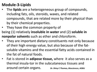
- 1. Module-3-Lipids • The lipids are a heterogeneous group of compounds, including fats, oils, steroids, waxes, and related compounds, that are related more by their physical than by their chemical properties. • They have the common property of being (1) relatively insoluble in water and (2) soluble in nonpolar solvents such as ether and chloroform. • They are important dietary constituents not only because of their high energy value, but also because of the fat- soluble vitamins and the essential fatty acids contained in the fat of natural foods. • Fat is stored in adipose tissue, where it also serves as a thermal insula-tor in the subcutaneous tissues and around certain organs. Dr. Shiny C Thomas, Biosciences, ADBU
- 2. Combinations of lipid and protein (lipoproteins) serve as the means of transporting lipids in the blood. LIPIDS ARE CLASSIFIED AS SIMPLE OR COMPLEX 1. Simple lipids: Esters of fatty acids with various alcohols. a. Fats: Esters of fatty acids with glycerol. Oils are fats in the liquid state. b. Waxes: Esters of fatty acids with higher molecular weight monohydric alcohols. 2.Complex lipids: Esters of fatty acids containing groups in addition to an alcohol and a fatty acid. a. Phospholipids: Lipids containing, in addition to fatty acids and an alcohol, a phosphoric acid residue. Dr. Shiny C Thomas, Biosciences, ADBU
- 3. They frequently have nitrogen containing bases and other substituents, eg, in glycerophospholipids the alcohol is glycerol and in sphingophospholipids the alcohol is sphingosine. b. Glycolipids (glycosphingolipids): Lipids containing a fatty acid, sphingosine, and carbohydrate. c. Lipoprteins: Macromolecular complexes of lipids with proteins d. Other complex lipids: Lipids such as sulfolipids and aminolipids. Lipoproteins may also be placed in this category. 3. Precursor and derived lipids: These include fatty acids, glycerol, steroids, other alcohols, fatty aldehydes, ketone bodies, hydrocarbons, lipid-soluble vitamins, and hormones.
- 4. • Because they are uncharged, acylglycerols (glycerides), cholesterol, and cholesteryl esters are termed neutral lipids. • Fatty acids occur in the body mainly as esters in natural fats and oils, but are found in the unesterified form as free fatty acids, a transport form in the plasma. • Fatty acids that occur in natural fats usually contain an even number of carbon atoms. • The chain may be saturated (containing no double bonds) or unsaturated (containing one or more double bonds).
- 5. Functions of lipids Lipids perform several important functions 1. They are the concentrated fuel reserve of the body (triacylglycerols). 2. Lipids are the constituents of membrane structure and regulate the membrane permeability (phospholipids and cholesterol ) . 3. They serve as a source of fat soluble vitamins (4, D, E and K). 4. Lipids are important as cellular metabolic regulators (steroid hormones and prostaglandins 5. Lipids protect the internal organs, serve as insulating materials and give shape and smooth appearance to the body.
- 6. • Other lipids, although present in relatively small quantities, play crucial roles as: • enzyme cofactors, • electron carriers, • light absorbing pigments, • hydrophobic anchors for proteins, • “chaperones” to help membrane proteins fold, • emulsifying agents in the digestive tract, • hormones, • and intracellular messengers.
- 7. The fatty acids are hydrocarbon derivatives. • Fatty acids are carboxylic acids with hydrocarbon chains ranging from 4 to 36 carbons long (C4 to C36). • In some fatty acids, this chain is unbranched and fully saturated (contains no double bonds); in others the chain contains one or more double bonds
- 9. A simplified nomenclature for these compounds specifies the chain length and number of double bonds, separated by a colon; for example, the 16-carbon saturated palmitic acid is abbreviated 16:0, and the 18-carbon oleic acid, with one double bond, is 18:1.
- 10. • The positions of any double bonds are specified by superscript numbers following Δ (delta); a 20-carbon fatty acid with one double bond between C-9 and C-10 (C-1 being the carboxyl carbon) and another between C-12 and C-13 is designated Δ delta 20:2(Δ 9,12). • There is also a common pattern in the location of double bonds; in most monounsaturated fatty acids the double bond is between C-9 and C-10 (Δ9), and the other double bonds of polyunsaturated fatty acids are generally Δ12 and Δ15 .
- 11. • The double bonds of polyunsaturated fatty acids are almost never conjugated (alternating single and double bonds, as in • but are separated by a methylene group: . Cis-trans isomerism • Because of the double bonds, unsaturated fatty acids exhibit cis-trans isomerism. • In the cis isomer bulky groups are located on the same side of double bond where as in trans isomer they are on the opposite side of double bond (Fig. 6.9b). • All the naturally occurring unsaturated fatty acids are cis- isomers.
- 13. Function Cis and trans isomers are not interchangeable in cells. Only cis isomers can fit into cell membrane because of bend at double bond. Nomenclature of Fatty Acids Saturated fatty acids Saturated fatty acids have both trivial names and systematic names. Systematic name • Systematic name of a saturated fatty acid consist of two parts. Name of hydrocarbon chain forms first part. ‘oic’ substituted in place of ‘e’ of hydrocarbon name forms second part. For example, systematic name for a saturated fatty acid containing 8 carbon atoms i.e., (octane + oic + acid) —→ octanoic acid.
- 14. • Usually saturated fatty acids end as anoic acids. Examples of saturated fatty acids with systematic names, trivial names and with sources are given in Table 6.1. The trivial name for octanoic acid is caprylic acid. Table 6.1 Sources, trivial and systematic names of saturated fatty acids
- 15. • In a fatty acid, the carbon atoms are numbered from the carboxyl carbon. • The carbon atom adjacent to the carboxyl carbon is known as the α-carbon. • Carbon atom adjacent to the α-carbon atom is known as β carbon atom and so on. • The end methyl carbon is called as ω-carbon (Fig. 6.9a). Unsaturated fatty acids They have trivial names, systematic names, ω-end names and short hand forms.
- 16. Systematic name • Like saturated fatty acids, the name of hydrocarbon forms first part of systematic name of unsaturated fatty acids. • But ‘enoic’ substituted in place of ‘ne’ of name of hydrocarbon forms second part. • Number of double bonds are written before ‘enoic’ and symbol showing position of double bonds and isomerism around double bond are written between two parts or in the beginning.
- 17. • For example, systematic name for a mono unsaturated fatty acid palmitoleic acid (trivial name) containing 16 carbon atoms and one double bond between 9 and 10 carbon atoms is monoenoic acid or cis-9-Hexadecaenoic acid. • Usually unsaturated fatty acids end as ‘enoic acids’.
- 18. Omega (Ѡ)-end series • Unsaturated fatty acids are also named according to the location of double bonds(s) from ω-end. • For example, palmitoleic acid containing a double bond between 9 and 10 carbon atoms is named as ω-7 fatty acid. Short hand forms • Number of carbon atoms, number of double bonds and location of double bonds of unsaturated fatty acid are represented with short form. • For example, palmitoleic acid is written as 16:1, Δ9 in short hand form. • First numeral indicates number of carbon atoms, later number indicates number of double bonds and Δ9 indicates position of double bond.
- 19. • Example of unsaturated fatty acids with trivial names, systematic names, ω-end names, short hand forms and along with sources are given Table 6.2.
- 20. Table 6.2 Unsaturated fatty acids with their sources.
- 21. ESSENTIAL FATTY ACIDS • They are not synthesized in the body. So they must be obtained from diet. • They are also called as poly unsaturated fatty acids (PUFA). • They are linoleic acid (LA), linolenic acid (LNA) and arachidonic acid (AA). Functions 1. They are essential for the synthesis of eicosanoids. 2. They are also required for membrane lipids.
- 22. Medical Importance 1. Dietary essential fatty acids has blood cholesterol lowering effect. 2. Deficiency status of essential fatty acids are rare with normal diet. However, deficiency of these in rats causes poor growth, reproductive disorders and dermatitis. 3. Lipid transport may be impaired. 4. Infants consuming formula diets are susceptible to deficiency of essential fatty acids. They may develop skin abnormalities.
- 23. 1. A simple lipid is a fatty acid ester of different alcohols and carries no other substance. • These lipids are nonpolar compounds, mostly insoluble in water, but soluble in nonpolar organic solvents such as chloroform and benzene. Simple lipids: esters of fatty acids with various alcohols. a. Fats: esters of fatty acids with glycerol. Oils are fats in the liquid state. Fats are also called triglycerides because all the three hydroxyl groups of glycerol are esterified. b. Waxes: Solid esters of long-chain fatty acids such as palmitic acid with aliphatic or alicyclic higher molecular weight monohydric alcohols. Waxes are water-insoluble due to the weakly polar nature of the ester group. Eg:Cetyl alcohol.
- 25. Compound Lipids or Heterolipids Heterolipids are esters of fatty acids with alcohol and possess additional groups also. • Compound (conjugated) lipids are lipids conjugated with other substances. They include: 1. Phospholipids formed of lipid, phosphoric acid and nitrogenous base. 2. Glycolipids, formed of lipid part and carbohydrate part 3. Sulpholipids, lipids containing sulphate. 4. Lipoproteins formed of lipid part and protein part
- 26. Phospholipids or Phosphatids are compound containing fatty acids and glycerol in addition to a phosphoric acid, nitrogen bases and other substituents. • They are a group of compound lipids formed of alcohol, fatty acids, phosphoric acid and nitrogenous base. • They usually possess one hydrophilic head and two non- polar tails. • They are called polar lipids and are amphipathic in nautre.
- 27. The parts of a phospholipid molecule This example is phosphatidylcholine, represented (A) schematically, (B) by a formula, (C) as a space-filling model, and (D) as a symbol. The kink resulting from the cis-double bond is exaggerated for emphasis.
- 28. They are classified according to the alcohol present into: A) Phosphoglycerides B) B) Sphingomyelin A- Phosphoglycerides • Phosphoglycerides are a group of phospholipids containing glycerol with N/P ratio = 1 • They include phosphatidic, lecithin, cephalins, phosphatidyl inositol, plasmalogen and cardiolipin.
- 29. 1- phosphatidic acid It is phosphoric acid ester of diglycerides Structure It is formed of: - Glycerol -Saturated fatty, attached to α -carbon of glycerol by ester bond. -Unsaturated fatty acid , attached to β- carbon of glycerol by ester bond. -Phosphoric acid attached to α’ carbon of glycerol by ester bond Function of phosphatidic acid It is an intermediate compound in biosynthesis of other phosphoglycerides and triglycerides
- 30. 2- Lecithin It is phosphatidyl choline Structure It is formed of: - Glycerol -Saturated fatty acid, attached to α carbon of glycerol by ester bond. - Unsaturated fatty acid, attached to β carbon of glycerol by ester bond. - Phosphoric acid attached to α’ carbon of glycerol by ester bond - Choline attached to phosphoric acid by ester bond
- 31. Function of lecithin 1. It is the most abundant phospholipid in the cell membrane 2. It acts as a lipotropic factor preventing accumulation of lipids in the liver. 3. Dipalmityl lecithin acts as a surfactant in the lung alveoli forming a layer at the interface of fluid lining the alveoli and air in side alveoli preventing lung collapse • In premature infants the lung alveoli do not secrete lecithin in sufficient amount so the lungs collapse this is called respiratory distress syndrome.
- 32. Lysolecithin • Snake venom contains lecithinase enzyme, which removes the unsaturated fatty acid from lecithin forming lysolecithin. • Lysolecithin is a strong surface-active substance that has a marked haemolytic action causing haemolysis of the red blood cells.
- 33. 3- Cephalins They are phosphatidyl ethanolamine and phosphatidyl serine Structure They are formed of: - Glycerol - Saturate d fatty acid, attached to α carbon of glycerol by ester bond. - Unsaturated fatty acid, attached to β carbon of glycerol by ester bond. - Phosphoric acid attached to α’ carbon of glycerol by ester bond - Ethanolamine or serine.
- 34. Functions of cephalins - They have a role in blood coagulation. - They accelerate blood clotting because they enter in the structure of thromboplastine, which is essential for blood clotting.
- 35. 4- Phosphatidyl inositol It is also called lipoinositol Structure It is formed of: - Glycerol -Saturate d fatty, attached to α carbon of glycerol by ester bond. -Unsaturated fatty acid , attached to β carbon of glycerol by ester bond. - Phosphoric acid attached to α’ carbon of glycerol by ester bond - Inositol, which is a cyclic alcohol derived from glucose
- 36. Function of phosphatidyl inositol • It is found in brain tissue. • It has a role in mechanism of hormone action. On hydrolysis by phospholipase C enzyme, it gives compounds, which act as second messengers in hormone action e.g. diacyl glycerol (DAG) and inositol triphosphate (IP3).
- 37. 5- Plasmalogens They are lecithin or cephalin in which the fatty acid attached to α carbon is replaced by fatty aldehyde in the enol form. Structure It is formed of: - Glycerol - Fatty aldehyde, enol form (unsaturated alcohol) - Unsaturated fatty acid - Phosphoric acid - Nitrogenous bas e, which may be choline, ethanolamine or serine Function of plasmalogens They are present in cardiac muscle, skeletal muscles and brain.
- 38. 6- Cardiolipin It is a diphosphatidyl glycerol formed of 2 phosphatidic acids attached together by glycerol. Structure It is formed of: - 3 glycerol molecules. - 2 saturated fatty acid s. - 2 unsaturated fatty acids. - 2 molecules of phosphoric acid. Functions of cardiolipin • Important component of the inner mitochondrial membrane, where it constitutes about 20% of the total lipid composition. Also found in the membranes of most bacteria • Cardiolipin is present in heart muscle. • It is used as antigen for detection of syphilis.(bacterial infection- painless sore on genitals, rectum or mouth, spread by sexual contact)
- 39. B- Sphingomyelins • Sphingomyelin is a phospholipid with N/P ratio = 2. In this type of phospholipids the alcohol is sphingol alcohol which is also called sphingosine base i.e. sphingomyelin contains sphingosine base instead of glycerol. • Sphingosine base contains 18 carbon atoms: • The first carbon contains hydroxyl group (- OH). • The second carbon contains amino group (-NH2) • The third carbon contains hydroxyl group. • There is a double bond between C4 and C5
- 40. Structure of sphingomyelin Sphingomyelin is formed of: • Sphingosine base • Unsaturated fatty acid attached to the amino group of sphingosine • Phosphoric acid attached to the first carbon of sphingosine. • Choline base attached to phosphoric acid.
- 41. Function of sphingomyelin • It is abundant in the nervous system in the myelin sheath also it is present to lesser extent in liver, spleen and bone marrow. N.B: Ceramide • It is formed of sphingosine base to which fatty acid is attached by amide linkage. • It differs from sphingomyelin, as it does not contain phosphoric acid or choline.
- 42. N.B: Nimann Pick disease • It is a disease caused by deficiency of sphingomyelinase enzyme, which catabolizes sphingomyelin. • This leads to accumulation of large amounts of sphingomyelin in liver, spleen and brain.
- 43. 2- Glycolipids • They are compound lipids that contain carbohydrates. They also contain sphingosine base They include cerebrosides and gangliosides 1- Cerebrosides • These are compound lipids formed of lipids and carbohydrates. • They are called cerebrosides because they are present mainly in the brain and nerves.
- 44. Structure It is formed of: - Sphingosine base. - Long chain fatty acid attached to the amino group of the sphingosine base by amide linkage. -Carbohydrate usually galactose but may be glucose. Functions of cerebrosides • They are present mainly in the nervous tissues i.e. brain and nerves. • They act as electric insulators of nerve impulses. Also, they are present in spleen, liver , adrenal gland, kidney and lungs.
- 45. 2- Gangliosides These are the most complex glycolipids. Structure - Sphingosine base. - Long chain fatty acid - One glucose molecule . - 2 galactose molecules. - N-acetyl galactosamine . *- N-acetyl neuraminic acid (siailic acid; NANA). Function Gangliosides are present in high concentration in brain. They act as receptors at cell membrane.
- 46. 3- Sulpholipids They are cerebrosides containing sulphate group attached to C3 of galactose. Structure of sulpholipids It is formed of: - Sphingosine base. - Long chain fatty ac id attached to the amino group of the sphingosine base by amide linkage. - Carbohydrate usually galactose but may be glucose. - Sulphate group attached to carbon number 3 of galactose. Function of sulpholipids They are present in brain and nervous tissues.
- 47. Derived Lipids Derived lipids are the substances derived from simple and compound lipids by hydrolysis. These includes fatty acids, alcohols, monoglycerides and diglycerides, steroids, terpenes, carotenoids. The most common derived lipids are steroids, terpenes and carotenoids. Steroids Steroids are complex molecules containing four fused rings. The four fused rings makeup ‘cyclopentanoperhydrophenanthrene’ or ‘sterane’ ring. Sterane ring is also called as steroid nucleus.
- 48. • The steroid nucleus, consisting of four fused rings, three with six carbons and one with five. • Steroids are called as non-saponifiable lipids because they contain no fatty acids and they can not form soaps. • The most abundant steroids are sterols which are steroid alcohols.
- 49. Sterols are structural lipids present in the membranes of most eukaryotic cells. 1. CHOLESTEROL Structure Cholesterol, 3-hydroxy-5,6-cholestene
- 50. • In animal tissue, cholesterol is the major sterol. Cholesterol is 3-hydroxy-5, 6-cholestene. It is found in bile (chol-bile). • In a normal 65 Kg adult, 200 gm of cholesterol is present. • Brain is rich in cholesterol. It is also present in spinal cord and neurons. • It is amphipathic, with a polar head group (the hydroxyl group at C-3) and a nonpolar hydrocarbon body (the steroid nucleus and the hydrocarbon side chain at C-17), about as long as a 16- carbon fatty acid in its extended form.
- 51. Other Noteworthy Steroids 1. Ergosterol Provitamin of vitamin D found in yeast and plants. 2. Sitosterol Present in plants. 3. Cardiac glycosides like quabain and streptomycin an antibiotic. 4. Coprostanol found in feces is derived from cholesterol. 5. Wool fat sterols like agnosterol and lanosterol.
- 52. 2. Ergosterol Is a Precursor of Vitamin D Ergosterol occurs in plants and yeast and is important as a precursor of vitamin D
- 53. Derived lipids They are compounds derived from simple and compound lipids by hydrolysis e.g. fatty acids glycerol glycerol. Also; the include substances related to lipids as steroids and isoprenoids. Steroids They are a large group of biologically important compounds. All of them contain a steroid nucleus, which is cyclopentano-perhydrophenanthrene nucleus.
- 55. Steroid compounds include: 1. Sterols 2. Steroid hormones 3. Vitamin D 4. Bile acids 5. Cardiac glycosides 6. Toad poisons
- 56. 1- Sterols They are complex alcohols that contain hydroxyl group and do not contain carbonyl or carboxyl groups e.g. cholesterol and ergosterol. They are present in animal and plant tissues; cholesterol is present in animals and ergosterol in plants. A- Cholesterol It is the best-known sterol. It is the most abundant animal sterol
- 57. Properties of cholesterol 1- Cholesterol is insolubl e in water but soluble in fat solvents. 2- It is present in every animal cell but mainly in adrenal cortex, liver, kidney, brain and nervous tissues. 3- It is present in human blood in concentration of 150 - 270 mg /dl 4- Reduction by intestinal bacteria converts cholesterol into dihydrocholesterol (coprastanol) by saturation of the double bond between C5 and C6. Coprastanol is present in faces.
- 58. Structure It is forme d of 27 carbon atoms. It is formed of: - Cyclopentano-perhydrophenanthrene (steroid) nucleus. - Alcoholic hydroxyl group (-OH) at C3. - Double bond between C5 and C6. - Side chain of 8 carbon atoms at C 17.
- 59. Functions of cholesterol 1. Cholesterol enters in the structure of all body cells 2. Cholesterol is the precursor of all steroid hormones. 3. It is oxidized in the liver to give bile acids. 4. It is oxidized to 7 dehydrocholesterol by introduction of double bond between C7 and C8. N.B. 7 dehydrocholesterol is provitamin D3 because it is converted to vitamin D3 when the skin is exposed to ultraviolet rays.
- 60. Colour reactions of cholesterol 1. Lieberman-Burchard test, on addition of acetic anhydride and concentrated sulphuric acid to cholesterol, it gives bluish green colour. 2. Salkowski test, on addition of chloroform and concentrated sulphuric acid to cholesterol it gives bluish red to purple colour.
- 61. B- Ergosterol It is plant sterol, which is poorly absorbed from small intestine. Structure It is forme d of 28 carbon atoms and contains: - Cyclopentano-perhydrophenanthrene nucleus . - Alcoholic hydroxyl group (-OH) at C3. - 2 double bonds, one between C5 and C6 and the other between C7 and C8. - Side chain of 9 carbon atoms at C17. It contains double bond between C22 and C23. - There is an extra methyl group at C24 in the side chain of ergosterol.
- 62. Function of ergosterol Ergosterol is provitam in D2 because it is converted to vitamin D2 when the skin is exposed to ultraviolet rays.
- 63. 2- Steroid Hormones These are hormones that contain steroid nucleus. They include: A- Male sex hormones: The main male sex hormone is called testosterone. B- Female sex hormon es: The female sex hormones in clude estrogens and progesterone C- Adrenal cortex hormones: They are secreted from the adre nal cortex. They are classified into: glucocorticoids, mineralocorticoids and aldosterone. D- Active forms of vitamin D: 1,25 dihydroxy vitamin D3 (calcitriol) and 1,25 dihydroxy vitamin D2 are the active forms of vitamin D.
- 64. • They are considered hormones as they are synthesized in an organ (skin), activated in other organs (liver and kidney) and exert function on other organs ( small intestine and bones). 3- Steroid vitamins These are vitamin D2 and vitamin D3. Structure Vitamin D 3 is derived from 7 dehydrocholesterol by rupture (opening) of ring B by ultraviolet rays Vitamin D2 is derived from ergosterol by rupture (opening) of ring B by ultraviolet rays.
- 65. 4- Bile acids These are acid s that contain 24 carbon atoms. They are formed from cholesterol in the liver by oxidation of the side chain. There are 4 bile acids: A- Cholic acid • It is the main bile acid in humans. • It is 3,7,12-trihydroxy cholanic acid. B- Deoxycholic acid • It is 3,12 dihydroxy cholanic acid. C- Chenodeoxycholic acid • It is 3,7 dihydroxy cholanic acid. D- Lithocholic acid • It is 3 hydroxy cholanic acid.
- 66. • Bile acids unite with glycine t o form glycocholic acid. Or they may unite with taurine (derived from amino acid cysteine) to form taurocholic acid. • Glyco cholic acid unites with sodium or potassium to form sodium glycocholate or potassium glycocholate. • Also, taurocholic acid forms sodium taurocholate and potassium taurocholate. • Sodium glycocholate, potassium glycocholate, sodium taurocholate and potassium taurocholate are bile salts. • Bile salts have a role in lipid digestion and absorption.
- 67. Isoprenoids They are unsaturated hydrocarbons. They are formed of repeated units of 5 carbon atoms called isoprene units. They include: 1. Ubiquinone, which is a member of the respiratory chain in mitochondria. 2. Dolichol, which is a long chain unsaturated alcohol which chairs in glycoprotein synthesis. 3. Carotenes, which are provitamin A. 4. Rubber 5. Camphor
- 68. Carotenes They are unsaturated hydrocarbons formed of 40 carbon atoms with the general formula C40H56. They are formed of repeated units of 5 carbon atoms called isoprene units. They are orange in colour. Sources of carotenes Carotenes are obtained from plant sources as carrots and plant leaves. Importance of carotenes • They are precursors of vitamin A. Types of carotenes There are 3 types o f carotenes α, β and γ
- 69. Structure They are formed of 2 ionone rings connected by a chain of repeated isoprene units.
- 70. Lipoproteins These are compound lipids formed of lipid part (which may be triglycerides, cholesterol or phospholipids) and protein part (which may be or globulin). Function of lipoproteins:- 1. They enter in the structure of cell membrane. 2. They are important for lipid transport in the blood. Lipids are insoluble in water so they cannot be transported alone. • Lipids bind to protein to make lipoproteins are water- soluble and can be transported in the blood. • The protein part of lipoprotein is called as apolipoprotein or apoprotein. The apoprotein and lipids are held together by non-covalent forces.
- 71. Classification of lipoproteins Lipoproteins are classified into 4 types according to the rate of floatation in sodium chloride solution after ultracentrifugation. • These types are chylomicrons, very low-density lipoproteins (VLDL), low density lipoproteins (LDL) and high-density lipoproteins (HDL)
- 72. Apoproteins of lipoproteins The proportion of protein part differs in various lipoproteins. Further the composition of apoprotein part also differs among various lipoproteins. There are five types of apoproteins. They are apoprotein A, apo B, apo C, apo D, and apo E. Some of the apoproteins have subtypes also.
- 73. Functions of Lipoproteins Lipoproteins are involved in the transportation of lipids in the body. 1. Chylomicrons They transport dietary or exogenous triglycerides from intestine to liver. 2. Very low density lipoproteins (VLDL) They are involved in the transport of endogenous triglycerides from liver to extra hepatic tissues. 3. Low density lipoproteins (LDL) LDL is the major vehicle for the transport of cholesterol from liver to extra hepatic tissues. 4. High density lipoproteins (HDL) HDL is the major vehicle for the transport of cholesterol from extra hepatic tissues to the liver.
- 74. EICOSANOIDS • These compounds, derived from eicosa (20-carbon) polyenoic fatty acids, comprise the prostanoids, leukotrienes (LTs), and lipoxins (LXs). • Prostanoids include prostaglandins (PGs), prostacyclins (PGIs), and thromboxanes (TXs). Often word prostaglandins is used to indicate all prostanoids.
- 75. Prostaglandins • Since they are initially found in prostate gland they are named as prostaglandins. • But later they are identified in all cells and tissues except erythrocytes. Structures • Chemically prostaglandins are derivatives of a 20 carbon prostanoic acid. • Prostanoic acid is a cyclic compound with two side chains • The cyclic ring present in prostanoic acid is a cyclopentane ring. • There are some six or more types of prostaglandins.
- 76. • They are prostaglandin A(PGA), PGB, PGC, PGD, PGE, PGF, PGG and PGH • Most widely distributed prostaglandins are PGA1, PGA2, PGE1, PGE2, PGE3, PGF1, PGF2, PGF3. Prostaglandin E2 (PGE2 )
- 78. Prostacylins Structure They contain a second five-numbered ring in addition to the one common to all prostaglandins. Thromboxane Structure They are so named because they are identified first in thrombocytes. They contain a six numbered heterocyclic oxane ring
- 79. Leukotriens and Lipoxins Structure • They are found in leukocytes. They are derivatives of arachidonic acid and contain no cyclic ring. • HPETE (5-hydroperoxy eicosa tetra enoic acid) derived from arachidonic acid serves as precursor for leukotriens and lipoxins. • They are characterized by the presence of three or four conjugated double bonds, respectively. • Leukotrienes cause bronchoconstriction as well as being potent proinflammatory agents, and play a part in asthma.
- 80. FUNCTIONS OF EICOSANOIDS They function as local hormones. They act on several organs and produce physiological as well as pharmacological effects. 1. Heart PGE class prostaglandins increases cardiac output and myocardial contraction. 2. Blood vessels Prostaglandins (PGE) maintain blood vessel tone and arterial pressure. 3. Blood pressure PGA and PGE class prostaglandins lower blood pressure. So they may be useful as anti hypertensive agents. 4. Brain PGE class prostaglandins produce sedation and tranquilizing effect in cerebral cortex.
- 81. 5. Kidney PGA and PGE class prostaglandins increases excretion of Na+, K+ and CI-. They may increase urine volume by increasing plasma flow. 6. Lungs Prostaglandins dilate bronchi, so they are useful in the treatment of asthma. 8. Stomach Prostaglandins decreases acid secretion in stomach. So they are useful in the treatment of peptic ulcers. 9. Uterus. Prostaglandins induces contraction of uterine muscle. So they are used in the termination of pregnancy. Prostaglandins also has role in fertility. 10. Metabolism Prostaglandins influences several metabolism by altering cAMP level. For example, they inhibit lipolysis in adipocyte by increasing cAMP level. 11. PGE class prostaglandins are involved in inflammation.
- 82. 12. Prostacylins inhibit platelet aggregation. 13. Thromboxanes causes platelet aggregation and clot formation. 14. Leukotreins are involved in the regulation of neutrophil and eosinophil function. They act as mediators of immediate hyper sensitivity reaction. The slow reacting substance of anaphylaxis (SRS-A) is a leukotriene. Some leukotriens act as chemotactic agents. Lipoxins are vasoactive and immuno regulatory substances. 15. Thromboxane A2 regulates acquired immunity. It causes construction of smooth muscle cells. It is a mitogen.
- 83. LIPID LAYERS, MICELLES AND LIPOSOMES • In general, lipids are insoluble in water since they contain a predominance of nonpolar (hydrocarbon) groups. • However, fatty acids, phospholipids, sphingolipids, bile salts, and, to a lesser extent, cholesterol contain polar groups. • Therefore, part of the molecule is hydrophobic, or water- insoluble; and part is hydrophilic, or water soluble. Such molecules are described as amphipathic
- 84. • They become oriented at oil:water interfaces with the polar group in the water phase and the nonpolar group in the oil phase. • A bilayer of such amphipathic lipids is the basic structure in biologic membranes
- 85. Lipid monolayer • When amphipathic molecules like phospholipids are present in water, their polar head groups orient towards water phase and hydrophobic tails towards air. • As a result, a unimolecular lipid layer is formed at water air interphase.
- 86. Micelles • When amphipathic lipids are present beyond a critical concentration in aqueons medium, they aggregate into spheres. • The sphere aggregates of amphipathic lipids are known as micelles • In the sphere shaped micelles polar head groups of amphipathic lipids are on the exterior whereas non-polar tails are in the interior. • Bile salts can form micelles.
- 87. Lipid Bilayer Structure • When phospholipids are present in water oil mixture, their polar head groups orient towards water and non- polar tails towards oil. • As result, a lipid bilayer is formed. Lipid bilayer is formed even in the absence of oil phase because of hydrophobic attraction. Function Lipid bilayer is the basic structure of cell membrane.
- 88. Liposomes Structure When a lipid bilayer closes on itself a spherical vesicle called as ‘liposome’ is formed. Liposomes may be formed by sonicating an amphipathic lipid in an aqueous medium. Functions 1. Liposomes are used as a carrier of certain drugs to specific site of body where they act. They can deliver drugs directly into cell because they easily fuses with cell membranes. 2. They are used in cancer therapy to deliver drugs only to cancer cells. 3. In gene therapy also they are used as vehicles for genes.
- 90. Emulsions are much larger particles, formed usually by nonpolar lipids in an aqueous medium. • These are stabilized by emulsifying agents such as amphipathic lipids (eg, lecithin), which form a surface layer separating the main bulk of the nonpolar material from the aqueous phase. Emulsification: It is the process by which a lipid mass is converted into a number of small lipid droplets. • The fats may be emulsified by shaking either with water or with emulsifying agents like soaps, gums, proteins etc.
- 91. Packing arrangements of lipid molecules in an aqueous environment (A) Wedge-shaped lipid molecules (above) form micelles, whereas cylinder- shaped phospholipid molecules (below) form bilayers. (B) A lipid micelle and a lipid bilayer seen in cross section. Lipid molecules spontaneously form one or other of these structures in water, depending on their shape.