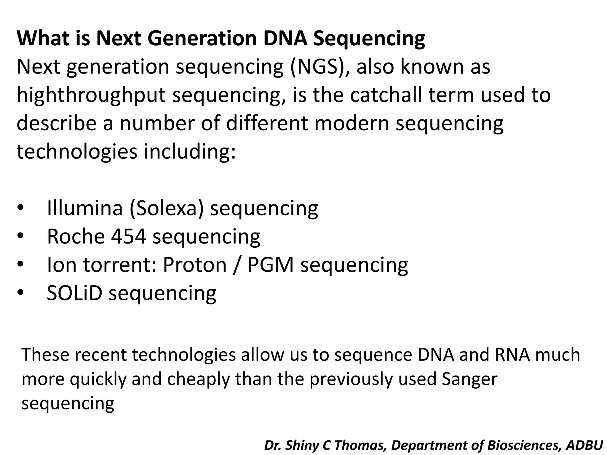
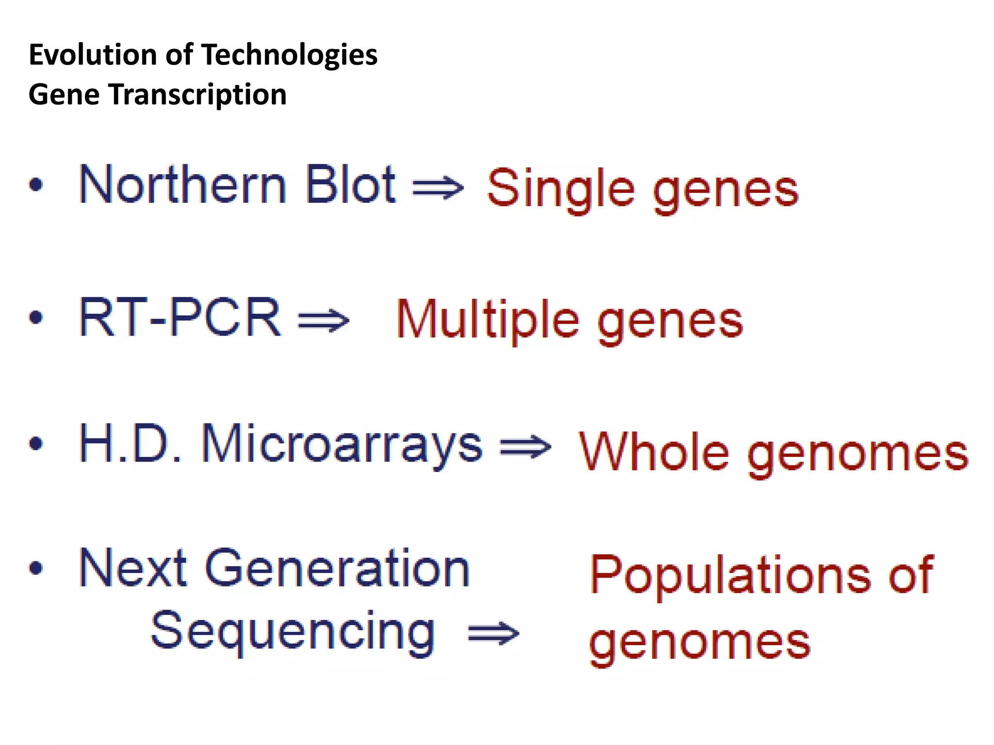
Sequencing genes and genomes in biology. The most important technique available to the molecular biologist is DNA sequencing, by which the precise order of nucleotides in a piece of DNA can be determined
Original Next Gen Seq Methods set of slides prepared for Technorazz Vibes 2016. There is also a shorter version.
This starts with an introduction to qPCR followed by an introduction to Library Complexity. Microarrays are discussed as well along with a very short introduction to FISH. Finally discussion of Next gen seq methods is done where generation of sequencers are discussed and a short discussion of the ILLUMINA protocol. Finally comparison of ILLUMINA amongst other 3rd gen sequencer, description of the standard pipeline and the omics technologies that have risen from this seq data.
whole genome analysis
history
needs
steps involved
human genome data
NGS
pyrosequencing
illumina
SOLiD
Ion torrent
PacBio
applications
problems
benefits
DNA Sequencing - DNA sequencing is like reading the instructions inside a cellAmitSamadhiya1
DNA sequencing is like reading the instructions inside a cell. It's figuring out the exact order of the building blocks that make up our DNA, represented by the letters A, T, C, and G. This order is like a code that tells our bodies how to function and grow.
By reading this code, scientists can understand genes, diagnose diseases, and even trace our ancestry. There are different ways to sequence DNA, kind of like having a few different ways to read a book. These techniques are constantly improving, making it faster and easier to unlock the secrets hidden in our DNA.
Deciphering DNA sequences is essential for virtually all branches of biological research. With the
advent of capillary electrophoresis (CE)-based Sanger sequencing, scientists gained the ability to
elucidate genetic information from any given biological system. This technology has become widely
adopted in laboratories around the world, yet has always been hampered by inherent limitations in
throughput, scalability, speed, and resolution that often preclude scientists from obtaining the essential
information they need for their course of study. To overcome these barriers, an entirely new technology
was required—Next-Generation Sequencing (NGS), a fundamentally different approach to sequencing
that triggered numerous ground-breaking discoveries and ignited a revolution in genomic science.
DNA sequencing is a laboratory technique used to determine the exact sequence of bases (A, C, G, and T) in a DNA molecule. The DNA base sequence carries the information a cell needs to assemble protein and RNA molecules. DNA sequence information is important to scientists investigating the functions of genes.
In medicine, DNA sequencing is used for a range of purposes, including diagnosis and treatment of diseases. In general, sequencing allows health care practitioners to determine if a gene or the region that regulates a gene contains changes, called variants or mutations, that are linked to a disorder.
DNA sequencing refers to the general laboratory technique for determining the exact sequence of nucleotides, or bases, in a DNA molecule. The sequence of the bases (often referred to by the first letters of their chemical names: A, T, C, and G) encodes the biological information that cells use to develop and operate. Establishing the sequence of DNA is key to understanding the function of genes and other parts of the genome. There are now several different methods available for DNA sequencing, each with its own characteristics, and the development of additional methods represents an active area of genomics research.
Herbal Drug Technology
Herbs as Raw Materials: Definition of herb, herbal medicine, herbal medicinal product and herbal drug preparation, source of herbs, selection, identification and authentication of herbal materials, processing of herbal raw material.
Herbal Excipients : Herbal Excipients – Significance of substances of natural origin as excipients, – colorants, sweeteners, binders, diluents, viscosity builders, dis-integrants, flavors & perfumes.
Herbal Formulations: Stages involved in herbal formulations, Orthodox formulations and methods of delivery of herbal extracts, Novel formulations of herbal extracts.
Introduction to proteomics, techniques to study proteomics such as protein electrophoresis, chromatography and mass spectrometry and protein database analysis, case studies derived from scientific literature including comparisons between healthy and diseased tissues, new approaches to analyse metabolic pathways, comprehensive analysis of protein-protein interactions in different cell types.
Metabolomics-Introduction, metabolism, intermediary metabolism, metabolic pathways, metabolites, metabolome, metabolic turnover, techniques used in metabolomics, metabolite profiling methods, data analysis, metabolomic resources, role of metabolomics in system biology.
Introduction to proteomics, techniques to study proteomics such as protein electrophoresis, chromatography and mass spectrometry and protein database analysis, case studies derived from scientific literature including comparisons between healthy and diseased tissues, new approaches to analyse metabolic pathways, comprehensive analysis of protein-protein interactions in different cell types.
The analysis of global gene expression and transcription factor regulation, global approaches to alternative splicing and its regulation, long noncoding RNAs, gene expression models of signalling pathways, from gene expression to disease phenotypes, introduction to isoform sequencing, systematic and integrative analysis of gene expression to identify feature genes underlying human diseases.
Genome projects
Definition of genome, history of genome projects, whole genome sequencing, Maxam Gilbert sequencing, sanger sequencing, explanation on the first sequenced organisms (Bacteriophage, bacteria, archaeon, virus, bakers yeast, nematode.
Model organism-Arabidopsis thaliana, Mus musculus, Oryza sativa, Pan troglodyte etc.
Human genome project, milestones and significance.
Epigenetics studies stably heritable traits that cannot be explained by changes in DNA sequence.
Covalent modifications in chromatin
DNA- DNA methylation (CpG); hydroxymethylation
Histone - lysine acetylation, lysine and arginine methylation, serine and threonine phosphorylation, and lysine ubiquitination and sumoylation
Epigenetic mechanisms:
Modified histones as post translational modification
DNA methylation – 5mC the 5th base, methyl transferases; genetic imprinting.
Epigenomics: complete set of epigenetic modifications on the genetic material of a cell.
Specific epigenetic regulation
RNA interference
X inactivation (Lyonization)
Genomic imprinting
Epigenetics in development and diseases.
Comparative genomics: Genomic features are compared, evolutionary relationship
The major principle of comparative genomics is that common features of two organisms will often be encoded within the DNA that is evolutionarily conserved between them. orthologous sequences,
Started as soon as the whole genomes of two organisms became available (that is, the genomes of the bacteria Haemophilus influenzae and Mycoplasma genitalium) in 1995, comparative genomics is now a standard component of the analysis of every new genome sequence. comparative genomics studies of small model organisms (for example the model Caenorhabditis elegans and closely related Caenorhabditis briggsae) are of great importance to advance our understanding of general mechanisms of evolution
Computational tools for analyzing sequences and complete genomes. Application of comparative genomics in agriculture and medicine.
Mapping and sequencing genomes: Genetic and physical mapping, Sequencing genomes different strategies, High-throughput sequencing, next-generation sequencing technologies, comparative genomics, population genomics, epigenetics, Human genome project, pharmacogenomics, genomic medicine, applications of genomics to improve public health.
Disorders of liver and kidney, Nitrogen metabolism.pdfshinycthomas
Disorders of liver and kidney – Jaundice, fatty liver, normal and abnormal functions of liver and kidney. Inulin and urea clearance.
Abnormalities of nitrogen metabolism
Lipid metabolism and its disorders.pdfshinycthomas
Disorders of Lipids – Plasma lipoproteins, cholesterol, triglycerides and phospholipids in health and disease, hyperlipidemia, hyperlipoproteinemia, Gaucher’s disease, Tay-Sach’s and Niemann-Pick disease, ketone bodies.
a) Definition, classification, structure, stereochemistry and reactions of amino acids;
b) Classification of proteins on the basis of solubility and shape, structure, and biological functions. Primary structure - determination of amino acid sequences of proteins, the peptide bond, Ramachandran plot.
c) Secondary structure - weak interactions involved - alpha helix and beta sheet and beta turns structure, Pauling and Corey model for fibrous proteins, Collagen triple helix, and super secondary structures - helix-loop-helix.
d) Tertiary structure - alpha and beta domains. Quaternary structure - structure of haemoglobin, Solid state synthesis of peptides, Protein-Protein interactions, Concept of chaperones.
Nucleic acid-DNA and RNA
Gene-part of DNA
Functions of DNA
RNA-Functions, different types of RNA-Ribosomal RNAs (rRNAs), Messenger RNAs (mRNAs), Transfer RNAs (tRNAs)-Other RNA-Small nuclear RNA (snRNA), Micro RNA (miRNA), Small interfering RNA (siRNA), Heterogenous RNA (hnRNA).
Nucleic acid-nucleotides-nucleoside
Components of nucleotide-a nitrogenous (nitrogen-containing) base (pyrimidine and purine), (2) a pentose, and (3) a phosphate
Structure of pentose sugar, and 5 major bases (cytosine, thymine, uracil-pyrimidine bases and adenine, guanine-purine bases).
Deoxyribonucleotides and Ribo nucleotides-Molecular structure of deoxyadenosine monophosphate (dAMP), deoxyguanosine monophosphate (dGMP), deoxythymidine monophosphate (dTMP), deoxycytidine monophosphate (dCMP) and Adenosine monophosphate (AMP), Guanosine monophosphate (GMP), Cytosine monophosphate (CMP) and Uridine monophosphate (UMP), Watson crick base pairing, Hoogsteen base pairing,
Helical structure-Heterocylic N -Glycosides, Syn and Anti Conformers, detailed structure of single strand and double stranded DNA.
DNA Nucleotides and Tautomeric Form-Tautomers of Adenine, Cytosine, Guanine, and Thymine
Template strand, non coding strand and coding strand
Hydrogen bonds, phosphodiester linkage, hydrolysis of DNA and RNA.
Different forms of DNA-A, B, and Z forms.
Palindrome sequence, Linear DNA, Cruciform DNA, H DNA (Triplex DNA), Denaturation of DNA, Hyperchromicity, Tm, Renaturation of DNA, Tertiary structure of DNA, Difference of DNA and RNA, RNA structural elements, primary. secondary and tertiary structure of RNA. Detailed structure and functions of tRNA, mRNA, rRNA, miRNA, siRNA, hn RNA, snRNA.
Nucleic acid hybridization, C0t analysis, Buoyant density of DNA, Isopycnic centrifugation.
Lipids-Introduction, properties and functions.
Classification-Simple lipids, complex lipids and derived lipids.
Lipids contain fatty acid and alcohol.
Saturated and Unsaturated fatty acids. Nomenclature of fatty acids, Cis-trans isomerism, essential fatty acids
Simple lipids-Fats, waxes
Compound lipids-Structure, function with examples of Phospholipids, Glycolipids, sulpholipids and lipoproteins.
Derived lipids: Structure, types, and functions of steroids, terpenes and carotenoids.
Lipoproteins-classified into chylomicrons, very low-density lipoproteins (VLDL), low density lipoproteins (LDL) and high-density lipoproteins (HDL) and their function.
Eicosanoids-prostanoids, leukotrienes (LTs), and lipoxins (LXs).
Functions of Eicosanoids
Lipids, micelles and liposomes.
Vitamins-Introduction, Water soluble and fat soluble vitamins.
Water soluble vitamins-B complex vitamins: thiamin (vitamin B1), riboflavin (vitamin B2), niacin (vitamin B3), vitamin B6 (pyridoxine), folate (folic acid), vitamin B12, biotin and pantothenic acid-their source, structure, properties, metabolism, physiological significance, deficiency disease and human requirements.
Fat soluble vitamins: Fat soluble vitamins, Vitamin A, D, E and K and their their source, structure, properties, metabolism, physiological significance, deficiency disease and human requirements.
Vitamin A-Carotene in plants-α-carotenes, β-carotenes and γ-carotenes, 3 forms of vitamin A-Retinol, Retinal, Retinoic acid.
Vitamin D3-cholecalciferol,
Vitamin E -Tocopherol, Vitamin K-Phylloquinone or Anti hemorrhagic Vitamin or Coagulation Vitamin
Seminar of U.V. Spectroscopy by SAMIR PANDASAMIR PANDA
Spectroscopy is a branch of science dealing the study of interaction of electromagnetic radiation with matter.
Ultraviolet-visible spectroscopy refers to absorption spectroscopy or reflect spectroscopy in the UV-VIS spectral region.
Ultraviolet-visible spectroscopy is an analytical method that can measure the amount of light received by the analyte.
Cancer cell metabolism: special Reference to Lactate PathwayAADYARAJPANDEY1
Normal Cell Metabolism:
Cellular respiration describes the series of steps that cells use to break down sugar and other chemicals to get the energy we need to function.
Energy is stored in the bonds of glucose and when glucose is broken down, much of that energy is released.
Cell utilize energy in the form of ATP.
The first step of respiration is called glycolysis. In a series of steps, glycolysis breaks glucose into two smaller molecules - a chemical called pyruvate. A small amount of ATP is formed during this process.
Most healthy cells continue the breakdown in a second process, called the Kreb's cycle. The Kreb's cycle allows cells to “burn” the pyruvates made in glycolysis to get more ATP.
The last step in the breakdown of glucose is called oxidative phosphorylation (Ox-Phos).
It takes place in specialized cell structures called mitochondria. This process produces a large amount of ATP. Importantly, cells need oxygen to complete oxidative phosphorylation.
If a cell completes only glycolysis, only 2 molecules of ATP are made per glucose. However, if the cell completes the entire respiration process (glycolysis - Kreb's - oxidative phosphorylation), about 36 molecules of ATP are created, giving it much more energy to use.
IN CANCER CELL:
Unlike healthy cells that "burn" the entire molecule of sugar to capture a large amount of energy as ATP, cancer cells are wasteful.
Cancer cells only partially break down sugar molecules. They overuse the first step of respiration, glycolysis. They frequently do not complete the second step, oxidative phosphorylation.
This results in only 2 molecules of ATP per each glucose molecule instead of the 36 or so ATPs healthy cells gain. As a result, cancer cells need to use a lot more sugar molecules to get enough energy to survive.
introduction to WARBERG PHENOMENA:
WARBURG EFFECT Usually, cancer cells are highly glycolytic (glucose addiction) and take up more glucose than do normal cells from outside.
Otto Heinrich Warburg (; 8 October 1883 – 1 August 1970) In 1931 was awarded the Nobel Prize in Physiology for his "discovery of the nature and mode of action of the respiratory enzyme.
WARNBURG EFFECT : cancer cells under aerobic (well-oxygenated) conditions to metabolize glucose to lactate (aerobic glycolysis) is known as the Warburg effect. Warburg made the observation that tumor slices consume glucose and secrete lactate at a higher rate than normal tissues.
This pdf is about the Schizophrenia.
For more details visit on YouTube; @SELF-EXPLANATORY;
https://www.youtube.com/channel/UCAiarMZDNhe1A3Rnpr_WkzA/videos
Thanks...!
Richard's aventures in two entangled wonderlandsRichard Gill
Since the loophole-free Bell experiments of 2020 and the Nobel prizes in physics of 2022, critics of Bell's work have retreated to the fortress of super-determinism. Now, super-determinism is a derogatory word - it just means "determinism". Palmer, Hance and Hossenfelder argue that quantum mechanics and determinism are not incompatible, using a sophisticated mathematical construction based on a subtle thinning of allowed states and measurements in quantum mechanics, such that what is left appears to make Bell's argument fail, without altering the empirical predictions of quantum mechanics. I think however that it is a smoke screen, and the slogan "lost in math" comes to my mind. I will discuss some other recent disproofs of Bell's theorem using the language of causality based on causal graphs. Causal thinking is also central to law and justice. I will mention surprising connections to my work on serial killer nurse cases, in particular the Dutch case of Lucia de Berk and the current UK case of Lucy Letby.
(May 29th, 2024) Advancements in Intravital Microscopy- Insights for Preclini...Scintica Instrumentation
Intravital microscopy (IVM) is a powerful tool utilized to study cellular behavior over time and space in vivo. Much of our understanding of cell biology has been accomplished using various in vitro and ex vivo methods; however, these studies do not necessarily reflect the natural dynamics of biological processes. Unlike traditional cell culture or fixed tissue imaging, IVM allows for the ultra-fast high-resolution imaging of cellular processes over time and space and were studied in its natural environment. Real-time visualization of biological processes in the context of an intact organism helps maintain physiological relevance and provide insights into the progression of disease, response to treatments or developmental processes.
In this webinar we give an overview of advanced applications of the IVM system in preclinical research. IVIM technology is a provider of all-in-one intravital microscopy systems and solutions optimized for in vivo imaging of live animal models at sub-micron resolution. The system’s unique features and user-friendly software enables researchers to probe fast dynamic biological processes such as immune cell tracking, cell-cell interaction as well as vascularization and tumor metastasis with exceptional detail. This webinar will also give an overview of IVM being utilized in drug development, offering a view into the intricate interaction between drugs/nanoparticles and tissues in vivo and allows for the evaluation of therapeutic intervention in a variety of tissues and organs. This interdisciplinary collaboration continues to drive the advancements of novel therapeutic strategies.
Slide 1: Title Slide
Extrachromosomal Inheritance
Slide 2: Introduction to Extrachromosomal Inheritance
Definition: Extrachromosomal inheritance refers to the transmission of genetic material that is not found within the nucleus.
Key Components: Involves genes located in mitochondria, chloroplasts, and plasmids.
Slide 3: Mitochondrial Inheritance
Mitochondria: Organelles responsible for energy production.
Mitochondrial DNA (mtDNA): Circular DNA molecule found in mitochondria.
Inheritance Pattern: Maternally inherited, meaning it is passed from mothers to all their offspring.
Diseases: Examples include Leber’s hereditary optic neuropathy (LHON) and mitochondrial myopathy.
Slide 4: Chloroplast Inheritance
Chloroplasts: Organelles responsible for photosynthesis in plants.
Chloroplast DNA (cpDNA): Circular DNA molecule found in chloroplasts.
Inheritance Pattern: Often maternally inherited in most plants, but can vary in some species.
Examples: Variegation in plants, where leaf color patterns are determined by chloroplast DNA.
Slide 5: Plasmid Inheritance
Plasmids: Small, circular DNA molecules found in bacteria and some eukaryotes.
Features: Can carry antibiotic resistance genes and can be transferred between cells through processes like conjugation.
Significance: Important in biotechnology for gene cloning and genetic engineering.
Slide 6: Mechanisms of Extrachromosomal Inheritance
Non-Mendelian Patterns: Do not follow Mendel’s laws of inheritance.
Cytoplasmic Segregation: During cell division, organelles like mitochondria and chloroplasts are randomly distributed to daughter cells.
Heteroplasmy: Presence of more than one type of organellar genome within a cell, leading to variation in expression.
Slide 7: Examples of Extrachromosomal Inheritance
Four O’clock Plant (Mirabilis jalapa): Shows variegated leaves due to different cpDNA in leaf cells.
Petite Mutants in Yeast: Result from mutations in mitochondrial DNA affecting respiration.
Slide 8: Importance of Extrachromosomal Inheritance
Evolution: Provides insight into the evolution of eukaryotic cells.
Medicine: Understanding mitochondrial inheritance helps in diagnosing and treating mitochondrial diseases.
Agriculture: Chloroplast inheritance can be used in plant breeding and genetic modification.
Slide 9: Recent Research and Advances
Gene Editing: Techniques like CRISPR-Cas9 are being used to edit mitochondrial and chloroplast DNA.
Therapies: Development of mitochondrial replacement therapy (MRT) for preventing mitochondrial diseases.
Slide 10: Conclusion
Summary: Extrachromosomal inheritance involves the transmission of genetic material outside the nucleus and plays a crucial role in genetics, medicine, and biotechnology.
Future Directions: Continued research and technological advancements hold promise for new treatments and applications.
Slide 11: Questions and Discussion
Invite Audience: Open the floor for any questions or further discussion on the topic.
1. What is Next Generation DNA Sequencing
Next generation sequencing (NGS), also known as
highthroughput sequencing, is the catchall term used to
describe a number of different modern sequencing
technologies including:
• Illumina (Solexa) sequencing
• Roche 454 sequencing
• Ion torrent: Proton / PGM sequencing
• SOLiD sequencing
These recent technologies allow us to sequence DNA and RNA much
more quickly and cheaply than the previously used Sanger
sequencing
Dr. Shiny C Thomas, Department of Biosciences, ADBU
2. The four main advantages of NGS over classical Sanger
sequencing are:
• speed
• cost
• sample size
• accuracy
• NGS is significantly cheaper, quicker, needs significantly
less DNA and is more accurate and reliable than Sanger
sequencing. Let us look at this more closely.
• For Sanger sequencing, a large amount of template DNA
is needed for each read.
• In NGS, a sequence can be obtained from a single strand
3. • NGS is quicker than Sanger sequencing in two ways.
Firstly, the chemical reaction may be combined with the
signal detection in some versions of NGS, whereas in
Sanger sequencing these are two separate processes.
• Secondly and more significantly, only one read
(maximum ~1kb) can be taken at a time in Sanger
sequencing, whereas NGS is massively parallel,
allowing 300Gb of DNA to be read on a single run on a
single chip.
4. • The reduced time, manpower and reagents in NGS mean that the
costs are much lower. The first human genome sequence cost in
the region of £300M.
Using modern Sanger sequencing methods, aided by data from the
known sequence, a full human genome would still cost £6M.
Sequencing a human genome with Illumina today would cost only
£6,000.
6. Sequencing for Transcriptomics?
• Sample cells
• Purify mRNA*
• mRNA ⇒ cDNA
• Cleaving into 35-400nt fragments
• Sequence fragments in parallel
• PCR amplification
* or DNA for genomic sequencing
7. Next Generation Sequencing
Technology Overview
1. (c)DNA is fragmented
2. Adaptors ligated to fragments
3. Several possible protocols yield array of PCR colonies.
4. Enzymatic extension with fluorescently tagged
nucleotides.
5. Cyclic readout by imaging the array.
8. • Fragmentation of DNA
• Primers are attached to the surface of a bead
Technology 1. (emulsion PCR)
9. 2. Bead preparation
• Fragments, with adaptors, are PCR amplified within a
water drop in oil.
• One primer is attached to the surface of a bead
• 3’ modification of fragments, to covalently bind bead to
chip surface.
12. Sequence of images
Sequence read over multiple cycles
Repeated cycles of sequencing to determine the
sequence of bases in a given fragment, a single
base at a time.
14. • While these “first-generation” instruments were
considered high throughput for their time, the Genome
Analyzer emerged in 2005 and took sequencing runs from
84 kilobase (kb) per run to 1 gigabase (Gb) per run.
• The short read, massively parallel sequencing technique
was a fundamentally different approach that
revolutionized sequencing capabilities and launched the
“next-generation” in genomic science.
• From that point forward, the data output of next-
generation sequencing (NGS) has—more than doubling
each year
15. Figure 1: Sequencing Cost and Data Output Since 2000—The dramatic
rise of data output and concurrent falling cost of sequencing since
2000. The Y-axes on both sides of the graph are logarithmic.
16. • In 2005, with the Genome Analyzer, a single sequencing
run could produce roughly one gigabase of data.
• By 2014, the rate climbed to a 1.8 terabases of data in a
single sequencing run—an astounding 1000× increase.
• It is remarkable to reflect on the fact that the first human
genome, famously co published in Science and Nature in
2001, required 15 years to sequence and cost nearly 3
billion dollars.
• In contrast, the HiSeq X™ Ten, released in 2014, can
sequence over 45 human genomes in a single day for
approximately $1000 each.
17. Human Genome Sequencing Over the Decades—The capacity to sequence all 3.2
billion bases of the human genome (at 30× coverage) has increased exponentially
since the 1990s.
In 2005, with the introduction of the Illumina Genome Analyzer System, 1.3 human
genomes could be sequenced annually. Nearly 10 years later, with the Illumina HiSeq
X Ten fleet of sequencing systems, the number has climbed to 18,000 human
genomes a year.
18. • Beyond the massive increase in data output, the
introduction of NGS technology has transformed the way
scientists think about genetic information.
• The $1000 dollar genome enables population-scale
sequencing and establishes the foundation for
personalized genomic medicine as part of standard
medical care.
• Researchers can now analyze thousands to tens of
thousands of samples in a single year.
• The rate of progress is stunning.
19. • As costs continue to come down, we are entering a period
where we are going to be able to get the complete catalog
of disease genes.
• This will allow us to look at thousands of people and see
the differences among them, to discover critical genes that
cause cancer, autism, heart disease, or schizophrenia.
20. The Basics of NGS Chemistry
• In principle, the concept behind NGS technology is
similar to CE (Capillary Electrophoresis) sequencing—
DNA polymerase catalyzes the incorporation of
fluorescently labeled deoxyribonucleotide triphosphates
(dNTPs) into a DNA template strand during sequential
cycles of DNA synthesis.
• During each cycle, at the point of incorporation, the
nucleotides are identified by fluorophore excitation.
• The critical difference is that, instead of sequencing a
single DNA fragment, NGS extends this process across
millions of fragments in a massively parallel fashion.
21. • Illumina sequencing by synthesis (SBS) chemistry is the
most widely adopted chemistry in the industry and
delivers the highest accuracy, the highest yield of error-
free reads, and the highest percentage of base calls above
Q30.
• The Illumina NGS workflows include 4 basic steps
22. 1. Library Preparation—
• The sequencing library is prepared by random
fragmentation of the DNA or cDNA sample, followed by
5’ and 3’ adapter ligation.
• Alternatively, “tagmentation” combines the
fragmentation and ligation reactions into a single step
that greatly increases the efficiency of the library
preparation process.
• Adapter-ligated fragments are then PCR amplified and
gel purified.
23. 2. Cluster Generation—
• For cluster generation, the library is loaded into a
flow cell where fragments are captured on a lawn
of surface-bound oligos complementary to the
library adapters.
• Each fragment is then amplified into distinct, clonal
clusters through bridge amplification.
• When cluster generation is complete, the
templates are ready for sequencing.
24. 3. Sequencing—
• Illumina SBS technology utilizes a proprietary
reversible terminator–based method that detects
single bases as they are incorporated into DNA
template strands.
• As all 4 reversible terminator-bound dNTPs are
present during each sequencing cycle, natural
competition minimizes incorporation bias and
greatly reduces raw error rates compared to other
technologies.
• The result is highly accurate base-by-base
sequencing that virtually eliminates sequence-
context-specific errors, even within repetitive
sequence regions and homopolymers.
25. 4. Data Analysis—
• During data analysis and alignment, the newly
identified sequence reads are then aligned to a
reference genome.
• Following alignment, many variations of analysis
are possible such as single nucleotide
polymorphism (SNP) or insertion-deletion (indel)
identification, read counting for RNA methods, phylogenetic
or metagenomic analysis, and more.
30. Advances in Sequencing Technology
Paired-End Sequencing
• A major advance in NGS technology occurred with the
development of paired-end (PE) sequencing.
• PE sequencing involves sequencing both ends of the DNA
fragments in a sequencing library and aligning the
forward and reverse reads as read pairs.
• In addition to producing twice the number of reads for
the same time and effort in library preparation,
sequences aligned as read pairs enable more accurate
read alignment and the ability to detect indels, which is
simply not possible with single-read data.
31. • Analysis of differential read-pair spacing also allows
removal of PCR duplicates, a common artifact resulting
from PCR amplification during library preparation.
• Furthermore, paired-end sequencing produces a higher
number of SNV calls following read-pair alignment.
• While some methods are best served by single-read
sequencing, such as small RNA sequencing, most
researchers currently use the paired-end approach.
32. Paired-End Sequencing and Alignment—Paired-end
sequencing enables both ends of the DNA fragment to be
sequenced.
• Because the distance between each paired read is known,
alignment algorithms can use this information to map the
reads over repetitive regions more precisely. This results in
much better alignment of the reads, especially across
difficult-to-sequence, repetitive regions of the genome.
33. What is the Illumina method of DNA sequencing?
Illumina sequencing has been used to sequence many
genomes and has enabled the comparison of DNA sequences
to improve understanding of health and disease.
Illumina sequencing generates many millions of highly
accurate reads making it much faster and cheaper than
other available
34. How does Illumina DNA sequencing work?
1. The first step in this sequencing technique is to break up
the DNA into more manageable fragments of around 200 to
600.
2. Short sequences of DNA called adaptors, are attached to
the DNA fragments.
3. The DNA fragments attached to adaptors are then made
single stranded. This is done by incubating the fragments
with sodium hydroxide.
4. Once prepared, the DNA fragments are washed across the
Flow cell. The complementary DNA binds to primers on the
surface of the flow cell and DNA that doesn’t attach is
washed away.
35. 5. The DNA attached to the flow cell is then replicated to
form small clusters of DNA with the same sequence.
• When sequenced, each cluster of DNA molecules will
emit a signal that is strong enough to be detected by a
camera.
6. Unlabelled nucleotides and DNA polymerase are then
added to lengthen and join the strands of DNA attached to
the flow cell.
• This creates ‘bridges’ of double-stranded DNA between
the primers on the flow cell surface.
7. The double-stranded DNA is then broken down into single
stranded DNA using heat, leaving several million dense
clusters of identical DNA sequences.
36. 8. Primers and fluorescently labelled terminators
(terminators are a version of nucleotide base – A, C, G or T -
that stop DNA synthesis) are added to the flowcell.
9. The primer attaches to the DNA being sequenced.
10. The DNA polymerase then binds to the primer and adds
the first fluorescently-labelled terminator to the new DNA
strand.
• Once a base has been added no more bases can be added
to the strand of DNA until the terminator base is cut from
the DNA.
37. 11. Lasers are passed over the flowcell to activate the
fluorescent label on the nucleotide base.
• This fluorescence is detected by a camera and recorded
on a computer. Each of the terminator bases (A, C, G and
T) give off a different colour.
12. The fluorescently-labelled terminator group is then
removed from the first base and the next fluorescently-
labelled terminator base can be added alongside.
• And so the process continues until millions of clusters
have been sequenced.
38. 13. The DNA sequence is analysed base-by-base during
Illumina sequencing, making it a highly accurate method.
• The sequence generated can then be aligned to a
reference sequence, this looks for matches or changes in
the sequenced DNA.
39. ➢ In Illumina sequencing, 100150bp reads are used.
➢ Somewhat longer fragments are ligated to generic
adaptors and annealed to a slide using the adaptors.
➢ PCR is carried out to amplify each read, creating a spot
with many copies of the same read.
➢ They are then separated into single strands to be
sequenced.
➢ The slide is flooded with nucleotides and DNA polymerase.
➢ These nucleotides are fluorescently labelled, with the
colour corresponding to the base.
➢ They also have a terminator, so that only one base is
added at a time.
40. ➢ An image is taken of the slide. In each read location, there
will be a fluorescent signal indicating the base that has
been added.
41. ➢ The slide is then prepared for the next cycle. The
terminators are removed, allowing the next base to be
added, and the fluorescent signal is removed, preventing
the signal from contaminating the next image.
➢ The process is repeated, adding one nucleotide at a time
and imaging in between.
➢ Computers are then used to detect the base at each site
in each image and these are used to construct a
sequence.
42.
43. ➢ All of the sequence reads will be the same length, as the
read length depends on the number of cycles carried out.
44. Ion Torrent
• The sequencing chemistry itself is remarkably simple.
Naturally, a proton is released when a nucleotide is
incorporated by the polymerase in the DNA molecule,
resulting in a detectable local change of pH.
• Each micro-well of the Ion Torrent semiconductor
sequencing chip contains approximately one million
copies of a DNA molecule.
• The Ion Personal Genome Machine (PGM™) sequencer
sequentially floods the chip with one nucleotide after
another.
45. •
• If a nucleotide complements the sequence of the DNA
molecule in a particular micro-well, it will be
incorporated and hydrogen ions are released.
• The pH of the solution changes in that well and is
detected by the ion sensor, essentially going directly
from chemical information to digital information.
• If there are two identical bases on the DNA strand, the
voltage is double, and the chip records two identical
bases.
• If the next nucleotide that floods the chip is not a match,
no voltage change is recorded and no base is called.
46. • Because this is direct detection—no scanning, no
cameras, no light—each nucleotide incorporation is
measured in seconds enabling very short run times.
• Naturally, a proton is released when a nucleotide is
incorporated by the polymerase in the DNA molecule,
resulting in a detectable local change of pH.
47.
48.
49.
50.
51.
52. Ion Torrent: Proton / PGM sequencing
• Unlike Illumina and 454, Ion torrent and Ion proton
sequencing do not make use of optical signals.
• Instead, they exploit the fact that addition of a dNTP
to a DNA polymer releases an H+ ion.
• As in other kinds of NGS, the input DNA or RNA is
fragmented, this time ~200bp. Adaptors are added and
one molecule is placed onto a bead.
• The molecules are amplified on the bead by emulsion
PCR.
• Each bead is placed into a single well of a slide.
Like 454, the slide is flooded with a single species of dNTP,
along with buffers and polymerase, one NTP at a time.
53. • The pH is detected is each of the wells, as each H+ ion
released will decrease the pH.
• The changes in pH allow us to determine if that base,
and how many thereof, was added to the sequence
read.
• The dNTPs are washed away, and the process is repeated
cycling through the different dNTP species.
54.
55. The pH change, if any, is used to determine how many bases
(if any) were added with each cycle.
56.
57. Pyrosequencing
• Pyrosequencing is a method of DNA sequencing
(determining the order of nucleotides in DNA) based on
the "sequencing by synthesis“ principle.
• It differs from Sanger sequencing, in that it relies on the
detection of pyrophosphate release on nucleotide
incorporation, rather than chain termination with
dideoxynucleotides.
58. • The desired DNA sequence is able to be determined by
light emitted upon incorporation of the next
complementary nucleotide
• Only one out of four of the possible A/T/C/G
nucleotides are added and available at a time
• So that only one letter can be incorporated on the
single stranded template (which is the sequence to be
determined).
59. • The intensity of the light determines if there are more
than one of these "letters" in a row.
• The previous nucleotide letter (one out of four possible
dNTP) is degraded before the next nucleotide letter is
added for synthesis: allowing for the possible revealing of
the next nucleotide(s) via the resulting intensity of light
(if the nucleotide added was the next complementary
letter in the sequence).
This process is repeated with each of the four letters until
the DNA sequence of the single stranded template is
determined.
61. • "Sequencing by synthesis" involves taking a single strand
of the DNA to be sequenced and then synthesizing its
complementary strand enzymatically.
• The pyrosequencing method is based on detecting the
activity of DNA polymerase (a DNA synthesizing enzyme)
with another chemoluminescent enzyme.
• Essentially, the method allows sequencing of a single
strand of DNA by synthesizing the complementary strand
along it, one base pair at a time, and detecting which base
was actually added at each step.
62. • The template DNA is immobile, and solutions of A, C, G,
and T nucleotides are sequentially added and removed
from the reaction.
Light is produced only when the nucleotide solution
complements the first unpaired base of the template.
• The sequence of solutions which produce
chemiluminescent signals allows the determination of the
sequence of the template.
63. • The single strand DNA (ssDNA) template is hybridized to
a sequencing primer and incubated with the enzymes
DNA polymerase, ATP sulfurylase, luciferase and apyrase,
and with the substrates adenosine 5´ phosphosulfate
(APS) and luciferin.
1. The addition of one of the four deoxynucleoside
triphosphates (dNTPs) (dATPαS, which is not a substrate for
a luciferase, is added instead of dATP to avoid noise)
initiates the second step.
• DNA polymerase incorporates the correct,
complementary dNTPs onto the template. This
incorporation releases pyrophosphate (PPi).
64.
65. 2. ATP sulfurylase converts PPi to ATP in the presence of
adenosine 5´ phosphosulfate.
• This ATP acts as a substrate for the luciferase mediated
conversion of luciferin to oxyluciferin that generates
visible light in amounts that are proportional to the
amount of ATP.
• The light produced in the luciferasecatalyzed reaction is
detected by a camera and analyzed in a pyrogram.
3. Unincorporated nucleotides and ATP are degraded by the
apyrase, and the reaction can restart with another
Nucleotide.
66. Limitation
• Currently, a limitation of the method is that the lengths
of individual reads of DNA sequence of 300-500
nucleotides, shorter than the 800-1000 obtainable with
chain termination methods (e.g. Sanger sequencing).
67. Pyrosequencing cycle
• Add dATP. If light is emitted, your sequence
starts with A. If not, the dATP is degraded (or
elutes past immobilized primer).
• Add dGTP. If light is emitted, the next base
must be a G.
• Then add T, then C. You now know at least
one (maybe more) base of the sequence.
• Repeat!
68. Pyrosequencing output
Runs of bases produce higher peaks – for instance, the sequence for (a)
is GGCCCTTG. Sample (c) comes from a heterozygous individual
(hence the heights in multiples of ½)
69. Roche 454
Roche 454 sequencing system is the first commercial
platforms for the next generation sequencing technology. Its
main principle of sequencing is illustrated as follows.
70. a. Preparation of DNA Library
• DNA Library construction in 454 sequencing system is
different from that of Illumina.
• It uses spray method to break DNA samples into small
fragments of 300-800bp, and adds different adapters at
both ends.
• Otherwise, use primers for amplification after DNA
denaturation, clone into specific vectors, and finally
constructing single stranded DNA library
71.
72. b. Emulsion PCR
• These single stranded DNAs would be fixed by 28um beads
which are buried in emulsion.
• The biggest feature of emulsion PCR is the formation of a
large number of independent reaction space for DNA
amplification.
• The key technology is to separate different beats using the
characteristics of emulsion.
73. • The basic process is as follows.
• Before sample DNA amplification, aqueous solution with
all components of PCR reaction will be infused into the
surface of mineral oil with high-speed rotation, and it
forms numerous small water droplets wrapped by
mineral oil.
• One small droplet forms an independent PCR reaction
space. Ideally, each small drop of water contains only
one DNA template and one bead.
74. • On the surface of beads, which are wrapped by small
water droplets, there are complementary oligos to match
those adapters, so the single stranded DNA can
specifically bind to the beads.
• At the same time, incubation system contains PCR
reagents to ensure that each small DNA fragment fixed on
the bead can be the unique template for amplification.
• Moreover, PCR products can be also combined with
magnetic beads.
• After the reaction accomplishment, emulsion system can
be destroyed and target DNAs would be accumulated.
75. • Finally, each small fragment will be amplified about 1
million times, so as to achieve the amount level required
by the sequencing process.
76. c. Pyrosequencing
• A polymerase and single strand DNA binding protein are
needed to process beads with DNAs before sequencing.
• Then these beads are put on PTP plate.
• This plate has many special nanopores of 44um
diameters.
• Each nanopore can only accommodate one bead, which
can fix the position of each bead through this method,
in order to be convenient for sequencing.
• The method used in this sequencing process is
pyrosequencing.
77. • Put a smaller bead into the nanopore, and start the
sequencing reaction.
• DNA sequencing reaction is based on the single stranded
DNAs which have been amplified and fixed.
• If one dNTP can pair with the template DNA, the
pyrophosphate group will be released after synthesis.
• The released pyrophosphate group reacts with ATP
sulfuric acid chemical enzymes to produce ATP.
• CO-oxidation of ATP and luciferase makes the fluorescein
molecule triggered and fluoresce, and the CCD camera on
the other side of the PTP board records the signal of
fluorescent.
78. • Finally the results are processed by computer software.
• Because each kind of dNTP produces unique fluorescence
color in the reaction, DNA sequence can be measured
according to the fluorescence colors.
• After the reaction, ATP are degraded by diphosphatase,
leading to fluorescence quenching, so that sequencing
reaction goes into the next cycle.
79. 454 sequencing
• Roche 454 sequencing can sequence much longer reads
than Illumina.
• Like Illumina, it does this by sequencing multiple reads at
once by reading optical signals as bases are added.
• As in Illumina, the DNA or RNA is fragmented into shorter
reads, in this case up to 1kb.
• Generic adaptors are added to the ends and these are
annealed to beads, one DNA fragment per bead.
• The fragments are then amplified by PCR using adaptor
specifc primers.
• Each bead is then placed in a single well of a slide. So
each well will contain a single bead, covered in many PCR
copies of a single sequence.
80. • The wells also contain DNA polymerase and sequencing
buffers.
• The slide is flooded with one of the four NTP species.
Where this nucleotide is next in the sequence, it is added
to the sequence read.
• If that single base repeats, then more will be added.
• So if we flood with Guanine bases, and the next in a
sequence is G, one G will be added, however if the next
part of the sequence is GGGG, then four Gs will be added.
81. • The addition of each nucleotide
releases a light signal.
• These locations of signals are
detected and used to determine
which beads the nucleotides are
added to.
82. • This NTP mix is washed away. The next
NTP mix is now added and the process
repeated, cycling through the four NTPs.
83. • This kind of sequencing generates graphs for each
sequence read, showing the signal density for each
nucleotide wash.
• The sequence can then be determined computationally
from the signal density in each wash.
84. • All of the sequence reads we get from 454 will be different
lengths, because different numbers of bases will be added
with each cycle.
85. SOLiD
• An open source sequencer that utilizes emulsion PCR to
immobilize the DNA library onto a solid support and
cyclic sequencing-by-ligation chemistry.
Sequencing Library Preparation and Immobilization
• The in vitro sequencing library preparation for SOLiD
involves fragmentation of the DNA sample to an
appropriate size range (400–850 bp), end repair and
ligation of “P1” and “P2” DNA adapters to the ends of the
library fragments
91. • Emulsion PCR is applied to immobilize the sequencing
library DNA onto “P1” coated paramagnetic beads.
• High-density, semi-ordered polony arrays are generated by
functionalizing the 3 ¢ ends of the templates and
immobilizing the modified beads to a glass slide.
• The glass slides can be segmented up to eight chambers to
facilitate up scaling of the number of analyzed samples.
92. • Sequencing by Ligation The SOLiD sequencing
chemistry is based on ligation (Fig)
• A sequencing primer is hybridized to
the “P1” adapter in the immobilized
beads.
• A pool of uniquely labeled
oligonucleotides contains all possible
variations of the complementary
bases for the template sequence
93. • SOLiD technology applies partially degenerate,
fluorescently labeled, DNA octamers with dinucleotide
complement sequence recognition core.
• These detection oligonucleotides are hybridized to the
template and perfectly annealing sequences are ligated
to the primer.
• After imaging, unextended strands are capped and
fluorophores are cleaved.
• A new cycle begins 5 bases upstream from the priming
site.
• After the seven sequencing cycles first sequencing
primer is peeled off and second primer, starting at n-1
site, is hybridized to the template.
94. • In all, 5 sequencing primers (n, n-1, n-2, n-3, and n-4)
are utilized for the sequencing.
• As a result, the 35-base insert is sequenced twice to
improve the sequencing accuracy.