Disorders of liver and kidney – Jaundice, fatty liver, normal and abnormal functions of liver and kidney. Inulin and urea clearance.
Abnormalities of nitrogen metabolism
This content is suitable for medical technologists/technicians/lab assistants/scientists writing the SMLTSA board exam. The content is also suitable for biomedical technology students and people also interested in learning about the liver. This chapter describes the liver and interpretation of the liver function tests. Please note that these notes are a collection I used to study for my board exam and train others who got distinctions using these.
Disclaimer: Credit goes to those who wrote the notes and the examiners of each exam question. Please use only as a reference guide and use your prescribed textbook for the latest and most accurate notes and ranges. The material here is not referenced as it is a collection of pieces of study notes from multiple people, and thus will not be held viable for any misinterpretations. Please use at your own discretion.
Liver is the largest gland and internal organ of the body.
The liver is a soft and reddish brown in colour and is shaped like a wedge.
Liver serve as our body’s internal chemical power plant.
It is located beneath the rib cage below the lungs in the right upper abdomen.
It weight between 3 and 3.5 pounds. (1.5 kg)
Liver is the most metabolically active and at any given movement , 36000 functions are being carried out in the liver.
The liver is consist of two lobes. Each lobe is made up of thousands of hexagonally shaped lobules.
Numerous liver lobules which are the smallest functional units of the liver.
Each lobule is itself made up of numerous liver cells, called hepatocytes.
The lobules are connected to small bile ducts that connect with larger ducts to ultimately from the hepatic duct.
Hepatic duct transports bile produced by the liver cells to the gallbladder & duodenum.
The gallbladder, a separate organ that attached to bile duct.
The gallbladder stores bile and releases it back into the duct on cues from the stomach.
Hepatic cells, hepatocytes or parenchymal calls are arranged in rows called Hepatic cord.
Spaces present between hepatic cord are filled with blood and Sinusoid.
There are specialise phagocytic macrophages called Kuffer cells in the wall of the sinusoids which are responsible for removing bacteria and worn out red blood cells and destroying them.
Oxygen rich blood from the hepatic artery and nutrient rich blood from the small intestine flow down the sinusoids towards the central vein.
Hepatocytes remove oxygen and nutrients from the blood and put in their waste products.
. Stores vitamins and minerals:
The liver stores significant amount of vitamins A, D, E, K and B12 as well as iron and copper.
2. Regulate blood clotting:
Blood clotting coagulants are created using vitamin K, which can only be absorbed with the help of bile, a fluid the liver production.
Fibrinogen and prothrombin produce by liver.
3. Resists infections:
As part of the filtering process, the liver also removes bacteria from the bloodstream.
4. Processes glucose:
The liver removes excess glucose from the bloodstream and stores it as glycogen.
As needed, it can convert glycogen back into glucose.
Bile production:
Hepatocytes produce a bile which is a fluid that helps to the digestion and absorption of fats in the small intestine.
6. Detoxification:
Liver is the body’s natural detoxifier, as it cleanses the body toxins and produces bile to support healthy digestion.
The liver filters toxns through the sinusoid channels, which are linked with immune calls called Kupffer cells. These engulf the toxin, digest it and excrete it.
7. Regulation of body temperature:
8. Deamination:
Deamination is the removal of amino group from the amino acid and converted to ammonia.
9. Formation and destruction of RBC:
In fetal, liver do the function of formation of RBC and after in adult stage liver destruction of RBC.
Dietary deficiency:
Fatty changes in kwashiorkor
Liver function tests and interpretation is a very important topic for students of medical and allied fields. It is essential for efficient practice of clinical and laboratory medicine.
Herbal Drug Technology
Herbs as Raw Materials: Definition of herb, herbal medicine, herbal medicinal product and herbal drug preparation, source of herbs, selection, identification and authentication of herbal materials, processing of herbal raw material.
Herbal Excipients : Herbal Excipients – Significance of substances of natural origin as excipients, – colorants, sweeteners, binders, diluents, viscosity builders, dis-integrants, flavors & perfumes.
Herbal Formulations: Stages involved in herbal formulations, Orthodox formulations and methods of delivery of herbal extracts, Novel formulations of herbal extracts.
Introduction to proteomics, techniques to study proteomics such as protein electrophoresis, chromatography and mass spectrometry and protein database analysis, case studies derived from scientific literature including comparisons between healthy and diseased tissues, new approaches to analyse metabolic pathways, comprehensive analysis of protein-protein interactions in different cell types.
More Related Content
Similar to Disorders of liver and kidney, Nitrogen metabolism.pdf
Liver is the largest gland and internal organ of the body.
The liver is a soft and reddish brown in colour and is shaped like a wedge.
Liver serve as our body’s internal chemical power plant.
It is located beneath the rib cage below the lungs in the right upper abdomen.
It weight between 3 and 3.5 pounds. (1.5 kg)
Liver is the most metabolically active and at any given movement , 36000 functions are being carried out in the liver.
The liver is consist of two lobes. Each lobe is made up of thousands of hexagonally shaped lobules.
Numerous liver lobules which are the smallest functional units of the liver.
Each lobule is itself made up of numerous liver cells, called hepatocytes.
The lobules are connected to small bile ducts that connect with larger ducts to ultimately from the hepatic duct.
Hepatic duct transports bile produced by the liver cells to the gallbladder & duodenum.
The gallbladder, a separate organ that attached to bile duct.
The gallbladder stores bile and releases it back into the duct on cues from the stomach.
Hepatic cells, hepatocytes or parenchymal calls are arranged in rows called Hepatic cord.
Spaces present between hepatic cord are filled with blood and Sinusoid.
There are specialise phagocytic macrophages called Kuffer cells in the wall of the sinusoids which are responsible for removing bacteria and worn out red blood cells and destroying them.
Oxygen rich blood from the hepatic artery and nutrient rich blood from the small intestine flow down the sinusoids towards the central vein.
Hepatocytes remove oxygen and nutrients from the blood and put in their waste products.
. Stores vitamins and minerals:
The liver stores significant amount of vitamins A, D, E, K and B12 as well as iron and copper.
2. Regulate blood clotting:
Blood clotting coagulants are created using vitamin K, which can only be absorbed with the help of bile, a fluid the liver production.
Fibrinogen and prothrombin produce by liver.
3. Resists infections:
As part of the filtering process, the liver also removes bacteria from the bloodstream.
4. Processes glucose:
The liver removes excess glucose from the bloodstream and stores it as glycogen.
As needed, it can convert glycogen back into glucose.
Bile production:
Hepatocytes produce a bile which is a fluid that helps to the digestion and absorption of fats in the small intestine.
6. Detoxification:
Liver is the body’s natural detoxifier, as it cleanses the body toxins and produces bile to support healthy digestion.
The liver filters toxns through the sinusoid channels, which are linked with immune calls called Kupffer cells. These engulf the toxin, digest it and excrete it.
7. Regulation of body temperature:
8. Deamination:
Deamination is the removal of amino group from the amino acid and converted to ammonia.
9. Formation and destruction of RBC:
In fetal, liver do the function of formation of RBC and after in adult stage liver destruction of RBC.
Dietary deficiency:
Fatty changes in kwashiorkor
Liver function tests and interpretation is a very important topic for students of medical and allied fields. It is essential for efficient practice of clinical and laboratory medicine.
Herbal Drug Technology
Herbs as Raw Materials: Definition of herb, herbal medicine, herbal medicinal product and herbal drug preparation, source of herbs, selection, identification and authentication of herbal materials, processing of herbal raw material.
Herbal Excipients : Herbal Excipients – Significance of substances of natural origin as excipients, – colorants, sweeteners, binders, diluents, viscosity builders, dis-integrants, flavors & perfumes.
Herbal Formulations: Stages involved in herbal formulations, Orthodox formulations and methods of delivery of herbal extracts, Novel formulations of herbal extracts.
Introduction to proteomics, techniques to study proteomics such as protein electrophoresis, chromatography and mass spectrometry and protein database analysis, case studies derived from scientific literature including comparisons between healthy and diseased tissues, new approaches to analyse metabolic pathways, comprehensive analysis of protein-protein interactions in different cell types.
Metabolomics-Introduction, metabolism, intermediary metabolism, metabolic pathways, metabolites, metabolome, metabolic turnover, techniques used in metabolomics, metabolite profiling methods, data analysis, metabolomic resources, role of metabolomics in system biology.
Introduction to proteomics, techniques to study proteomics such as protein electrophoresis, chromatography and mass spectrometry and protein database analysis, case studies derived from scientific literature including comparisons between healthy and diseased tissues, new approaches to analyse metabolic pathways, comprehensive analysis of protein-protein interactions in different cell types.
The analysis of global gene expression and transcription factor regulation, global approaches to alternative splicing and its regulation, long noncoding RNAs, gene expression models of signalling pathways, from gene expression to disease phenotypes, introduction to isoform sequencing, systematic and integrative analysis of gene expression to identify feature genes underlying human diseases.
Genome projects
Definition of genome, history of genome projects, whole genome sequencing, Maxam Gilbert sequencing, sanger sequencing, explanation on the first sequenced organisms (Bacteriophage, bacteria, archaeon, virus, bakers yeast, nematode.
Model organism-Arabidopsis thaliana, Mus musculus, Oryza sativa, Pan troglodyte etc.
Human genome project, milestones and significance.
Epigenetics studies stably heritable traits that cannot be explained by changes in DNA sequence.
Covalent modifications in chromatin
DNA- DNA methylation (CpG); hydroxymethylation
Histone - lysine acetylation, lysine and arginine methylation, serine and threonine phosphorylation, and lysine ubiquitination and sumoylation
Epigenetic mechanisms:
Modified histones as post translational modification
DNA methylation – 5mC the 5th base, methyl transferases; genetic imprinting.
Epigenomics: complete set of epigenetic modifications on the genetic material of a cell.
Specific epigenetic regulation
RNA interference
X inactivation (Lyonization)
Genomic imprinting
Epigenetics in development and diseases.
Comparative genomics: Genomic features are compared, evolutionary relationship
The major principle of comparative genomics is that common features of two organisms will often be encoded within the DNA that is evolutionarily conserved between them. orthologous sequences,
Started as soon as the whole genomes of two organisms became available (that is, the genomes of the bacteria Haemophilus influenzae and Mycoplasma genitalium) in 1995, comparative genomics is now a standard component of the analysis of every new genome sequence. comparative genomics studies of small model organisms (for example the model Caenorhabditis elegans and closely related Caenorhabditis briggsae) are of great importance to advance our understanding of general mechanisms of evolution
Computational tools for analyzing sequences and complete genomes. Application of comparative genomics in agriculture and medicine.
Mapping and sequencing genomes: Genetic and physical mapping, Sequencing genomes different strategies, High-throughput sequencing, next-generation sequencing technologies, comparative genomics, population genomics, epigenetics, Human genome project, pharmacogenomics, genomic medicine, applications of genomics to improve public health.
Lipid metabolism and its disorders.pdfshinycthomas
Disorders of Lipids – Plasma lipoproteins, cholesterol, triglycerides and phospholipids in health and disease, hyperlipidemia, hyperlipoproteinemia, Gaucher’s disease, Tay-Sach’s and Niemann-Pick disease, ketone bodies.
a) Definition, classification, structure, stereochemistry and reactions of amino acids;
b) Classification of proteins on the basis of solubility and shape, structure, and biological functions. Primary structure - determination of amino acid sequences of proteins, the peptide bond, Ramachandran plot.
c) Secondary structure - weak interactions involved - alpha helix and beta sheet and beta turns structure, Pauling and Corey model for fibrous proteins, Collagen triple helix, and super secondary structures - helix-loop-helix.
d) Tertiary structure - alpha and beta domains. Quaternary structure - structure of haemoglobin, Solid state synthesis of peptides, Protein-Protein interactions, Concept of chaperones.
Nucleic acid-DNA and RNA
Gene-part of DNA
Functions of DNA
RNA-Functions, different types of RNA-Ribosomal RNAs (rRNAs), Messenger RNAs (mRNAs), Transfer RNAs (tRNAs)-Other RNA-Small nuclear RNA (snRNA), Micro RNA (miRNA), Small interfering RNA (siRNA), Heterogenous RNA (hnRNA).
Nucleic acid-nucleotides-nucleoside
Components of nucleotide-a nitrogenous (nitrogen-containing) base (pyrimidine and purine), (2) a pentose, and (3) a phosphate
Structure of pentose sugar, and 5 major bases (cytosine, thymine, uracil-pyrimidine bases and adenine, guanine-purine bases).
Deoxyribonucleotides and Ribo nucleotides-Molecular structure of deoxyadenosine monophosphate (dAMP), deoxyguanosine monophosphate (dGMP), deoxythymidine monophosphate (dTMP), deoxycytidine monophosphate (dCMP) and Adenosine monophosphate (AMP), Guanosine monophosphate (GMP), Cytosine monophosphate (CMP) and Uridine monophosphate (UMP), Watson crick base pairing, Hoogsteen base pairing,
Helical structure-Heterocylic N -Glycosides, Syn and Anti Conformers, detailed structure of single strand and double stranded DNA.
DNA Nucleotides and Tautomeric Form-Tautomers of Adenine, Cytosine, Guanine, and Thymine
Template strand, non coding strand and coding strand
Hydrogen bonds, phosphodiester linkage, hydrolysis of DNA and RNA.
Different forms of DNA-A, B, and Z forms.
Palindrome sequence, Linear DNA, Cruciform DNA, H DNA (Triplex DNA), Denaturation of DNA, Hyperchromicity, Tm, Renaturation of DNA, Tertiary structure of DNA, Difference of DNA and RNA, RNA structural elements, primary. secondary and tertiary structure of RNA. Detailed structure and functions of tRNA, mRNA, rRNA, miRNA, siRNA, hn RNA, snRNA.
Nucleic acid hybridization, C0t analysis, Buoyant density of DNA, Isopycnic centrifugation.
Lipids-Introduction, properties and functions.
Classification-Simple lipids, complex lipids and derived lipids.
Lipids contain fatty acid and alcohol.
Saturated and Unsaturated fatty acids. Nomenclature of fatty acids, Cis-trans isomerism, essential fatty acids
Simple lipids-Fats, waxes
Compound lipids-Structure, function with examples of Phospholipids, Glycolipids, sulpholipids and lipoproteins.
Derived lipids: Structure, types, and functions of steroids, terpenes and carotenoids.
Lipoproteins-classified into chylomicrons, very low-density lipoproteins (VLDL), low density lipoproteins (LDL) and high-density lipoproteins (HDL) and their function.
Eicosanoids-prostanoids, leukotrienes (LTs), and lipoxins (LXs).
Functions of Eicosanoids
Lipids, micelles and liposomes.
Vitamins-Introduction, Water soluble and fat soluble vitamins.
Water soluble vitamins-B complex vitamins: thiamin (vitamin B1), riboflavin (vitamin B2), niacin (vitamin B3), vitamin B6 (pyridoxine), folate (folic acid), vitamin B12, biotin and pantothenic acid-their source, structure, properties, metabolism, physiological significance, deficiency disease and human requirements.
Fat soluble vitamins: Fat soluble vitamins, Vitamin A, D, E and K and their their source, structure, properties, metabolism, physiological significance, deficiency disease and human requirements.
Vitamin A-Carotene in plants-α-carotenes, β-carotenes and γ-carotenes, 3 forms of vitamin A-Retinol, Retinal, Retinoic acid.
Vitamin D3-cholecalciferol,
Vitamin E -Tocopherol, Vitamin K-Phylloquinone or Anti hemorrhagic Vitamin or Coagulation Vitamin
Earliest Galaxies in the JADES Origins Field: Luminosity Function and Cosmic ...Sérgio Sacani
We characterize the earliest galaxy population in the JADES Origins Field (JOF), the deepest
imaging field observed with JWST. We make use of the ancillary Hubble optical images (5 filters
spanning 0.4−0.9µm) and novel JWST images with 14 filters spanning 0.8−5µm, including 7 mediumband filters, and reaching total exposure times of up to 46 hours per filter. We combine all our data
at > 2.3µm to construct an ultradeep image, reaching as deep as ≈ 31.4 AB mag in the stack and
30.3-31.0 AB mag (5σ, r = 0.1” circular aperture) in individual filters. We measure photometric
redshifts and use robust selection criteria to identify a sample of eight galaxy candidates at redshifts
z = 11.5 − 15. These objects show compact half-light radii of R1/2 ∼ 50 − 200pc, stellar masses of
M⋆ ∼ 107−108M⊙, and star-formation rates of SFR ∼ 0.1−1 M⊙ yr−1
. Our search finds no candidates
at 15 < z < 20, placing upper limits at these redshifts. We develop a forward modeling approach to
infer the properties of the evolving luminosity function without binning in redshift or luminosity that
marginalizes over the photometric redshift uncertainty of our candidate galaxies and incorporates the
impact of non-detections. We find a z = 12 luminosity function in good agreement with prior results,
and that the luminosity function normalization and UV luminosity density decline by a factor of ∼ 2.5
from z = 12 to z = 14. We discuss the possible implications of our results in the context of theoretical
models for evolution of the dark matter halo mass function.
Introduction:
RNA interference (RNAi) or Post-Transcriptional Gene Silencing (PTGS) is an important biological process for modulating eukaryotic gene expression.
It is highly conserved process of posttranscriptional gene silencing by which double stranded RNA (dsRNA) causes sequence-specific degradation of mRNA sequences.
dsRNA-induced gene silencing (RNAi) is reported in a wide range of eukaryotes ranging from worms, insects, mammals and plants.
This process mediates resistance to both endogenous parasitic and exogenous pathogenic nucleic acids, and regulates the expression of protein-coding genes.
What are small ncRNAs?
micro RNA (miRNA)
short interfering RNA (siRNA)
Properties of small non-coding RNA:
Involved in silencing mRNA transcripts.
Called “small” because they are usually only about 21-24 nucleotides long.
Synthesized by first cutting up longer precursor sequences (like the 61nt one that Lee discovered).
Silence an mRNA by base pairing with some sequence on the mRNA.
Discovery of siRNA?
The first small RNA:
In 1993 Rosalind Lee (Victor Ambros lab) was studying a non- coding gene in C. elegans, lin-4, that was involved in silencing of another gene, lin-14, at the appropriate time in the
development of the worm C. elegans.
Two small transcripts of lin-4 (22nt and 61nt) were found to be complementary to a sequence in the 3' UTR of lin-14.
Because lin-4 encoded no protein, she deduced that it must be these transcripts that are causing the silencing by RNA-RNA interactions.
Types of RNAi ( non coding RNA)
MiRNA
Length (23-25 nt)
Trans acting
Binds with target MRNA in mismatch
Translation inhibition
Si RNA
Length 21 nt.
Cis acting
Bind with target Mrna in perfect complementary sequence
Piwi-RNA
Length ; 25 to 36 nt.
Expressed in Germ Cells
Regulates trnasposomes activity
MECHANISM OF RNAI:
First the double-stranded RNA teams up with a protein complex named Dicer, which cuts the long RNA into short pieces.
Then another protein complex called RISC (RNA-induced silencing complex) discards one of the two RNA strands.
The RISC-docked, single-stranded RNA then pairs with the homologous mRNA and destroys it.
THE RISC COMPLEX:
RISC is large(>500kD) RNA multi- protein Binding complex which triggers MRNA degradation in response to MRNA
Unwinding of double stranded Si RNA by ATP independent Helicase
Active component of RISC is Ago proteins( ENDONUCLEASE) which cleave target MRNA.
DICER: endonuclease (RNase Family III)
Argonaute: Central Component of the RNA-Induced Silencing Complex (RISC)
One strand of the dsRNA produced by Dicer is retained in the RISC complex in association with Argonaute
ARGONAUTE PROTEIN :
1.PAZ(PIWI/Argonaute/ Zwille)- Recognition of target MRNA
2.PIWI (p-element induced wimpy Testis)- breaks Phosphodiester bond of mRNA.)RNAse H activity.
MiRNA:
The Double-stranded RNAs are naturally produced in eukaryotic cells during development, and they have a key role in regulating gene expression .
Cancer cell metabolism: special Reference to Lactate PathwayAADYARAJPANDEY1
Normal Cell Metabolism:
Cellular respiration describes the series of steps that cells use to break down sugar and other chemicals to get the energy we need to function.
Energy is stored in the bonds of glucose and when glucose is broken down, much of that energy is released.
Cell utilize energy in the form of ATP.
The first step of respiration is called glycolysis. In a series of steps, glycolysis breaks glucose into two smaller molecules - a chemical called pyruvate. A small amount of ATP is formed during this process.
Most healthy cells continue the breakdown in a second process, called the Kreb's cycle. The Kreb's cycle allows cells to “burn” the pyruvates made in glycolysis to get more ATP.
The last step in the breakdown of glucose is called oxidative phosphorylation (Ox-Phos).
It takes place in specialized cell structures called mitochondria. This process produces a large amount of ATP. Importantly, cells need oxygen to complete oxidative phosphorylation.
If a cell completes only glycolysis, only 2 molecules of ATP are made per glucose. However, if the cell completes the entire respiration process (glycolysis - Kreb's - oxidative phosphorylation), about 36 molecules of ATP are created, giving it much more energy to use.
IN CANCER CELL:
Unlike healthy cells that "burn" the entire molecule of sugar to capture a large amount of energy as ATP, cancer cells are wasteful.
Cancer cells only partially break down sugar molecules. They overuse the first step of respiration, glycolysis. They frequently do not complete the second step, oxidative phosphorylation.
This results in only 2 molecules of ATP per each glucose molecule instead of the 36 or so ATPs healthy cells gain. As a result, cancer cells need to use a lot more sugar molecules to get enough energy to survive.
introduction to WARBERG PHENOMENA:
WARBURG EFFECT Usually, cancer cells are highly glycolytic (glucose addiction) and take up more glucose than do normal cells from outside.
Otto Heinrich Warburg (; 8 October 1883 – 1 August 1970) In 1931 was awarded the Nobel Prize in Physiology for his "discovery of the nature and mode of action of the respiratory enzyme.
WARNBURG EFFECT : cancer cells under aerobic (well-oxygenated) conditions to metabolize glucose to lactate (aerobic glycolysis) is known as the Warburg effect. Warburg made the observation that tumor slices consume glucose and secrete lactate at a higher rate than normal tissues.
Slide 1: Title Slide
Extrachromosomal Inheritance
Slide 2: Introduction to Extrachromosomal Inheritance
Definition: Extrachromosomal inheritance refers to the transmission of genetic material that is not found within the nucleus.
Key Components: Involves genes located in mitochondria, chloroplasts, and plasmids.
Slide 3: Mitochondrial Inheritance
Mitochondria: Organelles responsible for energy production.
Mitochondrial DNA (mtDNA): Circular DNA molecule found in mitochondria.
Inheritance Pattern: Maternally inherited, meaning it is passed from mothers to all their offspring.
Diseases: Examples include Leber’s hereditary optic neuropathy (LHON) and mitochondrial myopathy.
Slide 4: Chloroplast Inheritance
Chloroplasts: Organelles responsible for photosynthesis in plants.
Chloroplast DNA (cpDNA): Circular DNA molecule found in chloroplasts.
Inheritance Pattern: Often maternally inherited in most plants, but can vary in some species.
Examples: Variegation in plants, where leaf color patterns are determined by chloroplast DNA.
Slide 5: Plasmid Inheritance
Plasmids: Small, circular DNA molecules found in bacteria and some eukaryotes.
Features: Can carry antibiotic resistance genes and can be transferred between cells through processes like conjugation.
Significance: Important in biotechnology for gene cloning and genetic engineering.
Slide 6: Mechanisms of Extrachromosomal Inheritance
Non-Mendelian Patterns: Do not follow Mendel’s laws of inheritance.
Cytoplasmic Segregation: During cell division, organelles like mitochondria and chloroplasts are randomly distributed to daughter cells.
Heteroplasmy: Presence of more than one type of organellar genome within a cell, leading to variation in expression.
Slide 7: Examples of Extrachromosomal Inheritance
Four O’clock Plant (Mirabilis jalapa): Shows variegated leaves due to different cpDNA in leaf cells.
Petite Mutants in Yeast: Result from mutations in mitochondrial DNA affecting respiration.
Slide 8: Importance of Extrachromosomal Inheritance
Evolution: Provides insight into the evolution of eukaryotic cells.
Medicine: Understanding mitochondrial inheritance helps in diagnosing and treating mitochondrial diseases.
Agriculture: Chloroplast inheritance can be used in plant breeding and genetic modification.
Slide 9: Recent Research and Advances
Gene Editing: Techniques like CRISPR-Cas9 are being used to edit mitochondrial and chloroplast DNA.
Therapies: Development of mitochondrial replacement therapy (MRT) for preventing mitochondrial diseases.
Slide 10: Conclusion
Summary: Extrachromosomal inheritance involves the transmission of genetic material outside the nucleus and plays a crucial role in genetics, medicine, and biotechnology.
Future Directions: Continued research and technological advancements hold promise for new treatments and applications.
Slide 11: Questions and Discussion
Invite Audience: Open the floor for any questions or further discussion on the topic.
Richard's aventures in two entangled wonderlandsRichard Gill
Since the loophole-free Bell experiments of 2020 and the Nobel prizes in physics of 2022, critics of Bell's work have retreated to the fortress of super-determinism. Now, super-determinism is a derogatory word - it just means "determinism". Palmer, Hance and Hossenfelder argue that quantum mechanics and determinism are not incompatible, using a sophisticated mathematical construction based on a subtle thinning of allowed states and measurements in quantum mechanics, such that what is left appears to make Bell's argument fail, without altering the empirical predictions of quantum mechanics. I think however that it is a smoke screen, and the slogan "lost in math" comes to my mind. I will discuss some other recent disproofs of Bell's theorem using the language of causality based on causal graphs. Causal thinking is also central to law and justice. I will mention surprising connections to my work on serial killer nurse cases, in particular the Dutch case of Lucia de Berk and the current UK case of Lucy Letby.
Seminar of U.V. Spectroscopy by SAMIR PANDASAMIR PANDA
Spectroscopy is a branch of science dealing the study of interaction of electromagnetic radiation with matter.
Ultraviolet-visible spectroscopy refers to absorption spectroscopy or reflect spectroscopy in the UV-VIS spectral region.
Ultraviolet-visible spectroscopy is an analytical method that can measure the amount of light received by the analyte.
A brief information about the SCOP protein database used in bioinformatics.
The Structural Classification of Proteins (SCOP) database is a comprehensive and authoritative resource for the structural and evolutionary relationships of proteins. It provides a detailed and curated classification of protein structures, grouping them into families, superfamilies, and folds based on their structural and sequence similarities.
Disorders of liver and kidney, Nitrogen metabolism.pdf
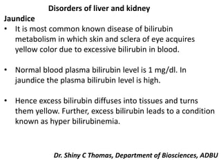
1. Disorders of liver and kidney
Jaundice
• It is most common known disease of bilirubin
metabolism in which skin and sclera of eye acquires
yellow color due to excessive bilirubin in blood.
• Normal blood plasma bilirubin level is 1 mg/dl. In
jaundice the plasma bilirubin level is high.
• Hence excess bilirubin diffuses into tissues and turns
them yellow. Further, excess bilirubin leads to a condition
known as hyper bilirubinemia.
Dr. Shiny C Thomas, Department of Biosciences, ADBU
2. • Thus the characteristic signs of jaundice are
hyperbilirubinemia and yellow colored skin and sclera.
• Based on clinical causes jaundice is classified into pre
hepatic jaundice, hepatic jaundice and post hepatic
jaundice.
• However jaundice (Icterus) may be due to several
underlying diseases.
3. Pre hepatic or hemolytic jaundice
• It is due to excessive breakdown of erythrocytes. This
leads to increased production of bilirubin.
• But liver cells are unable to conjugate all bilirubin formed.
Hence unconjugatedbilirubin level of plasma is elevated.
• Excessive breakdown of RBC occurs in
hemglobinopathies, hereditary spherocytosis,
incompatible blood transfusion and in malaria.
• Administration of sulfonamides, aspirin and primaquine
can cause excessive breakdown of RBC in glucose-6-
phosphate dehydrogenase deficiency.
4. Hepatic or hepatocellular jaundice
• It is due to damaged hepatocytes. Poisons like
chloroform, carbon tetrachloride, phosphorus,
antibiotics, amanita mushroom poison and hepatitis virus
can damage parenchymal cells of liver. In cirrhosis also
liver cell damage occurs.
• Damaged hepatocytes are unable to perform functions.
So in hepatic jaundice liver cells are unable to conjugate or
secrete bilirubin though the production of bilirubin is as
usual.
• If conjugation of bilirubin is impaired unconjugated
bilirubin in plasma is elevated .
5. • If secretion of conjugated bilirubin is impaired
conjugated bilirubin in plasma is elevated.
• Therefore in hepatic jaundice appreciable amounts of
conjugated as well as unconjugated bilirubin are present
in plasma.
Post hepatic or obstructive jaundice
• It is due to obstruction of bile duct.
• Gall stones and cancer of head of pancreas can cause
obstruction of bile duct.
• Due to blockage of bile duct conjugated bilirubin
secreted by liver returns to blood.
• Hence in obstructive jaundice conjugated bilirubin in
plasma is elevated.
Cholestatic jaundice is term used to indicate all forms of
extrahepatic or post hepatic obstructive jaundice.
8. Vanden Bergh reaction and Jaundice
• Since bilirubin level is elevated in all forms of jaundice
measurement of serum bilirubin is useful in the diagnosis
and management of jaundice.
• Vanden Bergh devised a method based on Ehrlichs
reaction for measurement of bilirubin in plasma. It
involves coupling of diazotized sulphanilic acid (diazo
reagent) and bilirubin to produce a reddish purple azo
compound.
• It consists of two parts (a) Direct Vanden Bergh reaction
and (b) Indirect Vanden Bergh reaction.
9. Urine bilirubin in Jaundice
• Normal urine does not contain bilirubin because normal blood
contains water insoluble unconjugated bilirubin which can not be
filtered at glomerulus.
• Bilirubin is excreted in urine in hepatic and obstructive jaundice
because conjugated bilirubin level in plasma is above renal
threshold value in these conditions.
• (Obstructive jaundice is a specific type of jaundice where symptoms develop
due to a narrowed or blocked bile duct or pancreatic duct, preventing the
normal drainage of bile from the bloodstream into the intestines)
• However bilirubin is absent in urine in hemolytic jaundice.
Excretion of bilirubin urine is called as choluria.
• So hepatic and obstructive jaundice are called as choluric jaundice
where as hemolytic jaundice is called as acholuric jaundice.
10. Urine Urobilinogen in Jaundice
• About 4 mg of urobilinogen is excreted in urine per day.
• The excretion of urobilinogen depends on amount of
bilirubin entering intestine which in turn depends on
amount of bilirubin formed.
• In obstructive jaundice urobilinogen is not found in urine
because bilirubin can not enter intestine.
• In hemolytic jaundice urine urobilinogen is more
because of increased production of bilirubin.
Urine bilirubin and Urobilinogen in Jaundice
Combination of urine bilirubin and urobilinogen is useful in
differential diagnosis of jaundice.
11. Urobilinogen is a colorless by-product of bilirubin reduction.
• It is formed in the intestines by bacterial action on
bilirubin. About half of the urobilinogen formed is
reabsorbed and taken up via the portal vein to the liver,
enters circulation and is excreted by the kidney.
• Increased amounts of bilirubin are formed in hemolysis,
which generates increased urobilinogen in the gut.
• In liver disease (such as hepatitis), the intrahepatic
urobilinogen cycle is inhibited also increasing
urobilinogen levels.
• Urobilinogen is converted to the yellow
pigmented urobilin apparent in urine
12. Fatty livers
1. Liver contains about 5% lipid. Of this, about 1/4 is
triglyceride.
2. Extensive accumulation of lipid in the liver leads to
condition known as fatty liver. In the fatty livers, the lipid
content increases to 25-30%. Further, triglycerides and fatty
acid may occupy entire cytoplasm of hepatocyte.
• Presence of bilirubin in urine without urobilinogen
suggests obstructive jaundice.
• Absence of bilirubin in urine with increased urobilinogen
suggest hemolytic jaundice.
13. Several factors causes accumulation of lipid in liver
1. Raised plasma free fatty acid level
• When there is a mobilization of fat from adipose and
extrahepatic tissues plasma free fatty acid level
increases. Liver takes up increasing amounts of fatty
acids and esterifies them.
• Hence rate of triglyceride synthesis is more.
• However, the synthesis of VLDL occurs only at normal
rate. As a result triglycerides accumulate and cause fatty
liver.
• The plasma free fatty acid level is elevated in (a)
starvation (b) diabetes (c) high fat diet (d) carnitine
deficiency. Hence, in these conditions fatty liver occurs.
14. Carnitine is the generic term for a number of compounds that include L-carnitine,
acetyl-L-carnitine, and propionyl-L-carnitine [1,2]. Carnitine plays a critical role in
energy production. It transports long-chain fatty acids into the mitochondria so they
can be oxidized ("burned") to produce energy
15. 2. Metabolic block in the production of lipoproteins
• Block in VLDL formation causes fatty liver even though
the rate of triglyceride synthesis is normal because VLDL
transports triglyceride from liver to extra hepatic tissues.
• VLDL formation may be blocked if substance (s) required
for its formation are deficient. However, when deficient
substances are supplied fat accumulation cease.
16. 3. Lipotropic factors
• They are compounds that relieve or prevent excess
accumulation of lipids in the liver. They are choline,
methionine and betaine.
• These lipotropic factors cure fatty liver due to choline or
methionine deficiency. Choline deficiency may result
from impaired transmethylation reactions associated
with methionine catabolism.
• Choline deficiency leads to block in choline dependent
phospholipid biosynthesis. This in turn impairs formation
of membranes needed for lipoprotein synthesis.
17. • Thus choline deficiency results in block in VLDL
formation and causes accumulation of fat in liver.
• Other noteworthy lipotropic factors are PUFA, vit E,
pyridoxine and pantothenic acid.
• The deficiency of any one of these substances causes
fatty liver. However, they can not prevent occurrence of
fatty liver due to choline deficiency.
18. 4. Toxic substances
• Several hepato toxic agents like carbon tetrachloride,
chloroform, phosphorus, lead, arsenic, alcohol and orotic
acid causes fatty liver.
• Substances, which inhibit protein synthesis like puromycin
and ethionine, a methionine analog also cause fatty liver.
Medical Importance
Effect of fatty liver When accumulation of lipid in liver
becomes chronic fibrotic changes takes place in hepatocytes,
which progress to cirrhosis and finally impaired liver function.
19. Functions of liver
1. Liver is an essential organ. It has diverse functions.
2. Liver is involved in secretion or excretion of several
components like bilirubin and bile
acids.
3. It is involved in the synthesis of plasma proteins and
blood clotting factors.
4. It is involved in metabolism of carbohydrates, lipids and
proteins.
5. It is sensitive to actions of several hormones.
6. It is involved in xenobiotics metabolism.
20. Functions of Kidney
1. Kidney is an essential organ. It has several diverse
functions.
2. Nephron is functional unit of kidney. It consists of
glomerulus and renal tubules
3. Kidney maintains water, electrolyte and acid base balance
of the body through filtration and reabsorption process.
Glomerulus is responsible for filtration and renal tubules are
involved in reabsorption. In addition renal tubules secretes
some solute molecules.
4. Kidney clears several non-protein metabolic waste products
like urea, uric acid, creatinine etc., from circulation.
5. Kidney produces erythropoietin, calcitriol, renin and
prostaglandins.
21. Since glomerulus and renal tubules are major functional
units of kidney most of the kidney function tests done to
assess renal damage are based on either function of
glomerulus or renal tubules.
22. Inulins and Urea clearance
• Inulin are a group of naturally occurring polysaccharides produced
by many types of plants, industrially most often extracted
from chicory.
• The inulins belong to a class of dietary fibers known as fructans.
Inulin is used by some plants as a means of storing energy and is
typically found in roots or rhizomes.
• Most plants that synthesize and store inulin do not store other
forms of carbohydrate such as starch. I
• n the United States in 2018, the Food and Drug
Administration approved inulin as a dietary fiber ingredient used to
improve the nutritional value of manufactured food products.
• Using inulin to measure kidney function is the "gold standard" for
comparison with other means of estimating glomerular filtration
rate.
23. Abnormalities of Nitrogen metabolism
Uraemia
It is the condition of a raised plasma urea concentration and
is almost always accompanied by an elevated creatinine
concentration.
Urea is derived in the liver from amino acids and from
protein, either from the diet or from tissues.
The normal kidney can excrete large amounts of urea. If the
rate of production exceeds the rate of clearance, plasma
concentrations rise.
24. The rate of production is accelerated by:
• a high-protein diet,
• absorption of amino acids and peptides from digested
blood
• increased catabolism due to starvation, tissue damage,
sepsis or steroid treatment.
• Renal failure- causes retention of water and salt
25. Effects of Renal Failure on the
Body Fluids—Uremia
The effect of complete renal
failure on the body fluids
depends on (1) water and food
intake and (2) the degree of
impairment of renal function.
Assuming that a person with
complete renal failure
continues to ingest the same
amounts of water and food, the
concentrations of different
substances in the extracellular
fluid are approximately those
shown in Figure.
26. Important effects include
(1) generalized edema resulting from water and salt
retention,
(2) acidosis resulting from failure of the kidneys to rid the
body of normal acidic products,
(3) high concentration of the nonprotein nitrogens—
especially urea, creatinine, and uric acid—resulting from
failure of the body to excrete the metabolic
end products of proteins, and
(4) high concentrations of other substances excreted by the
kidney, including phenols, sulfates, phosphates, potassium,
and guanidine bases.
This total condition is called uremia because of the high
concentration of urea in the body fluids.
27. Hyperuricemia is an excess of uric acid in the blood. Uric
acid passes through the liver, and enters your bloodstream.
Most of it is excreted (removed from your body) in your
urine, or passes through your intestines to regulate
"normal" levels.
Normal Uric acid levels are 2.4-6.0 mg/dL (female) and 3.4-
7.0 mg/dL (male). Normal values will vary from laboratory
to laboratory.
Also important to blood uric acid levels are purines. Purines
are nitrogen-containing compounds, which are made inside
the cells of your body (endogenous), or come from outside
of your body, from foods containing purine (exogenous).
28. Purine breaks down into uric acid. Increased levels of uric
acid from excess purines may accumulate in your tissues,
and form crystals. This may cause high uric acid levels in the
blood.
Uric acid formation may occur when the blood uric acid
level rises above 7 mg/dL. Problems, such as kidney stones,
and gout (collection of uric acid crystals in the joints,
especially in your toes and fingers), may occur.
What causes hyperuricemia?
Causes of high uric acid levels (hyperuricemia) can be
primary (increased uric acid levels due to purine), and
secondary (high uric acid levels due to another disease or
condition). Sometimes, the body produces more uric acid
than it is able to excrete.
29. Causes of high uric acid levels include:
Primary hyperuricemia
Increased production of uric acid from purine
Your kidneys cannot get rid of the uric acid in your
blood, resulting in high levels
Secondary hyperuricemia
• Certain cancers, or chemotherapy agents may cause an
increased turnover rate of cell death.
30. • Kidney disease - not be able to clear the uric acid out
of body system, thus causing hyperuricemia.
• Medications - can cause increased levels of uric acid in
the blood
• Endocrine or metabolic conditions -certain forms of
diabetes, or acidosis can cause hyperuricemia
• Elevated uric acid levels may produce kidney problems
31. What are some symptoms of hyperuricemia to look for?
• kidney problems, or
• gouty arthritis (an inflammation of a joint (called "gout")
• fever, chills, fatigue if you have certain forms of cancer,
and your uric acid levels are elevated (caused by tumor
lysis syndrome)
• You may have kidney problems (caused by formation of
kidney stones), or problems with urination
32. Porphyria a rare hereditary disease in which there is
abnormal metabolism of the blood pigment haemoglobin.
Porphyrins are excreted in the urine, which becomes dark;
other symptoms include mental disturbances and extreme
sensitivity of the skin to light.
Porphyria is a group of rare diseases in which chemical
substances called porphyrins accumulate, leading to either
skin changes or neurological symptoms or sometimes both.
33. • The body requires porphyrins to produce heme, which
carries oxygen in the blood, but in the porphyrias there is
a deficiency (inherited or acquired) of the enzymes that
transform the various porphyrins into others, leading to
abnormally high levels of one or more of these
substances.
34. Porphyrias are classified in two ways, by symptoms and by
pathophysiology. Symptomatically,
(1) acute porphyrias primarily cause brain and nerve
involvement, often with severe abdominal
pain, vomiting, neuropathy, and mental disturbances.
(2) Cutaneous porphyrias cause skin problems, often after
exposure to sunlight, because porphyrins react with light.
Physiologically, porphyrias are classified as hepatic or
erythropoietic based on the sites of accumulation
of heme precursors, either in the liver or in the bone
marrow and red blood cells.
35. Change in urine color before and after sun exposure Left
figure is urine of the first day. Right figure is urine after sun
exposure for 3 days. Urine color changed to “port wine”
color after sun exposure.
36. • Skin disease is encountered where excess porphyrins
accumulate in the skin.
• Porphyrins are photoactive molecules, and exposure to
light results in promotion of electrons to higher energy
levels.
• When these return to the resting energy level or ground
state, energy is released. This accounts for the property
of fluorescence typical of the porphyrins. This causes
local skin damage.
37. Nitrogen Balance
• Nitrogen is a main body component and is required for
both tissue protein synthesis and the production of
several nitrogenous compounds involved in a variety of
functions (hormones, immune mediators,
neurotransmitters, antioxidant defences, etc.).
• Thus, the body nitrogen content should be both
quantitatively and qualitatively normal, as well as
normally maintained, to ensure normal body functions.
38. Nitrogen balance is a measure of nitrogen input minus
nitrogen output.
Nitrogen Balance = Nitrogen intake - Nitrogen loss
Sources of nitrogen intake include meat, dairy, eggs, nuts
and legumes, and grains and cereals.
Examples of nitrogen losses include urine, faeces, sweat,
hair, and skin.
Blood urea nitrogen can be used in estimating nitrogen
balance, as can the urea concentration in urine.
Nitrogen is a fundamental component of amino acids, which
are the molecular building blocks of protein. Therefore,
measuring nitrogen inputs and losses can be used to study
protein metabolism.
39. Positive nitrogen balance is associated with periods of
growth, hypothyroidism, tissue repair, and pregnancy.
This means that the intake of nitrogen into the body is
greater than the loss of nitrogen from the body, so there is an
increase in the total body pool of protein.
Negative nitrogen balance is associated with burns, serious
tissue injuries, fevers, hyperthyroidism, wasting diseases, and
during periods of fasting.
This means that the amount of nitrogen excreted from the
body is greater than the amount of nitrogen ingested.
A negative nitrogen balance can be used as part of a clinical
evaluation of malnutrition.
40. • Nitrogen balance is the traditional method of
determining dietary protein requirements.
• Determining dietary protein requirements using nitrogen
balance requires that all nitrogen inputs and losses are
carefully collected, to ensure that all nitrogen exchange is
accounted for.
• In order to control nitrogen inputs and losses, nitrogen
balance studies usually require participants to eat very
specific diets (so total nitrogen intake is known) and stay
in the study location for the duration of the study (to
collect all nitrogen losses).
• Because of these conditions, it can be difficult to study
the dietary protein requirements of certain populations
using the nitrogen balance technique (e.g. children).
41. Nitrogen Balance
Since protein is the main source of nitrogen in body the
dietary protein must make up nitrogen lost from body to
maintain nitrogen balance.
If an individuals total nitrogen content of the urine and
faeces equals the amount of dietary nitrogen then the
individual is said to be in nitrogen balance or equilibrium
Faecal nitrogen (N) + Urinary nitrogen (N) = Dietary nitrogen
(N)
N output = N intake
i.e.,
N intake
-------------- =1
N output
42. In other words if the ratio of nitrogen intake to nitrogen
output is one then the individual is in nitrogen balance or
equilibrium.
43. Positive nitrogen balance
• If the ratio of N intake to N output is greater than one
then it is called as positive nitrogen balance or if the N
output is less than N intake then the individual is in
positive nitrogen balance.
• In the positive nitrogen balance most of dietary nitrogen
is retained in the body and less is eliminated from body.
• More over in positive nitrogen balance the tissue protein
content increases due to increased protein synthesis.
• Usually it occurs during growth, pregnancy, lactation and
post operative recovery.
44. Negative nitrogen balance
• If the nitrogen output is more than the N intake then the
individual is in negative nitrogen balance or if the ratio of
N intake to N output is less than one then the individual is
in negative nitrogen balance.
• In the negative nitrogen balance nitrogen lost is not
replaced by dietary nitrogen. It occurs in malnutrition and
starvation, uncontrolled diabetes mellitus and cancer.
• Menstruating women may have transient negative
nitrogen balance if proper replacement for nitrogen lost is
not possible.
• Physical exercise trainee may also have transient negative
nitrogen balance because of atrophy of muscle.
45. Protein minimum
It is the minimum amount of dietary protein required to
maintain nitrogen balance.
It is 1 gm/kg body weight per day.
However protein requirement also depends on the (a)
Protein quality (b) Carbohydrate and fat contents (c) Physical
activity.
Protein quality
• Essential amino acid content determines quality of a
protein.
• An ideal or a good quality protein is the one which has
amino acid composition of body protein synthesized at
any given time.
• Further an ideal protein must meet essential amino acid
requirement.
46. Carbohydrate and fat content
• If diet contains sufficient amounts of carbohydrate and
fat then use of protein for energy production is reduced.
• Hence protein requirement in diet is minimum.
• In contrast if the diet contains inadequate amounts of
carbohydrates and fats then use of protein for energy
production is more. This increases protein requirement in
diet.
Physical activity
Protein requirement increases with increases in physical
activity due to retention of nitrogen or increased muscle
protein in the body.
47. • If a protein is unable to maintain nitrogen balance then it
is a poor quality protein.
• However nitrogen balance does not indicate anything
about digestibility, essential amino acid content and
assimilation of products of digestion.
• Usually good quality protein maintain nitrogen balance if
taken in adequate amounts.
48. Nitrogen Balance:
A healthy adult eating a varied and plentiful diet is generally
in “Normal Nitrogen Balance” a state where the amount of
nitrogen ingested each day is balanced by the amount
excreted resulting no net change in the amount of the body
Nitrogen. In a well fed condition, excreted nitrogen comes
from digestion of excess protein or from normal turnover.
Protein turnover (Synthesis and degradation)
Under some conditions. The body is either in negative or
positive nitrogen balance. In negative nitrogen balance more
nitrogen is excreted than ingested. This occurs in starvation
and certain diseases.
49. During starvation the carbon skeleton of most amino acids
from proteins fed in to gluconeogenesis to maintain the
blood glucose level ; in this process ammonia is released and
excreted mostly as urea and is not reincorporated in to
protein.
Positive nitrogen balance occurs in pregnancy and during
feeding after starvation.
A diet deficient in an essential amino acid also leads to a
negative nitrogen balance since body proteins are degraded
to provide the deficient essential amino acid.
Positive nitrogen balance occurs in growing children who are
increasing their body weight and incorporating more amino
acids in to protein than they breakdown.
50. Cysteine and Arginine are not essential in adults but
essential in children because they are synthesized from
Methionine and ornithine. These amino acids are readily
available in adults but limited in children.
Negative Nitrogen balance occurs in injury when there is net
destruction of tissue and in major trauma or illness.