
Recommended
More Related Content
Similar to Unit-2_first_year_bkg_final[1].pptx
Similar to Unit-2_first_year_bkg_final[1].pptx (20)
More from ChevallaMaheshwari
More from ChevallaMaheshwari (12)
Recently uploaded
Recently uploaded (20)
Unit-2_first_year_bkg_final[1].pptx
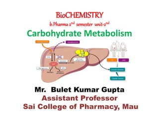
- 1. BioCHEMISTRY b.Pharma 2nd semester unit-2nd Carbohydrate Metabolism Mr. Bulet Kumar Gupta Assistant Professor Sai College of Pharmacy, Mau
- 2. Krebs Cycle Introduction The Krebs cycle or TCA cycle (tricarboxylic acid cycle) or Citric acid cycle is a series of enzyme catalysed reactions occurring in the mitochondrial matrix, where acetyl-CoA is oxidised to form carbon dioxide and coenzymes are reduced, which generate ATP in the electron transport chain. Krebs Cycle is a part of Cellular Respiration Cellular respiration is a four-stage process. In the process, glucose is oxidised to carbon dioxide and oxygen is reduced to water. The energy released in the process is stored in the form of ATPs. 36 to 38 ATPs are formed from each glucose molecule.
- 3. The four stages are: 1) Glycolysis 2) Formation of Acetyl Co-A Note-This is linked reaction because it links Glycolysis with Krebs cycle. 3) Krebs cycle 4) Electron Transport Chain (ETC)
- 5. Krebs Cycle Steps It is an eight-step process. Krebs cycle or TCA cycle takes place in the matrix of mitochondria under aerobic condition. Step 1: The first step is the condensation of acetyl CoA with 4-carbon compound oxaloacetate to form 6C citrate, coenzyme A is released. The reaction is catalysed by citrate synthase. Step 2: Citrate is converted to its isomer, isocitrate. The enzyme aconitase catalyses this reaction. Step 3: Isocitrate undergoes dehydrogenation and decarboxylation to form 5C 𝝰-ketoglutarate. A molecular form of CO2 is released. Isocitrate dehydrogenase catalyses the reaction. It is an NAD+ dependent enzyme. NAD+ is converted to NADH.
- 6. Step 4: 𝝰-ketoglutarate undergoes oxidative decarboxylation to form succinyl CoA, a 4C compound. The reaction is catalyzed by the 𝝰- ketoglutarate dehydrogenase enzyme complex. One molecule of CO2 is released and NAD+ is converted to NADH. Step 5: Succinyl CoA forms succinate. The enzyme succinyl CoA synthetase catalyses the reaction. This is coupled with substrate-level phosphorylation of GDP to get GTP. GTP transfers its phosphate to ADP forming ATP. Step 6: Succinate is oxidised by the enzyme succinate dehydrogenase to fumarate. In the process, FAD is converted to FADH2. Step 7: Fumarate gets converted to malate by the addition of one H2O. The enzyme catalysing this reaction is fumarase.
- 7. Step 8: Malate is dehydrogenated to form oxaloacetate, which combines with another molecule of acetyl CoA and starts the new cycle. Hydrogens removed, get transferred to NAD+ forming NADH. Malate dehydrogenase catalyses the reaction. Location: Krebs cycle occurs in the mitochondrial matrix. Note that 2 molecules of Acetyl Co-A are produced from oxidative Decarboxylation of 2 Pyruvates so two cycles are required per glucose molecule. To summarize, for complete oxidation of a glucose molecule, Krebs cycle yields 4 CO2, 6NADH, 2 FADH2 and 2 ATPs.
- 8. Significance of Krebs Cycle 1) Krebs cycle or Citric acid cycle is the final pathway of oxidation of glucose, fats and amino acids 2) Many animals are dependent on nutrients other than glucose as an energy source. 3) Amino acids (metabolic product of proteins) are deaminated and get converted to pyruvate and other intermediates of the Krebs cycle. 4) Fatty acids undergo 𝞫-oxidation to form acetyl CoA, which enters the Krebs cycle. 5) It is the major source of ATP production in the cells. 6) It plays an important role in gluconeogenesis and lipogenesis and interconversion of amino acids 7) Many intermediate compounds are used in the synthesis of amino acids & nucleotides. 8) The genetic defects of the Krebs cycle enzymes are associated with neural damage.
- 9. Energetic of Aerobic Respiration Glycolysis Produced -------- 8 ATP Oxidation of Pyruvic Acid -------- 6 ATP Krebs Cycle -------- 24 ATP Total -------- 38 ATP Note- 1 FADH (Flavin adenine dinucleotide) =2 ATP 1 NADP (Nicotinamide adenine dinucleotide) = 3 ATP
- 10. GLUCONEOGENESIS The synthesis of glucose from non-carbohydrate compounds is known as Gluconeogenesis. The major substrates/precursors for Gluconeogenesis are lactate, Pyruvate, Glucogenic amino acids, propionate and glycerol. Location of Gluconeogenesis- Gluconeogenesis occurs mainly in the Cytosol Gluconeogenesis works in the opposite direction of glycolysis, which creates glucose from pyruvate, lactate, and glucogenic amino acids. It’s also known as Neoglucogenesis. It’s a universal pathway found in humans, animals, plants, fungus, and other living species. Gluconeogenesis Functions Human systems create glucose to keep blood sugar levels in check. Because cells use glucose to create the energy component adenosine triphosphate, blood glucose levels must be maintained (ATP).
- 11. When a person hasn’t eaten in a while, such as during a starvation, gluconeogenesis takes place.
- 12. Importance of Gluconeogenesis 1) During deprivation, the gluconeogenesis cycle is important for blood glucose regulation. 2) Many cells and tissues, including RBCs, neurons, skeletal muscle, the medulla of the kidney, and embryonic tissue, rely on glucose to meet their energy needs. 3) The Neoglucogenesis cycle removes metabolites such as lactate (produced by muscles and RBCs) and glycerol from the bloodstream (produced from adipose tissue).
- 13. Glycogen Metabolism Glycogenolysis is a metabolic process that converts glycogen from the muscles and liver to its monosaccharide form, glucose. Animals store glucose as glycogen, which is broken down in a process called Glycogenolysis. Glycogen is a glucose polysaccharide stored in the muscles and liver. The body utilises this to produce ATP (adenosine triphosphate), an organic substance that provides energy to drive various activities in living cells. Low levels of ATP within live cells trigger the glycogenolysis process. When the cells detect a low level of ATP, the liver and muscles liberate glycogen and break it down into glucose or simple sugar.
- 14. Which are then used to produce ATP. Location Glycogenolysis occurs in the cytoplasm of cells in the liver, muscles, and adipose tissue. The liver breaks down the glycogen to maintain the glucose level in the blood. The muscle cells break down the glycogen to conserve the energy required for the contraction of muscles.
- 15. Functions and Significance 1) The liver and muscle tissue cells undergo glycogenolysis in response to neurological and hormonal impulses. Glycogenolysis, in particular, is crucial for controlling blood glucose levels and the fight-or-flight response. 2) Glycogen breakdown in the muscle cells (myocytes) serves as an instant resource of glucose-6-phosphate for the process of glycolysis, which produces energy for muscle contraction. 3) The process of glycogenolysis differs in liver cells or hepatocytes. The liver does not immediately utilize the glucose that is created during glycogenolysis in the liver. 4) Glycogen is a form of energy storage in animals similar to starch.
- 16. Glycogen storage disease (GSD) Glycogen storage disease (GSD) is a rare metabolic disorder where the body is not able to properly store or break down glycogen, a form of sugar or glucose. GSD affects the liver, muscles and other areas of the body, depending on the specific type. Children with GSD are missing one of the several enzymes that break down glycogen, and glycogen can build up in the liver, causing problems in the liver, muscles or other parts of the body. When the enzyme deficiency affects the liver, it leads to low blood glucose levels (also called Hypoglycemia) during periods of fasting. GSD is hereditary, meaning it is passed down from parents to children.
- 19. The most common types of GSD include:- 1. Glycogen storage disease type I (GSD I), also known as Von Gierke disease, accounts for about 25 percent of all children with GSD. Symptoms typically appear when an infant is 3 to 4 months of age and may include hypoglycemia (low blood sugar, which can cause fatigue, constant hunger. The liver and sometimes the kidneys swell due to built-up glycogen. 1. Glycogen storage disease type III (GSD III), also known as Cori disease or Forbes disease, causes glycogen to build up in the liver and muscles. Symptoms typically appear within the first year of life. Children with this type of GSD may have a swollen belly, delayed growth, and weak muscles.
- 20. 3. Glycogen storage disease type IV (GSD IV), also known as Andersen disease, is one of the most serious types of GSD. Symptoms typically appear in a child’s first month of life and include failure to gain weight or grow at an expected rate. This type of GSD often leads to cirrhosis of the liver and can affect the heart and other organs as well.
- 21. Hexose Monophosphate (HMP) Shunt The Hexose Monophosphate (HMP) shunt, also known as the pentose phosphate pathway or Phosphogluconate pathway, is a metabolic pathway that runs parallel to Glycolysis. This pathway produces NADPH and intermediates required for the synthesis of nucleic acids and amino acids. Features of the HMP Shunt •It is an anabolic pathway that takes place in the cytosol for most organisms. •The pathway takes place in two distinct phases: oxidative and non- oxidative phases. •The reactions of this pathway are enzyme catalysed.
- 22. •The products obtained from the pathway include NADPH, which is used in biosynthesis in cells, ribose-5-phosphate, which is used in the synthesis of nucleic acid and nucleotides, and erythrose-4-phosphate, which is used for the synthesis of aromatic amino acids. •The HMP shunt is a tightly controlled metabolic pathway that is connected with other pathways, such as glycolysis and Gluconeogenesis, depending upon the metabolic needs of the body. •The pathway takes place in two distinct phases: oxidative and non- oxidative phases.
- 23. Phases of the HMP Shunt Oxidative Phase In this phase, NADPH is produced by the reduction of two molecules of NADP+. The energy for this production is utilised by the conversion of glucose-6-phosphate to ribulose-5-phosphate. The three steps of the oxidative phase of the HMP shunt are: Non-Oxidative Phase The non-oxidative phase can be summarised in five steps:
- 25. Significance of HMP Shunt The HMP shunt pathway is significant because it provides NADPH and important intermediated products for the synthesis of biomolecules. Some of the points of significance are: NADPH performs several functions in the body, such as: It takes part in the synthesis of steroids and fatty acids. It is an important component within phagolysosomes in the immune response. Glutathione is reduced by NADPH in the presence of glutathione reductase. The glyceraldehyde 3-phosphate and fructose 6-phosphate produced in the pathway are intermediates for glycolysis and gluconeogenesis.
- 26. Clinical Significance of HMP Shunt Deficiency of Glucose-6-Phosphate Dehydrogenase: The deficiency of this enzyme which is required in the initial steps for the production of NADPH, affects the red blood cells. Conversely, this deficiency can provide resistance to the malarial parasite Plasmodium falciparum. This is possible because the cell membranes of the RBCs are weakened, and the parasite cannot continue its life cycle. Diagnosis of Thiamine Deficiency: It is diagnosed by administering thiamine to suspected patients and observing the activity of transketolase enzymes in the RBCs. If the enzyme shows high activity, the deficiency of thiamine (vitamin B1) is confirmed.
- 27. Hormonal regulation of blood glucose level and Diabetes mellitus. Diabetes mellitus is a disorder related to carbohydrate metabolism caused due to deficient secretion or utilization of insulin and is characterized by many symptoms collectively called Syndrome X. The pancreas secretes insulin and glucagon. Both hormones work in balance to play a vital role in regulating blood sugar levels. If the level of one hormone is higher or lower than the ideal range, blood sugar levels may spike or drop. The secreted insulin then activates the enzyme glycogen synthase which proceeds to form Glycogen, the polymer of glucose -------> Blood Glucose level falls.
- 29. •Once blood glucose levels start following the Alpha cells of islets of Langerhans of Pancreas start secreting the “Glucagon” which has inhibitory effect on glycogenesis and also causes breakdown of the Glycogen (Glycogenolysis) stored in liver hence increasing the blood sugar levels. •Delta cells of islets of Langerhans of Pancreas secrete “Somatostatin” which is a local hormone and controls secretion of other hormones of Pancreas. •Together, insulin and glucagon help maintain a state called homeostasis in which conditions inside the body remain steady. When blood sugar is too high, the pancreas secretes more insulin. When blood sugar levels drop, the pancreas releases glucagon to raise them.
- 30. Complications of Diabetes mellitus: The symptoms of high blood sugar include: •Frequent urination. •Excessive thirst (Polydipsia) •Feeling excessively hungry. •unexplained weight loss •slow healing •itchy, dry skin •increased likelihood of infections •headaches •fatigue
- 31. Electron transport chain An electron transport chain (ETC) is a series of protein complexes and other molecules that transfer electrons from electron donors to electron acceptors via redox reactions (both reduction and oxidation occurring simultaneously) and couples this electron transfer with the transfer of protons (H+ ions) across a membrane. The electrons that transferred from NADH and FADH2 to the ETC involves four multi-subunit large enzymes complexes and two mobile electron carriers. Many of the enzymes in the electron transport chain are membrane-bound. Electron Transport Chain is a series of compounds where it makes use of electrons from electron carrier to develop a chemical gradient.
- 32. Large amounts of ATP could be produced through a highly efficient method termed oxidative Phosphorylation. ATP is a fundamental unit of metabolic process. The electrons are transferred from electron donor to the electron acceptor leading to the production of ATP. It is one of the vital phases in the electron transport chain. Compared to any other part of cellular respiration the large amount of ATP is produced in this phase. When electrons are passed from one component to another until the end of the chain the electrons reduce molecular oxygen thus producing water.
- 34. Electron Transport Chain in Mitochondria Complex 1- It comprises enzymes consisting of iron-sulfur and FMN. Here two electrons are carried out to the first complex aboard NADH. FMN is derived from vitamin B2. Complex 2- FADH2 that is not passed through complex 1 is received directly from complex 2. The first and the second complexes are connected to a third complex through compound ubiquinone (Q). The Q molecule is soluble in water and moves freely in the hydrophobic core of the membrane. In this phase, an electron is delivered directly to the electron protein chain. The number of ATP obtained at this stage is directly proportional to the number of protons that are pumped across the inner membrane of the mitochondria.
- 35. Complex 3- The third complex is comprised of Fe-S protein, Cytochrome b, and Cytochrome c proteins. Cytochrome proteins consist of the heme group. Complex 3 is responsible for pumping protons across the membrane. It also passes electrons to the cytochrome c where it is transported to the 4th complex of enzymes and proteins. Here, Q is the electron donor and Cytochrome C is the electron acceptor. Complex 4- The 4th complex is comprised of cytochrome c, a and a3. There are two heme groups where each of them is present in cytochromes c and a3. The cytochromes are responsible for holding oxygen molecule between copper and iron until the oxygen content is reduced completely. In this phase, the reduced oxygen picks two hydrogen ions from the surrounding environment to make water.
- 36. Oxidative Phosphorylation Oxidative Phosphorylation is the process of ATP formation, when electrons are transferred by electron carriers from NADH or FADH2 to oxygen. Oxidative phosphorylation is the final step in cellular respiration. It occurs in the mitochondria. It is linked to a process known as electron transport chain. The electron transport system is located in the inner mitochondrial membrane. The electrons are transferred from one member of the transport chain to another through a series of redox reactions.
- 38. Substrate Level Phosphorylation Phosphorylation involves the transfer of phosphate group from one compound to other. Substrate level Phosphorylation is a direct Phosphorylation of ADP with a Phosphatase group by using the energy obtain from a coupled reaction. Occurs in cytoplasm (Glycolysis - due to aerobic and anaerobic condition) and in mitochondrial matrix (Krebs cycle).
- 39. Inhibitors of Elecron transport chain •Retinone •Piericidin-A •Barbiturates •Antimycins •Dimercaprol •Cyanides •Azide •Hydrogen sulphide •Carbon mono-oxide
- 40. Inhibitors of Oxidative Phosphorylation •Oligomycin •Rutamycin •Atractylate