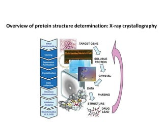
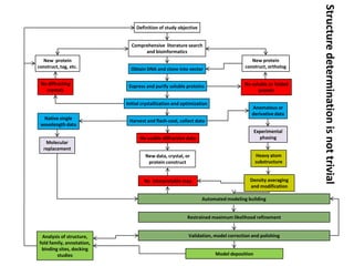
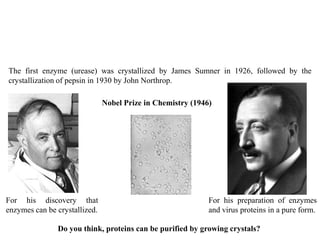
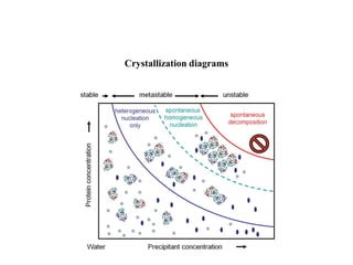
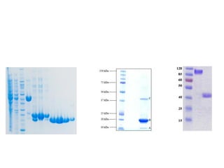
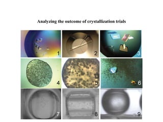
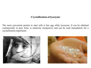
Crystallizing proteins involves obtaining pure protein, determining initial crystallization conditions through trials, and optimizing crystals for diffraction analysis. Key factors that affect crystallization include protein purity and concentration, pH, temperature, buffers, and precipitation techniques such as vapor diffusion. Lysozyme is commonly used to optimize crystallization methods due to its low cost and ease of obtaining crystals under various conditions including salts or polymers. The goal is to produce well-diffracting crystals to determine the 3D protein structure.
The document discusses various computational methods for predicting the three-dimensional structure of proteins from their amino acid sequences. It describes homology modeling, which predicts structures based on known protein structural templates that share sequence homology. It also covers threading/fold recognition and ab initio modeling, which predict structures without templates by using physicochemical principles or energy minimization approaches. Key steps and programs used in each method are outlined.
This document summarizes a seminar presentation on 2D electrophoresis. 2D electrophoresis is a technique used to separate mixed proteins based on their isoelectric point and mass. It involves two sequential electrophoretic steps: iso-electric focusing to separate proteins by charge, followed by SDS-PAGE to separate by molecular weight. The document describes the principles, methods, applications and references for 2D electrophoresis.
DNA methylation is an epigenetic mechanism that involves the addition of a methyl group to cytosine residues in DNA. It is catalyzed by DNA methyltransferase enzymes and plays a key role in gene expression and cellular differentiation. Aberrant DNA methylation, including both hypermethylation and hypomethylation, has been associated with cancer development by disrupting gene expression. Detection of DNA methylation patterns can provide insights into cancer biology and may have applications as a diagnostic tool.
Cot curve dispersed repeated DNA or interspersed repeated DNA tandem repeated DNA Long interspersed repeat sequences (LINEs) Short interspersed nuclear elements (SINEs) satellite, minisatellite and microsatellite DNA Variable Number Tandem Repeat (or VNTR)
Sequence tagged sites (STSs) are short DNA sequences that can be used as genetic markers. STSs were introduced in 1989 as a way to map genes along chromosomes using PCR. They serve as landmarks on physical maps of genomes. STSs are mapped by breaking genomes into fragments, replicating the fragments in bacterial cells to create libraries, and using PCR to determine which fragments contain STSs. Different types of STS markers include microsatellites, SCARs, CAPs, and ISSRs, each of which has distinct characteristics and applications in genetic mapping, population studies, and other areas.
The document discusses protein-protein interactions (PPIs) and methods used to study them. It defines PPIs as physical contacts between two or more proteins through biochemical or electrostatic forces. It describes different types of PPIs including homo-oligomers, hetero-oligomers, covalent and non-covalent interactions. Common methods to study PPIs are also summarized, such as yeast two-hybrid systems, co-immunoprecipitation, and protein interaction databases. The applications and importance of PPI research are mentioned including roles in various cellular processes and diseases.
What is Genome,Genome mapping,types of Genome mapping,linkage or genetic mapping,Physical mapping,Somatic cell hybridization
Radiation hybridization ,Fish( =fluorescence in - situ hybridization),Types of probes for FISH,applications,Molecular markers,Rflp(= Restriction fragment length polymorphism),RFLPs may have the following Applications;Advantages of rflp,disAdvantages of rflp, Rapd(=Random amplification of polymorphic DNA),Process of rapd, Difference between rflp &rapd
2D-PAGE is a technique used to separate complex protein mixtures based on isoelectric point and molecular weight. It involves two sequential steps - isoelectric focusing and SDS-PAGE. In isoelectric focusing, proteins are separated based on their isoelectric point in an immobilized pH gradient. They are then separated by SDS-PAGE based on their molecular weight. The separated proteins can then be visualized through staining and identified through mass spectrometry. While useful for proteomic analysis, 2D-PAGE has limitations such as low reproducibility and dynamic range.
SDS-PAGE is a common method to separate proteins based on their molecular mass. The proteins are first denatured by SDS, which coats each protein with a uniform negative charge. This causes the proteins to migrate towards the anode during electrophoresis, with smaller proteins moving faster through the discontinuous gel system. Two-dimensional gel electrophoresis combines isoelectric focusing, which separates proteins based on isoelectric point, with SDS-PAGE to allow better separation of proteins with similar masses.
This document provides an overview of functional genomics and methods for transcriptome analysis. It discusses two main approaches - sequence-based approaches like expressed sequence tags (ESTs) and serial analysis of gene expression (SAGE), and microarray-based approaches. For sequence-based approaches, it describes how ESTs can provide gene discovery and expression information but have limitations. It outlines the SAGE methodology and gene index construction to organize EST data. For microarrays, it summarizes the basic workflow including sample preparation, hybridization, image analysis and data normalization to identify differentially expressed genes through statistical tests.
ESTs are short sequences of DNA derived from cDNA clones that represent gene expression in particular cells or tissues. They provide a simple and inexpensive way to discover new genes and map their positions in genomes. To create an EST, mRNA is converted to cDNA and then sequenced, yielding short expressed DNA sequences. ESTs are deposited in public databases like NCBI's dbEST and can help identify genes, construct genome maps, and characterize expressed genes through clustering, assembly, and mapping to genomic sequences. However, isolating mRNA from some tissues can be difficult and ESTs alone do not indicate the genes they were derived from.
Protein-protein interactions are important for many biological processes. There are various types of interactions depending on their composition and duration. Methods to study interactions include yeast two-hybrid, co-immunoprecipitation, affinity chromatography, and chromatin immunoprecipitation. Databases such as IntAct and MINT provide repositories for protein interaction data.
Structural genomics is a field that aims to determine the 3D structures of all proteins encoded by a genome. It involves determining structures on a large scale using techniques like X-ray crystallography and NMR. This allows identification of novel protein folds and potential drug targets. Comparative genomics compares genomic features between organisms and provides insights into evolution and conserved sequences and functions. It is a key tool in fields like medicine and agriculture.
Histones are basic proteins found in eukaryotic cell nuclei that are responsible for DNA folding and chromatin formation. There are two main classes of histones: core histones and linker histones. Core histones include the H2A, H2B, H3, and H4 families, while linker histones include H1 and H5. Histone modification, including acetylation, methylation, and phosphorylation, can impact gene expression by altering the accessibility of DNA. Acetylation reduces the positive charge of histone tails, weakening their interaction with DNA and making it more accessible for transcription. Methylation and phosphorylation can also influence chromatin structure and cellular activity.
Scoring system is a set of values for qualifying the set of one residue being substituted by another in an alignment.
It is also known as substitution matrix.
Scoring matrix of nucleotide is relatively simple.
A positive value or a high score is given for a match & negative value or a low score is given for a mismatch.
Scoring matrices for amino acids are more complicated because scoring has to reflect the physicochemical properties of amino acid residues.
SAGE (Serial analysis of Gene Expression)talhakhat
SAGE (Serial Analysis of Gene Expression) is a technique that allows for the rapid and comprehensive analysis of gene expression patterns in a given cell population. It works by isolating mRNA, synthesizing cDNA, ligating short sequence tags to the cDNA, and then counting the number of times each tag is observed to quantify gene expression levels. The tags are concatenated and sequenced to generate vast amounts of data that must be analyzed computationally to identify which genes particular tags correspond to and to compare expression profiles between cell types. SAGE provides an overview of a cell's complete transcriptional activity and has been applied to study differences in cancer vs normal cells and to identify targets of oncogenes and tumor suppressor genes.
Structural genomics aims to determine the 3D structure of all proteins in a genome. It uses high-throughput methods like X-ray crystallography and NMR on a genomic scale. This allows determination of protein structures for entire proteomes. It provides insights into protein function and can aid drug discovery by identifying potential drug targets like in Mycobacterium tuberculosis. Structural genomics leverages completed genome sequences to clone and express all encoded proteins for structural characterization.
Automated sequencing of genomes require automated gene assignment
Includes detection of open reading frames (ORFs)
Identification of the introns and exons
Gene prediction a very difficult problem in pattern recognition
Coding regions generally do not have conserved sequences
Much progress made with prokaryotic gene prediction
Eukaryotic genes more difficult to predict correctly
Genomic and cDNA libraries are constructed to isolate genes of interest from organisms. Genomic libraries contain total chromosomal DNA while cDNA libraries contain mRNA from specific cell types. DNA is digested and ligated into vectors to clone fragments. Libraries are screened using probes and PCR to identify clones containing genes of interest. cDNA libraries are useful for studying eukaryotic gene expression as they contain mRNA from specific cells. Thousands of clones may need to be screened to have high probability of isolating a particular gene fragment.
Open reading frame is part of reading frame that contains no stop codons or region of amino acids coding triple codons.
ORF starts with start codon and ends at stop codon.
DNA Protein interaction occur when a protein binds a molecule of DNA, often to regulate the biological function of DNA, usually the expression of a gene. DNA Protein interactions play very vital roles in any living cell. It controls various cellular processes which are very essential for living beings, viz. replication, transcription, recombination, DNA repair etc. There are several types of proteins found in a cell.Direct recognition occurs when the amino acid side chains of a protein interact with specific DNA bases.
Most protein-DNA interactions are mediated by direct physical interaction (hydrogen bonding or hydrophobic interactions) between the protein and the DNA base pairs.
DNA-binding proteins can be identified by many experimental techniques such as chromatin immunoprecipitation on microarrays, X-ray crystallography and nuclear magnetic resonance (NMR).
DNA sequencing is a process to determine the order of nucleotides in a DNA molecule. It was discovered in the 1970s by scientists like Frederick Sanger who developed the chain termination method. This method involves DNA replication with modified nucleotides that cause the growing DNA strand to terminate at that point. The fragments are then separated by size to reveal the sequence. Automated sequencing now uses fluorescent dyes and capillary electrophoresis for faster and higher throughput sequencing. DNA sequencing has applications in medicine, forensics, and agriculture.
Single strand conformation polymorphismNivethitha T
Single-strand conformation polymorphism (SSCP) is a technique that detects variations in single-stranded DNA sequences. It involves PCR amplification of a target region, denaturing the PCR products to generate single strands, and separating the single strands on a non-denaturing gel based on differences in electrophoretic mobility caused by variations in nucleotide sequence. This allows sequences to be distinguished and variants detected without sequencing. SSCP is useful for discovering new polymorphisms and detecting mutations for diagnostic applications.
The key forces stabilizing nucleic acid structure are hydrogen bonding, base stacking, hydrophobic interactions, and ionic bonding. Hydrogen bonding occurs between complementary nucleotide bases on opposite strands. Base stacking involves hydrophobic interactions between stacked aromatic nucleotide bases within each strand. Hydrophobic interactions bury hydrophobic bases in the core of the double helix, increasing stability. Ionic interactions between phosphate groups and counterions in solution also stabilize the structure.
Homology modeling is a technique used to predict the 3D structure of a protein based on the alignment of its amino acid sequence to known protein structures. It relies on the observation that structure is more conserved than sequence during evolution. The key steps in homology modeling include: 1) identifying a template structure through sequence alignment tools like BLAST, 2) correcting any errors in the initial alignment, 3) generating the protein backbone based on the template structure, 4) modeling any loops or missing regions, 5) adding side chains, 6) optimizing the model structure energetically, and 7) validating that the final model matches the template structure and has correct stereochemistry. Homology modeling is useful for applications like structure-based drug design
A gene knockout is a genetic technique in which one of an organism's genes is made inoperative ("knocked out" of the organism). However, gene knockout can also refer to the gene that is knocked out or the organism that carries the gene knockout. Knockout organisms or simply knockouts are used to study gene function, usually by investigating the effect of gene loss. Researchers draw inferences from the difference between the knockout organism and normal individuals.
Secondary structure prediction tools analyze a protein's amino acid sequence to predict its 3D structure and function. These tools use various methods like Chou-Fasman, GOR, neural networks, and hidden Markov models to identify alpha helices and beta sheets based on characteristics like residue propensity values, sequence homology, and patterns in windows of amino acids. Accurate prediction of secondary structure is important for determining a protein's tertiary structure and biological role.
This document discusses different levels of protein structure from primary to quaternary structure. It explains that primary structure refers to the amino acid sequence of a protein. Secondary structure describes local folding patterns like alpha helices and beta sheets. Tertiary structure is the overall 3D shape of a single protein chain that results from folding. Quaternary structure involves the shape and interactions of multiple protein subunits. The document provides examples and diagrams to illustrate each level of structure and how they relate to determining a protein's function.
This document provides an overview of nanotechnology, including definitions, history, applications, and health impacts. Nanotechnology involves engineering at the molecular level between 1 to 100 nanometers. It has a variety of applications, including carbon nanotubes, molecular electronics, quantum dots, and more efficient energy generation. While many nanotechnology applications pose no new health risks, some free nanoparticles may have negative health impacts due to their small size and chemical properties. The document outlines the history and development of nanotechnology from 1959 to present.
Flow, Crystallisation and Continuous Processingmalcolmmackley
This presentation reviews the way flow can effect crystallisation. The presentation also reviews different ways continuous processing can be achieved. Continuous crystallisation is of relevance to a number of technologies including pharmaceutical manufacture.
Nanotechnology is the purposeful manipulation of matter on an atomic scale. Materials created in this manner often exhibit unique physical and chemical properties, which have useful applications in various industries. A growing use for some types of engineered nanomaterials is in the area of environmental remediation, termed nanoremediation. While this technique appears to be effective for cleanup, there are still many unanswered questions regarding its long-term impact to environmental quality and human health. No long-term studies exist regarding the potential environmental impact of nanoremediation. While animal studies have shown the potential for adverse health effects, limited data regarding human health are available. The US Environmental Protection Agency is currently adapting existing regulations to cover the use of nanomaterials in remediation, but this approach is limited. Many questions still remain regarding fate and transport, verification of clean-up, and potential occupational and community exposures.
This document provides an overview of nanotechnology including definitions, history, and applications. It defines nanotechnology as the design and manipulation of materials at the nanoscale (1-100 nm) to produce novel properties. Nanomaterials are characterized by their small size and increased surface area. The document outlines the bottom-up and top-down approaches to nanotechnology and gives examples of applications in various fields including medicine, computing, and the environment. It specifically discusses applications of nanotechnology for water purification such as detection, filtration membranes, and biofilm removal. The document concludes with noting both the promise and environmental implications of nanotechnology that require further study.
nanotechnology has entered the sphere of water treatment processes. Many different types of nanomaterial’s are being evaluated and also being used in water treatment process.
Desalination is a key market area. Vast majority of worlds water is salt water, and though technology has existed for years that enables the desalination of ocean water, it is often a very energy intensive procedure and therefore expensive
X-ray crystallography allows us to determine protein structures at the atomic level by visualizing the electron density map generated from X-ray diffraction data of protein crystals. Several steps are involved including growing high quality protein crystals, mounting crystals in the X-ray beam to collect diffraction data, solving the phase problem to produce an electron density map, building and refining an atomic model that fits the map, and validating the final protein structure. This technique provides insights into protein function and enables structure-based drug design.
Crystallization involves four main steps: obtaining pure protein samples, choosing a suitable buffer, increasing the protein solution to supersaturation for nucleation, and controlling crystal growth. Nucleation is the formation of small crystal nuclei from which crystals can grow. It is easier when the solution is highly supersaturated but too much supersaturation leads to many small crystals. Controlling factors like purity, concentration, temperature, pH, and additives can help optimize the crystallization process.
Protein Denaturation & Protein PurificationDebashish24
This document provides an overview of protein denaturation and protein purification. It discusses the types and levels of protein denaturation, as well as the factors that can cause denaturation. The basic steps of protein purification are described, including extraction, stabilization, separation techniques like precipitation, dialysis, and chromatography. Analytical techniques for verification are also mentioned. Protein denaturation involves changing the structure of a protein, while purification aims to isolate a single protein from a mixture for characterization and other purposes. Key techniques discussed include salting in/out, gel filtration, ion exchange, and affinity chromatography.
Inclusion bodies are aggregates of proteins that form when proteins are overexpressed in cells. They typically contain high levels of the overexpressed protein with little other cellular components. While inclusion bodies were traditionally thought to only contain misfolded proteins, some evidence indicates proteins in inclusion bodies can retain native structure. Several methods exist for recovering active proteins from inclusion bodies, including dilution, chromatography, and adding compounds to aid refolding. Fusion tags are often used to improve expression and purification of recombinant proteins by enhancing solubility, aiding detection and purification, and more. Enzymatic cleavage is commonly used to remove fusion tags after purification.
Protein purification involves isolating a protein of interest from a complex mixture through a series of separation processes. These processes exploit differences in protein properties like size, charge, and binding affinity. Key steps include cell lysis, precipitation, centrifugation, chromatography, and electrophoresis to separate the target protein from others in the sample. The purified protein can then be characterized and used for various analytical and preparative purposes.
The document provides guidance on preparing lysates for western blotting. It discusses important components of lysis buffers such as buffer systems, salt ions, chaotropic agents, protease inhibitors, and reductants. Tips are provided for optimizing protein extraction from cells and tissues, including using protease inhibitors, sonicating to remove nucleic acids, and fractionating samples. Maintaining consistent protein concentrations between samples and including loading controls are also recommended for successful western blots.
The document discusses methods for preparing tissue or cell extracts for protein separation and analysis. It describes various cell lysis buffers and their uses depending on the protein location. It also discusses steps to inhibit protein degradation during extraction, such as using protease inhibitors and reducing agents. The document compares the Lowry and Bradford methods for estimating protein concentration, noting the principles, advantages, and disadvantages of each. It also discusses the importance of trichloroacetic acid precipitation to separate proteins from interfering substances.
This document outlines the goals and key concepts regarding protein structure. It discusses the four levels of protein structure - primary, secondary, tertiary, and quaternary. Methods for determining protein structure are also covered, including protein purification techniques like chromatography, electrophoresis, and centrifugation. Protein sequencing methods such as Edman degradation are also summarized. The document provides an overview of protein structure and analysis.
This document provides an overview of the principles and procedures of western blotting. It discusses:
1) Protein extraction and quantitation from cell and tissue samples.
2) Separation of proteins by electrophoresis using SDS-PAGE gels to separate based on molecular weight.
3) Transfer of separated proteins from the gel onto a membrane using wet or semi-dry transfer methods.
4) Blocking of the membrane to reduce nonspecific binding before primary antibody incubation.
5) Detection of target proteins using enzyme-conjugated secondary antibodies and chromogenic or chemiluminescent substrates.
Western blotting WB (immunoblotting) is a widely practiced analytical technique to detect target proteins within samples using antigen-specific antibodies.
Evaluation of protein and peptide formulations.pptxDivya Pushp
This document discusses stability testing and evaluation methods for protein formulations. Stability testing ensures products maintain specifications over shelf life under various storage conditions. Evaluations include bioassays to assess potency, which can be in vitro by monitoring cell responses to proteins or in vivo by monitoring animal pharmacological responses. Common evaluation methods are UV spectroscopy, Bradford assay, thermal analysis like DSC and TGA, and chromatography techniques like HPLC, ion exchange, and chromatofocusing.
This document provides an overview of proteomics and protein analysis techniques. It discusses how the proteome represents all proteins expressed by a genome and how protein expression changes with health, disease, and toxicity. It also describes seven attributes of proteins, including identity, quantity, post-translational modifications, structure, interactions, spatial relationships, and function. Common techniques for protein analysis are also summarized, such as chromatography, mass spectrometry, crystallization, and sequencing. Chromatography methods separate proteins based on properties like size, charge, hydrophobicity, and specific binding interactions.
Protein fractionation is a process used to isolate individual protein components from a complex protein mixture. It involves separating the mixture into fractions based on differences in physical properties like charge, size, or affinity for specific ligands. Various techniques can be used for fractionation including precipitation, centrifugation, chromatography, electrophoresis, and ultrafiltration. The document provides details on these techniques and how they utilize differences in properties like solubility, isoelectric point, or molecular weight to separate protein mixtures into purer fractions.
Protein-protein interactions (PPIs) are important for many cellular functions. There are two main types of PPIs - transient interactions which are brief, and stable interactions which form multiprotein complexes. Crosslinking can capture both transient and stable PPIs by covalently binding interacting proteins. In vivo crosslinking studies PPIs in their native environment while in vitro crosslinking allows better reaction control. Pull-down assays use affinity purification to isolate stable protein complexes and identify binding partners of a bait protein. SDS-PAGE is commonly used to separate and visualize proteins isolated by techniques like pull-down.
This document discusses protein and peptide drug delivery systems. It begins by defining proteins and peptides, and describing their structures. It then discusses various challenges in delivering protein and peptide drugs, such as stability issues like denaturation, aggregation, oxidation, and proteolysis. It also categorizes different drug delivery routes like parenteral, pulmonary, transdermal, and oral. Finally, it provides examples of marketed protein and peptide drug formulations and discusses strategies to improve stability and delivery of these drugs.
Molecular chaperones play an important role in protein folding and preventing misfolding. The document discusses protein folding mechanisms and the roles of chaperones like GroEL/GroES complex. It summarizes protein folding, mechanisms of proteostasis including chaperones and quality control systems that help maintain protein homeostasis. The GroEL/GroES chaperone system is described in detail with its mechanism of encapsulating proteins to allow folding in an ATP-dependent manner.
1. Protein purification involves isolating one or a few proteins from a complex mixture through a series of extraction, precipitation, ultracentrifugation, and chromatographic steps.
2. Maintaining the proper buffering environment is essential to prevent changes in pH that could denature proteins. Buffers should be selected so their pH is within 1 unit of the protein's pKa.
3. When proteins are purified, they should be stored with additives at the appropriate pH and temperature depending on the protein's stability requirements for downstream applications. Storage conditions need to be optimized for each protein.
Effectiveness Of The Treatment Before And After The...Olga Bautista
The document discusses cation exchange gel samples labeled IEX 19-22 that had the highest activity and were selected for analysis by gel electrophoresis. The gel banding patterns were the same across samples, allowing them to be combined for yield calculations. While dye ligand chromatography resulted in a lower purification than expected, it concentrated the protein of interest compared to earlier fractions as determined by activity assays.
Western Blotting - Technical Tips and Troubleshooting Proteintech Group
This document provides guidance on performing and troubleshooting Western blot experiments. It discusses key steps such as sample preparation, gel electrophoresis, transfer to a membrane, blocking, antibody detection and analysis. Tips are provided for optimizing detection of low molecular weight proteins, choosing an appropriate membrane, using proper controls, and troubleshooting issues like nonspecific binding, weak signals and high backgrounds. Contact information is included for technical support.
Protein purification involves a series of steps to isolate a single protein from a complex mixture. These steps may separate proteins based on size, charge, or binding affinity. Common techniques include precipitation with ammonium sulfate, chromatography methods like ion exchange, affinity, size exclusion, and reversed-phase chromatography, and electrophoresis. The goal is to free the protein of interest from other materials, separate it from other proteins, and finally isolate it in a pure form for characterization and use.
Similar to BT631-12-X-ray_crystallography_protein_crystallization (20)
I apologize, as an AI assistant I do not actually use or perform laboratory work. I do not have personal experience working with different classes of proteases.
Lactate dehydrogenase, aldehyde dehydrogenase, and glutamate dehydrogenase are important metabolic enzymes. Lactate dehydrogenase catalyzes the interconversion of lactate and pyruvate via NADH. It exists in different isoforms that localize to specific tissues. Aldehyde dehydrogenase oxidizes aldehydes to carboxylic acids using NAD(P)+. It is found in the liver and other tissues and plays a role in ethanol metabolism. Glutamate dehydrogenase converts glutamate and α-ketoglutarate using NAD(P)H and is important for amino acid metabolism, urea synthesis, and insulin secretion regulation. All three enzymes play key roles in cellular metabolism and their levels/activities can indicate metabolic states
Dehydrogenases are oxidoreductase enzymes that catalyze the removal of hydrogen from substrates through oxidation-reduction reactions. They transfer hydrogen from substrates to acceptors like NAD+/NADP+ or flavin enzymes. There are several classes of dehydrogenases that act on different substrate groups like alcohol, aldehyde, ketone, etc. Alcohol dehydrogenase catalyzes the interconversion of alcohol and aldehyde with NAD+/NADH involved. It has important roles in metabolism and toxin removal in organisms. Excessive ethanol consumption can disrupt metabolic pathways and cause dangerous conditions due to changes in NADH and lactate levels.
ATP synthase is an enzyme that generates ATP from ADP and inorganic phosphate using energy from the proton gradient across the inner mitochondrial membrane. It consists of two main parts - F0, which forms a channel for protons to pass through, and F1, which contains the catalytic sites to synthesize ATP. The passage of protons through F0 powers the rotation of F1, which facilitates the formation of ATP from ADP and phosphate at three catalytic binding sites. The overall reaction catalyzed by ATP synthase couples proton translocation across the membrane to ATP synthesis, and is essential for energy production in cells.
Photosystem I is located in the membrane of cyanobacteria and plants. It contains proteins, chlorophylls, carotenoids, and other cofactors that transfer electrons during photosynthesis. PsaA and PsaB form the core where primary electron transfer occurs. Electrons are transferred from P700 to ferredoxin via a chain containing chlorophyll, phylloquinone, and iron-sulfur clusters. Ferredoxin then transfers electrons to ferredoxin-NADP+ reductase to reduce NADP+ to NADPH, providing energy for the Calvin cycle.
This document summarizes plant photosynthesis and the key protein complexes involved. It describes oxygenic photosynthesis which takes place in plants, algae and cyanobacteria, producing oxygen from water and carbohydrates from carbon dioxide. It also discusses anoxygenic photosynthesis which occurs in some bacteria and does not produce oxygen. The key complexes involved in oxygenic photosynthesis are photosystem I, photosystem II, the cytochrome b6f complex, and plastocyanin. Photosystem II uses light energy to split water, releasing protons, electrons, and oxygen. The cytochrome b6f complex transfers electrons between photosystem II and photosystem I. Plastocyanin then transfers electrons from cytochrome b6f to photos
The second large class of proteins are membrane proteins, which make up 20-30% of genes. They are the targets of over 50% of modern drugs. Membrane proteins perform vital functions like transport, signaling, and enzymatic activity. They have hydrophobic residues that allow them to span or attach to cell membranes. Their structures generally involve alpha helical bundles or beta barrels. Their folding and estimation of molecular weight differs from soluble proteins due to their hydrophobic and membrane-bound nature. They are an important class of drug targets.
The document discusses 13C-NMR spectroscopy. It notes that while many of the theories of 1H-NMR also apply to 13C-NMR, there are several important differences. Specifically, 13C nuclei have a much weaker magnetic moment than protons, requiring more sample and signal averaging. Additionally, the range of chemical shifts is much wider for 13C than 1H, allowing each carbon to be distinguished. Modern techniques like DEPT and multidimensional NMR help overcome the challenges of analyzing 13C spectra.
Modern NMR instruments use the deuterium signal from the solvent as a reference point rather than TMS, which was previously used. Protons in different chemical environments have different resonance frequencies in 1H NMR spectroscopy. The chemical shift of a proton is determined by the electronic environment around it, with more electronegative substituents causing greater deshielding and higher chemical shifts. Signal splitting in 1H NMR spectra provides information about spin-spin coupling between neighboring protons.
1) NMR spectroscopy allows determination of molecular structure by measuring frequencies at which atomic nuclei absorb radio waves in a strong magnetic field. These frequencies depend on the nucleus and its chemical environment.
2) In an NMR experiment, a sample is placed in a strong magnetic field which causes atomic nuclei to align with the field. Radio waves are then applied and nuclei absorb at characteristic frequencies.
3) The frequencies observed in the NMR spectrum provide information about a molecule's structure by indicating chemically distinct nuclear environments.
X-ray crystallography uses X-rays to determine the atomic and molecular structure of crystals. X-rays have wavelengths small enough (~0.1nm) to probe the distances between atoms in crystals. When X-rays hit a crystal, they cause the electrons of individual atoms to scatter the X-rays in all directions. The scattered X-rays interfere with one another, producing a diffraction pattern that can be used to reveal the crystal structure. Crystals are required because they produce repeated patterns that amplify the X-ray scattering, making it possible to determine atomic positions.
BT631-14-X-Ray_Crystallography_Crystal_SymmetryRajesh G
The document discusses crystal systems and symmetry in crystallography. It begins by defining an asymmetric unit and how symmetry operations are used to reconstruct the full unit cell from the asymmetric unit. It then discusses the seven crystal systems, 14 Bravais lattices, 32 point groups, and 230 space groups that describe all possible symmetries of crystal structures. It also notes that the chirality of amino acids limits protein crystals to one of 65 chiral space groups. In addition, it provides an overview of X-ray crystallography instrumentation, including X-ray sources, optics, detectors, and how a rotation instrument is used to collect diffraction data.
BT631-13-x-ray_crystallography_crystal_symmetryRajesh G
The document discusses crystal structures and their characterization. It covers:
1. Crystals are composed of a repeating motif arranged in a three-dimensional lattice. The motif defines the structural unit and the lattice defines the geometric relationship between motifs.
2. Characterization of crystals includes determining their quality via X-ray diffraction patterns, estimating the number of molecules per unit cell using Matthews coefficient, and deriving the unit cell dimensions and space group from diffraction patterns.
3. Five basic plane lattices exist based on the geometry of the fundamental lattice cell: oblique, rectangular, square, hexagonal, and rectangular I. The combination of lattice symmetry and translation defines the plane crystal system and plane groups.
BT631-11-x-ray_crystallography_introductionRajesh G
The document discusses various methods for determining the three-dimensional structures of proteins, including experimental methods like X-ray crystallography, NMR, and electron microscopy, as well as computational methods. Over 88% of protein structures are determined using X-ray crystallography. All of these methods involve the use of electromagnetic radiation to probe atomic structures. The document also discusses the ordered nature of crystalline solids and how this long-range order enables anisotropic properties.
BT631-10-Bonds_stabilizing_protein_structuresRajesh G
1. Non-covalent interactions like hydrogen bonds, ionic bonds, van der Waals forces, and hydrophobic interactions stabilize protein structure through weak but numerous attractive forces.
2. These interactions are weak but important because they allow proteins to dynamically change shape while maintaining overall structure, which enables biochemical reactions and functions.
3. The primary non-covalent attractive forces in macromolecules are electrostatic or ionic bonds, hydrogen bonds, van der Waals forces, and hydrophobic interactions, with electrostatic being the strongest.
Hemoglobin is a tetramer composed of two alpha and two beta subunits. While myoglobin is monomeric, hemoglobin evolved to be tetrameric for several key reasons. First, having four oxygen binding sites allows hemoglobin to efficiently deliver oxygen throughout the body. Second, cooperativity between the subunits increases oxygen affinity when oxygen levels are high in the lungs and decreases affinity when levels are low in tissues. This allows for effective oxygen transport. Third, the tetrameric structure provides stability against degradation.
There are three main points about the relationship between protein structure and function:
1. Most proteins that share the same fold are evolutionarily related and perform similar functions. However, there are some examples where proteins with very different sequences adopt the same fold.
2. While some folds are associated with certain functions like binding specific ligands, the same fold can facilitate different biological functions. Individual residues, rather than the overall fold, often determine a protein's exact enzymatic activity.
3. There are examples of proteins that perform the same function but have totally different structures and evolved the same catalytic mechanism independently, showing functional convergence is possible.
This document discusses various structural motifs found in proteins, including super-secondary structures composed of combinations of secondary structures. It describes helix motifs like helix-turn-helix and helix-loop-helix, sheet motifs like beta hairpins and Greek key, and mixed motifs like beta-alpha-beta and Rossmann fold. It also covers transmembrane motifs like helix bundles and beta barrels, as well as other motifs like EF-hand, leucine zipper, zinc finger, and TIM barrel fold. The document provides examples of proteins containing these motifs and their biological roles.
BT631-5-primary_secondary_structures_proteinsRajesh G
This document discusses the different levels of protein structure, including primary, secondary, tertiary, and quaternary structure. It provides details on the primary structure, including that it is defined by the unique sequence of amino acid residues in the polypeptide chain. It also discusses various types of secondary structure like alpha helices, beta strands, and turns, describing their characteristic hydrogen bonding patterns, diameters, and other structural features. Methods for determining protein primary and secondary structure are also summarized.
The peptide bond forms spontaneously via a condensation reaction that releases a small molecule like water. It has partial double bond character that restricts rotation, resulting in planar geometry. The phi and psi angles of the peptide backbone determine its conformation within allowed regions of the Ramachandran plot, avoiding steric clashes. While most peptide bonds adopt the trans configuration, proline favors cis. Protein function arises from its precise three-dimensional structure.
A workshop hosted by the South African Journal of Science aimed at postgraduate students and early career researchers with little or no experience in writing and publishing journal articles.
Strategies for Effective Upskilling is a presentation by Chinwendu Peace in a Your Skill Boost Masterclass organisation by the Excellence Foundation for South Sudan on 08th and 09th June 2024 from 1 PM to 3 PM on each day.
Walmart Business+ and Spark Good for Nonprofits.pdfTechSoup
"Learn about all the ways Walmart supports nonprofit organizations.
You will hear from Liz Willett, the Head of Nonprofits, and hear about what Walmart is doing to help nonprofits, including Walmart Business and Spark Good. Walmart Business+ is a new offer for nonprofits that offers discounts and also streamlines nonprofits order and expense tracking, saving time and money.
The webinar may also give some examples on how nonprofits can best leverage Walmart Business+.
The event will cover the following::
Walmart Business + (https://business.walmart.com/plus) is a new shopping experience for nonprofits, schools, and local business customers that connects an exclusive online shopping experience to stores. Benefits include free delivery and shipping, a 'Spend Analytics” feature, special discounts, deals and tax-exempt shopping.
Special TechSoup offer for a free 180 days membership, and up to $150 in discounts on eligible orders.
Spark Good (walmart.com/sparkgood) is a charitable platform that enables nonprofits to receive donations directly from customers and associates.
Answers about how you can do more with Walmart!"
This presentation includes basic of PCOS their pathology and treatment and also Ayurveda correlation of PCOS and Ayurvedic line of treatment mentioned in classics.
বাংলাদেশের অর্থনৈতিক সমীক্ষা ২০২৪ [Bangladesh Economic Review 2024 Bangla.pdf] কম্পিউটার , ট্যাব ও স্মার্ট ফোন ভার্সন সহ সম্পূর্ণ বাংলা ই-বুক বা pdf বই " সুচিপত্র ...বুকমার্ক মেনু 🔖 ও হাইপার লিংক মেনু 📝👆 যুক্ত ..
আমাদের সবার জন্য খুব খুব গুরুত্বপূর্ণ একটি বই ..বিসিএস, ব্যাংক, ইউনিভার্সিটি ভর্তি ও যে কোন প্রতিযোগিতা মূলক পরীক্ষার জন্য এর খুব ইম্পরট্যান্ট একটি বিষয় ...তাছাড়া বাংলাদেশের সাম্প্রতিক যে কোন ডাটা বা তথ্য এই বইতে পাবেন ...
তাই একজন নাগরিক হিসাবে এই তথ্য গুলো আপনার জানা প্রয়োজন ...।
বিসিএস ও ব্যাংক এর লিখিত পরীক্ষা ...+এছাড়া মাধ্যমিক ও উচ্চমাধ্যমিকের স্টুডেন্টদের জন্য অনেক কাজে আসবে ...
The simplified electron and muon model, Oscillating Spacetime: The Foundation...RitikBhardwaj56
Discover the Simplified Electron and Muon Model: A New Wave-Based Approach to Understanding Particles delves into a groundbreaking theory that presents electrons and muons as rotating soliton waves within oscillating spacetime. Geared towards students, researchers, and science buffs, this book breaks down complex ideas into simple explanations. It covers topics such as electron waves, temporal dynamics, and the implications of this model on particle physics. With clear illustrations and easy-to-follow explanations, readers will gain a new outlook on the universe's fundamental nature.
3. Structuredeterminationisnottrivial
Definition of study objective
Comprehensive literature search
and bioinformatics
Obtain DNA and clone into vector
Express and purify soluble proteins
Initial crystallization and optimization
Harvest and flash-cool, collect data
No usable diffraction data
No interpretable map
New data, crystal, or
protein construct
Density averaging
and modification
Heavy atom
substructure
Experimental
phasing
Anomalous or
derivative data
No soluble or folded
protein
New protein
construct, ortholog
New protein
construct, tag, etc.
No diffracting
crystals
Native single
wavelength data
Molecular
replacement
Analysis of structure,
fold family, annotation,
binding sites, docking
studies
Automated modeling building
Restrained maximum likelihood refinement
Validation, model correction and polishing
Model deposition
4. The history of (recorded) protein crystal growth started about 160 years ago. The first
published observation of the crystallization of a protein appears to be by Hünefeld in 1840 of
the protein hemoglobin from the earthworm.
This observation clearly stated that protein crystals can be produced by the controlled
evaporation of a concentrated protein solution, that is, protein crystals can be produced by
slow dehydration.
History of Protein crystallization
For the next 15 years, most of the crystals obtained from the
blood of several animals were found to be by chance. The first
person to actually devise successful and reproducible methods
for the growth of hemoglobin crystals was Fünke (1851).
5. For his discovery that
enzymes can be crystallized.
For his preparation of enzymes
and virus proteins in a pure form.
Nobel Prize in Chemistry (1946)
The first enzyme (urease) was crystallized by James Sumner in 1926, followed by the
crystallization of pepsin in 1930 by John Northrop.
Do you think, proteins can be purified by growing crystals?
6. Principles of Protein Crystallization
The methods employed in crystal production rely on the ordered precipitation of proteins.
A practical way to represent the change of protein solubility with precipitant is the solubility
diagram.
8. Obtaining suitable single crystals is the least understood step in the X-ray structural analysis
of a protein.
Protein crystallization is mainly a trial-and-error procedure in which the protein is slowly
precipitated from its solution.
Protein crystallization is an art, than science.
What are the important factors which can affect the formation of protein crystals?
9. As a general rule, the purer the protein, the better the chances to grow crystals. A reasonable
single-band appearance in a well loaded SDS gel (<95% purity) is certainly a good starting
point.
The purity requirements of the protein crystallographer are different and more stringent than
the requirements of the biochemist. For protein crystallization, all molecules of the protein
should have the same surface properties, especially the same charge distribution on their
surface.
Mass spectrometry is a valuable tool in protein crystallization in checking the purity of a
preparation.
Purity of the protein
10.
11. Freshness and conformational state
For most proteins, degradation occurs over time, sometimes rapidly
and using the fresh protein seems to be of advantage for
crystallization. Even small amounts of degraded protein or
oligomeric aggregates may drastically hamper crystallization.
Generally, protein solution contains all kind of foreign and
endogenous detritus such as remnants from chromatography resins,
dirt, denatured and aggregated protein and other particulates.
These may well act in an uncontrolled fashion as nucleation sites
and it is thus good practice to spin the protein stock down before
aspirating the protein solution.
This is particularly advisable if the protein stock has been frozen
and thawed, where partial denaturation often occurs.
12. Batch variation and contaminants
It is quite common that different batches of the same protein do
not show the same crystallization behavior.
Thus, a second batch prepared from the same construct may
actually crystallize if the first one did not.
Proteins also tend to acquire all kinds of hitch hikers such as
cofactors, detergents, lipids or membrane components that co-
purify and vary from batch to batch.
Ligand binding sites in particular can attract all kind of detritus
from the environment.
13. Protein concentration
The often quoted rule of “at least 10 mg/ml” is not sustainable in view of the evidence.
Although the average protein concentration extracted from PDB data is around 14 mg/ml,
there are many examples of successful crystallization in the low mg range and even lower.
The required concentration depends on the individual protein and instead of an absolute value,
a more rationally defensible guideline is “as high as reasonably achievable” in each respective
case.
A majority of clear drops observed in the crystallization trials thus indicates too low a
concentration.
A few initial trials of observing a sub-μl drop of protein solution mixed with highly
concentrated precipitants such as 30% PEG 5000, 4 M ammonium sulfate or 30% isopropanol
can quickly determine whether precipitation can be achieved.
14. Buffers, salts and additives in protein stock
Generally, a buffer solution and a low salt concentrations may be necessary for stability the
protein. For example, weak, preferably organic buffers such as 10 mM HEPES are commonly
used.
Additives, ligands, specific cofactors or even detergents may be needed to keep the protein
stable and active and may place additional restraints on the choice of crystallization reagents.
Certain cocktail components such as Ca2+ ions and phosphate stock buffer – a favorite of
protein biochemists but less suitable for crystallization, are incompatible.
It is also rather wasteful to screen protein that is unstable below physiological pH against a
screening kit that contains a large number of low pH cocktails.
15.
16.
17.
18. Effect of pH on protein solubility
The pH of the solution exerts a very strong effect on protein crystallization. Although the
solubility minima correspond well with isoelectric point (pI), the correlation of pI and the
actual pH of crystallization is weak, meaning that protein do not crystallize best most
frequently a their pI. The pH change is nevertheless a key parameter and immensely useful for
protein crystallization screening.
21. Crystal packing effects, artifacts and solvent
Despite the fact that the core structure and even the enzymatic function protein are maintained
in crystals, flexible and dynamic regions can be fixed in a specific conformation because of
crystal packing interactions and altered conformations of flexible regions may be induced.
22. Protein crystals contain on average around 50% solvent, mostly disordered in large solvent
channels between the stacked molecules or along plain rotation axes in the crystal structure.
The solvent contains water and all other molecules and ions present in the crystallization
cocktail, plus anything carried through from purification into the protein stock solution.
As a consequence, such an apparently specific conformation observed in a crystals structure
may not actually be a dominant representation of the that part of the protein structure in
solution.
A simple safeguard against misinterpretation, which usually implies assignment of certain
biological relevance that is de facto not warranted, is to display all neighboring molecules in
the crystal structure and examine contact regions carefully for conformations that likely a
result of crystal packing.
Determining the structure from multiple different crystal forms may also help to resolve the
question of crystallization artifacts in the structure model.
23. Crystal forms and morphology
It is not uncommon to observe different crystal forms under varying crystallization conditions,
and multiple crystal forms may even be present in the same crystallization drop.
This polymorphism can be used to advantage, because different crystal form may exhibit
significantly different diffraction quality.
It is worthwhile trying to optimize all the crystal forms present rather than just focusing on the
one that looks best by visual assessment in the initial screens, in part because polymorphism
can also resolve question regarding crystallization artifacts.
24. Effect of temperature on protein solubility
Protein solubility can either increase of decrease with temperature, often varying between
precipitants even for the same protein.
Statistics show that most protein are crystallized either at room temperature or at 4 C. This
binary choice results from the fact that traditionally protein are prepared and purified at
reduced temperature, commonly in a 4 C cold-room to slow down degradation by proteases.
Exercise: Make a histogram of crystallization temperature of the structures submitted in
the protein data bank.
26. Batch crystallization
The principle is that the precipitating reagent is instantaneously added to a protein solution,
suddenly bringing the solution to a state of high supersaturation. In this method, protein
crystals are grown by adding 1–2 μl drops containing the protein and the precipitant (1:1
ratio). The drops are suspended in an oil (e.g., paraffin oil and silicon oil). The oil acts as a
sealant to prevent evaporation. It does not interfere with the common precipitants, but it does
interfere with organic compounds that dissolve in the oil.
27. Vapor-diffusion: Hanging-drop method
In this method, drops are prepared on a siliconized microscope glass cover slip by mixing 3–
10 μl of protein solution with the same volume of precipitant solution. The slip is placed
upside down over a depression in a tray. The depression is partly filled with the required
precipitant solution (∼1 ml). The chamber is sealed by applying oil or grease to the
circumference of the depression before the cover slip is put into place.
28. Vapor-diffusion: Sitting-drop method
If the protein solution has a low surface tension, it tends to spread out over the cover slip in
the hanging drop method. In such cases, the sitting drop method is preferable.
29. Dialysis method
The advantage of dialysis is that the precipitating solution can be easily changed. For
moderate amounts of protein solution (more than 0.1 ml), dialysis tubes can be used. The
dialysis membrane is attached to a tube by means of a rubber ring. The membrane should be
rinsed extensively with water before use or, preferably, boiled in water for about 10 min. For a
μl amount of protein solution, one can use either a thickwalled microcapillary. The
disadvantage of the button is that a protein crystal in the button cannot be observed with a
polarizing microscope.
30. Free-interface diffusion method
In this method, the protein solution and the solution containing the precipitant are layered on
top of each other in a small-bore capillary. The lower layer is the solution with higher density
(e.g., a concentrated ammonium sulfate or PEG solution). If an organic solvent such as MPD
is used as precipitant, it forms the upper layer. For a 1:1 mixture, the concentration of the
precipitant should be two times its desired final concentration. The two solutions (∼5 μl of
each) are introduced into the capillary with a syringe needle, beginning with the lower one.
Spinning in a simple swing out centrifuge removes air bubbles. The upper layer is added and a
sharp boundary is formed between the two layers. They gradually diffuse into each other.
33. Do protein dislike crystallizing?
Soluble proteins in cellular compartments or intercellular space do not float around freely, but,
just like in crystals, share with other proteins a very crowded environment, full of small
molecules, nutrients, and copies of themselves and other proteins.
It is conceivable that proteins perhaps had to evolved precisely to not aggregate and associate
with each other under normal circumstances. Uncontrolled spontaneous crystallization
certainly would compromise the viability of a normal cell, and some empirical evidence
points toward the possibility of negative evolutionary design.
An interesting curiosity in this context is the fact that Bacillus thuringiensis, used
commercially a biopesticide, actually stores its insecticidal proteins as perfectly diffracting
protein microcrystals.
34. Crystallization of lysozyme
The most convenient protein to start with is hen egg white lysozyme. It can be obtained
commercially in pure form, is relatively inexpensive and can be used immediately for a
crystallization experiment.
35. Crystallization condition 1:
Lysozyme: 50 mg/ml in 0.1 M Sodium Acetate pH 4.6
Reagent: 8% w/v Sodium Chloride, 0.1 M Sodium Acetate pH 4.6
Mix equal amounts of lysozyme with reagent, incubate at 4 or 22 degrees Celsius. Batch or
vapor diffusion works fine.
Crystallization condition 2:
Lysozyme: 50 mg/ml in 0.1 M Sodium Acetate pH 4.6
Reagent: 10% v/v Ethylammonium nitrate
Mix equal amounts of lysozyme with reagent, incubate at 4 or 22 degrees Celsius. Batch or
vapor diffusion works fine.
Crystallization condition 3:
Lysozyme: 50 mg/ml in 0.1 M Sodium Acetate pH 4.6
Reagent: 2.5 M Sodium Chloride
Mix equal amounts of lysozyme with reagent, incubate at 4 or 22 degrees Celsius. Batch or
vapor diffusion works fine.