SlideShare a Scribd company logo
1 of 29
Download to read offline
ACDEFGHIKLMNPQRSTVWY
Primary structure
Secondary structure
Tertiary structure
Quaternary structure
• Refers to the organization of subunits in a protein with multiple subunits
• Subunits may be identical or different
• Associate to form dimers (TIM, HIV protease, DNA binding proteins), trimers (MS2 viral
capsid protein), tetramers (Haemoglobin, Proteasome, Bacterial photosynthetic reaction
center), etc.
• Subunits have a defined stoichiometry and arrangement
• Subunits are held together by weak, noncovalent interactions (hydrophobic, electrostatic)
• Typical Kd for two subunits: 10-8 to 10-16 M (tight association)
– Entropy loss due to association - unfavorable
– Entropy gain due to burying of hydrophobic groups - very favourable
Why do proteins attain quaternary structures?
• Morphological function:
 Many proteins have functions that require creation of large, stable structures. These
include long, thin structural elements and large, hollow capsids and rings.
• Bringing catalytic sites together
 More complex scaffolds may better support function e.g. by the introduction of a new
active site at the interface between subunits. It has been estimated that roughly one
sixth of oligomeric enzymes has an active site located at the inter-subunit interface.
• Stability
 Larger proteins are more resistant to degradation and denaturation. Indeed, an increase
in oligomerization state is one of the protein stabilization strategies observed in
thermophilic organisms. Protein stability involves a fine balance between the
enthalpic stabilization by many weak nonbonded interactions and the competing effect
of various entropic factors of conformational mobility and solvation.
• Cooperativity (allostery):
 Allostery and multivalent associations are other functions that create an evolutionary
force selecting large proteins with several identical active sites rather than monomeric
proteins with a single active site.
• Reduction of surface area:
 In general, it is preferable to reduce the protein surface area that is exposed to solvent,
by creating a large protein with several identical active sites. Reduction of surface
area reduces the amount of solvent needed to hydrate proteins. The reduced surface
area provided by an oligomeric protein provides protection from degradation.
Reduced surface area also improves the diffusion of substrates to enzyme active sites.
Assume that the aqueous cytoplasm is composed entirely of 20 kDa subunits and that all
oligomers are spherical in shape. Thus, a monomer would be a sphere of radius 1.8 nm, a
dimer would have a radius of 2.2 nm, and so on.
Assume that the cytoplasm is 20% protein and that the bound water of hydration is ~1.4 g/g of
protein or a hydration shell ~0.6 nm thick.
If the aqueous cytoplasm is composed entirely of monomers, the hydrated proteins occupy
47% of the total volume, over twice the 20% volume occupied by the protein alone.
Assuming this same 0.6 nm layer of hydration, the volume of the hydrated protein drops to
40% for dimers, 35% for tetramers and 30% for dodecamers. Thus, oligomerization can
significantly reduce the amount of water bound to protein surfaces.
An analysis of surface area to volume ratio of oligomeric proteins
How is large proteins built?
The large proteins may be constructed in one of following ways:
1. As long single chains
2. As heterooligomers of several smaller chains
3. As homooligomers of identical chains
1. Error control: By building a large complex from many small subunits, translation errors
may be reduced by discarding subunits with defects, providing an extra step for
proofreading.
2. Coding efficiency: Homooligomers provide a genetically compact way to encode the
information to build a large protein. It reduces the genetic space such as in viruses.
3. Genetic efficiency: Oligomeric proteins may be subjected to amplified evolutionary
pressures, as deleterious mutations may be more pronounced and thus removed sooner
from the gene pool. Conversely, the advantages of beneficial mutations may also be made
evident sooner.
4. Regulation of assembly: Large assemblies built of many identical subunits have
attractive regulatory properties, because they are subject to sensitive phase transitions. For
instance, actin is involved in many dynamic processes at the cell surface. A collection of
actin-binding proteins control the nucleation, growth, termination and disassembly of actin
filaments allowing fine spatial and temporal control.
What are the advantages of having multimers than large monomers?
Protein surface is irregular. What does enable proteins to bind specific molecules?
How does oligomerization of proteins occur in the cell?
Shape complementarity is necessary for large number of weak interactions and to maximize
the strength of interactions ((H-bonds and van der Waals).
Finally, oligomerization can arise via fusion of a gene encoding a dimerization or
oligomerization domain, such as a coiled-coil domain, onto a previously monomeric protein.
Quaternary Structure: Geometry
Protein assemblies built of identical subunits are usually symmetric
The homooligomeric proteins found in modern cells are also highly symmetrical with soluble
oligomers forming closed complexes related by simple point groups and extended polymers
showing helical symmetry.
The human growth hormone-receptor complex
Asymmetric complexSymmetric complex
Why build symmetrical oligomeric proteins?
1. Stability of association: The stability of closed, symmetrical oligomers is a consequence
of two factors: (a) the specificity of protein-protein interfaces favors symmetrical
complexes, and (b) the maximum numbers of inter subunit interactions are formed in
closed complexes.
2. Finite assembly: Proteins must avoid unwanted aggregation. Point group symmetry
provides a method to create oligomers of defined copy number. Several disease states
seem to be the result of pathological aggregation of mutant proteins such as sickle-cell
anemia, Alzheimer’s disease and prion-related diseases.
3. Folding efficiency: Symmetric protein structures provide fewer kinetic barriers to folding
than do asymmetric structures.
Where does oligomerization of proteins occur inside the cell?
The primary sites for protein synthesis and folding are the cytosol and the endoplasmic
reticulum. Cytosolic proteins are synthesized, folded and oligomerized in the cytosol.
Membrane and secretory proteins are synthesized in the endoplasmic reticulum (ER) and
oligomerization typically occurs within the ER, although, in some cases, oligomerization
takes place in the intermediate compartment and Golgi apparatus.
What are the factors which can affect the oligomer formation?
1. Ligand binding: Many receptors undergo dimerization upon ligand binding.
2. Polymerization: Proteins such as actin can polymerize. Other proteins can polymerize
after undergoing a conformational change giving rise to amyloid fibrils.
3. Concentration: The oligomerization state of a protein depends on the concentration of
protein. At nM concentration the tendency to be in the monomeric state will be much
higher than in the μM or mM range.
4. Environmental condition: Weak associations may happen due to conditions such as
concentration, temperature, pH, solvent conditions (the ionic strength, metal cofactors and
effectors concentrations) and have higher Kd values in the μM or mM range.
5. Domain linker: it is known that variation of inter-domain linker lengths can result in
variations in oligomeric state. Some examples are the legume lectins, which can dimerize
by various modes as well as tetramerize, the cystine-knot growth factors and lumazine
synthase.
How to determine the oligomeric states of proteins experimentally?
In general, the following in vitro biophysical techniques can be used:
1. Size exclusion chromatography
2. Cross-linking
3. Analytical ultracentrifugation
4. Isothermal titration calorimetry
5. Mass spectrometry
6. Förster resonance energy transfer (FRET)
7. Scattering techniques
8. Yeast two hybrid assays
9. Fluorescence anisotropy
10. NMR spectroscopy
Characteristics of oligomeric interfaces
Interfacial residues tend to protrude from the surface of the protein and the interaction surface
tends to be circular in shape. Protein–protein interaction interfaces are relatively planar as are
many hetero-oligomer interfaces.
The buried surface area in obligate homodimeric proteins is usually greater than 1400 Å2.
In nonobligate complexes, the interface buried surface area is usually less than 2500 Å2,
whereas for weak and transient associations the buried surface area of the interface is less than
1000 Å2 .
It has been found that certain conserved residues or hot spots generally at the center of an
interface are responsible for most of the binding energy of an oligomeric interaction.
Inter-subunit interfaces are less non-polar, and have a greater proportion of hydrophilic and
polar residues, than a typical protein hydrophobic core. Approximately one-fifth of the
residues at oligomeric interfaces are polar, a greater proportion than is found in buried
hydrophobic cores.
Hydrogen bonds and salt bridges are important for the stabilization of oligomeric interfaces,
as suggested by the prevalence of polar hot spot residues. Early studies suggested that there is
about one hydrogen bond per 200 Å2 of subunit interface.
Oligomeric interfaces often have significant electrostatic and geometrical shape
complementarity that gives rise to the specificity of the interaction.
It has been calculated that the average oligomeric state of cellular proteins is tetrameric and a
survey suggests that 35% or more of the proteins in a cell are oligomeric.
Most oligomeric proteins are homo-oligomers.
Higher-order oligomers are less prevalent and a relatively small fraction of oligomeric
structures have odd numbered stoichiometries.
Most oligomeric proteins and essentially all homo-oligomeric proteins, are symmetrical. This
symmetry is most frequently cyclic, dihedral, or cubic.
Observations from the databases analysis
(A) Isologous dimer (B) heterologous tetrame (C)
Heterologous polymer.
Oligomeric state No. of
homo
oligomers
No. of
hetero
oligomers
Percent
Monomer 72 - 19.4
Dimer 115 27 38.2
Trimer 15 5 5.4
Tetramer 62 16 21.0
Pentamer 1 1 0.1
Hexamer 20 1 5.6
Heptamer 1 1 0.1
Goodsell and Olson, 2000, Annu. Rev. Biophys. Biomol. Struct. 29, 105-153.
Natural occurrence of oligomeric proteins in E. coli
Oligomeric state No. of
homo
oligomers
No. of
hetero
oligomers
Percent
Octamer 3 6 2.4
Nonamer 0 0 0.0
Decamer 1 0 0.0
Undecamer 0 1 0.0
Dodecamer 4 2 1.6
Higher oligomers 8 - 2.2
Polymers 10 - 2.7
So, do you think that all proteins should be multimer?
In most cases, evolution appears to drive proteins to larger size and thus to symmetric,
oligomeric complexes. In some specialized classes of proteins, however, functional
considerations have the opposite effect, favoring small, monomeric proteins:
1. Rapid diffusion: Cytochrome c, ferredoxin, plastocyanin, and other soluble electron
transport proteins must be small and streamlined to diffuse rapidly to their sites of action
in the crowded environment inside cells. Extracellular hydrolases, hormones, and many
toxins are small for the same reason.
2. Stability at low concentrations: Oligomeric proteins are unstable at very low
concentrations, so secreted proteins are commonly monomeric. Apparently, the disulfide
bridge serves primarily to hold the subunits together at the low concentrations found as the
toxin diffuses to its target.
Inappropriate quaternary interactions induce disease
Sickel-cell hemoglobin: Hydrophobic patch from the mutation in b2 subunit (Gln  Val)
Thick fiber
153 aa
Alpha – 144 and Beta – 146 aa
Hemoglobin Vs Myoglobin
Hemoglobin is a dimer or tetramer?
RMSD (α,β) of hemoglobin = 1.244 Å
(43% sequence identity)
RMSD (Hem_β, myoglobin) = 1.301
Å (no significant similarity)
RMSD (Hem_α, myoglobin) = 1.397 Å
(27% sequence identity)
Myoglobin (dimer) interface area = 399 Å2.
Why myoglobin itself is not multimeric? Why hemoglobin is not homotetramer?
Hemoglobin (alpha, alpha) interface area
= 276 Å2.
Hemoglobin (α,β) interface area = 814 Å2.Hemoglobin (β,β) interface area = 240 Å2.
Now, can you tell me whether hemoglobin is a dimer or tetramer?
BT631-9-quaternary_structures_proteins

More Related Content

What's hot

What's hot (20)

Enzyme inhibitors, reversible_and_irreversible
Enzyme inhibitors, reversible_and_irreversibleEnzyme inhibitors, reversible_and_irreversible
Enzyme inhibitors, reversible_and_irreversible
 
Protein Chemistry and Functions
Protein Chemistry and FunctionsProtein Chemistry and Functions
Protein Chemistry and Functions
 
Maple syrup urine disease (msud)
Maple syrup urine disease (msud)Maple syrup urine disease (msud)
Maple syrup urine disease (msud)
 
Polypeptides
PolypeptidesPolypeptides
Polypeptides
 
Biochemisrty
BiochemisrtyBiochemisrty
Biochemisrty
 
Oxidative Phosphorylation
Oxidative PhosphorylationOxidative Phosphorylation
Oxidative Phosphorylation
 
Amino acids and proteins
Amino acids and proteinsAmino acids and proteins
Amino acids and proteins
 
Anfinsen's Experiment
Anfinsen's ExperimentAnfinsen's Experiment
Anfinsen's Experiment
 
Rbc membrane
Rbc membraneRbc membrane
Rbc membrane
 
Industrial and clinical (medical) applications of enzymes ppt dr. r. mallika
Industrial and clinical (medical) applications of enzymes ppt dr. r. mallikaIndustrial and clinical (medical) applications of enzymes ppt dr. r. mallika
Industrial and clinical (medical) applications of enzymes ppt dr. r. mallika
 
Protein folding
Protein foldingProtein folding
Protein folding
 
Fad – Flavin Adenine Dinucleotide
Fad – Flavin Adenine DinucleotideFad – Flavin Adenine Dinucleotide
Fad – Flavin Adenine Dinucleotide
 
Enzymes
EnzymesEnzymes
Enzymes
 
Amino acid degradation 1
Amino acid degradation 1Amino acid degradation 1
Amino acid degradation 1
 
Biomolecules - Glycoside, Amino sugar, Deoxy sugar
Biomolecules - Glycoside, Amino sugar, Deoxy sugarBiomolecules - Glycoside, Amino sugar, Deoxy sugar
Biomolecules - Glycoside, Amino sugar, Deoxy sugar
 
Denaturation of proteins
Denaturation of proteinsDenaturation of proteins
Denaturation of proteins
 
AMINOACIDURIA
AMINOACIDURIAAMINOACIDURIA
AMINOACIDURIA
 
CYSTEINE METABOLISM
CYSTEINE METABOLISMCYSTEINE METABOLISM
CYSTEINE METABOLISM
 
Factors affecting enzyme activity SlideShare
Factors affecting enzyme activity SlideShareFactors affecting enzyme activity SlideShare
Factors affecting enzyme activity SlideShare
 
Protein structural organisation
Protein structural organisationProtein structural organisation
Protein structural organisation
 

Viewers also liked

Lec 2 level 3-de(chemistry of proteins)
Lec 2  level 3-de(chemistry of proteins)Lec 2  level 3-de(chemistry of proteins)
Lec 2 level 3-de(chemistry of proteins)dream10f
 
BEL110 presentation
BEL110 presentationBEL110 presentation
BEL110 presentationvariable_orr
 
Proteinstructure
ProteinstructureProteinstructure
Proteinstructuremreve
 
BT631-5-primary_secondary_structures_proteins
BT631-5-primary_secondary_structures_proteinsBT631-5-primary_secondary_structures_proteins
BT631-5-primary_secondary_structures_proteinsRajesh G
 
Structure of proteins
Structure of proteinsStructure of proteins
Structure of proteinsDevyani Joshi
 
Protein structure: details
Protein structure: detailsProtein structure: details
Protein structure: detailsdamarisb
 
Classification and properties of protein
Classification and properties of proteinClassification and properties of protein
Classification and properties of proteinMark Philip Besana
 

Viewers also liked (10)

Lec 2 level 3-de(chemistry of proteins)
Lec 2  level 3-de(chemistry of proteins)Lec 2  level 3-de(chemistry of proteins)
Lec 2 level 3-de(chemistry of proteins)
 
BEL110 presentation
BEL110 presentationBEL110 presentation
BEL110 presentation
 
Proteinstructure
ProteinstructureProteinstructure
Proteinstructure
 
BT631-5-primary_secondary_structures_proteins
BT631-5-primary_secondary_structures_proteinsBT631-5-primary_secondary_structures_proteins
BT631-5-primary_secondary_structures_proteins
 
Protein structure
Protein structureProtein structure
Protein structure
 
The Quaternary Structure of Protein
The Quaternary Structure of Protein The Quaternary Structure of Protein
The Quaternary Structure of Protein
 
Structure of proteins
Structure of proteinsStructure of proteins
Structure of proteins
 
Food proteins
Food proteinsFood proteins
Food proteins
 
Protein structure: details
Protein structure: detailsProtein structure: details
Protein structure: details
 
Classification and properties of protein
Classification and properties of proteinClassification and properties of protein
Classification and properties of protein
 

Similar to BT631-9-quaternary_structures_proteins

integral membrane protein.pdf
integral membrane protein.pdfintegral membrane protein.pdf
integral membrane protein.pdfLEKHANAGOWDA7
 
Electrosomes preparation and application
Electrosomes preparation and applicationElectrosomes preparation and application
Electrosomes preparation and applicationmahesh745
 
Protein folding & its relation to function; biochemistry - April 2014
Protein folding & its relation to function; biochemistry - April 2014Protein folding & its relation to function; biochemistry - April 2014
Protein folding & its relation to function; biochemistry - April 2014Kareem Alnakeeb
 
introduction physiology.pptx
introduction physiology.pptxintroduction physiology.pptx
introduction physiology.pptxNusratJahan25636
 
CELL MEMBRANE , RECEPTOR , DRUG RECEPTOR INTERACTION
CELL MEMBRANE , RECEPTOR , DRUG RECEPTOR INTERACTIONCELL MEMBRANE , RECEPTOR , DRUG RECEPTOR INTERACTION
CELL MEMBRANE , RECEPTOR , DRUG RECEPTOR INTERACTIONSATYAM ASATI
 
Synthetic peptides as models for intrinsic membrane proteins
Synthetic peptides as models for intrinsic membrane proteinsSynthetic peptides as models for intrinsic membrane proteins
Synthetic peptides as models for intrinsic membrane proteinsLadislav Šigut
 
presentation. (1).pptx
presentation. (1).pptxpresentation. (1).pptx
presentation. (1).pptxMISSCOM1
 
Biomembrane and its composition
Biomembrane and its compositionBiomembrane and its composition
Biomembrane and its compositionPranay Garlapati
 
Membrane structure and membrane chemistry.pptx
Membrane structure and membrane chemistry.pptxMembrane structure and membrane chemistry.pptx
Membrane structure and membrane chemistry.pptxrajashri101
 
Structure, Chemical Properties, and Function of Proteins, Intracellular Traff...
Structure, Chemical Properties, and Function of Proteins, Intracellular Traff...Structure, Chemical Properties, and Function of Proteins, Intracellular Traff...
Structure, Chemical Properties, and Function of Proteins, Intracellular Traff...OMEED AKBAR
 
Membrane Proteins & its types
Membrane Proteins & its typesMembrane Proteins & its types
Membrane Proteins & its typesSabahat Ali
 
chapter 3 receptors and signaling pathway.ppt
chapter 3 receptors and signaling pathway.pptchapter 3 receptors and signaling pathway.ppt
chapter 3 receptors and signaling pathway.pptNabdNabd
 

Similar to BT631-9-quaternary_structures_proteins (20)

integral membrane protein.pdf
integral membrane protein.pdfintegral membrane protein.pdf
integral membrane protein.pdf
 
Membrane binding proteins
 Membrane binding proteins Membrane binding proteins
Membrane binding proteins
 
report
reportreport
report
 
219103 lecture 8
219103 lecture 8219103 lecture 8
219103 lecture 8
 
Electrosomes preparation and application
Electrosomes preparation and applicationElectrosomes preparation and application
Electrosomes preparation and application
 
Electrosomes
ElectrosomesElectrosomes
Electrosomes
 
Protein folding & its relation to function; biochemistry - April 2014
Protein folding & its relation to function; biochemistry - April 2014Protein folding & its relation to function; biochemistry - April 2014
Protein folding & its relation to function; biochemistry - April 2014
 
5 protein
5 protein5 protein
5 protein
 
introduction physiology.pptx
introduction physiology.pptxintroduction physiology.pptx
introduction physiology.pptx
 
CELL MEMBRANE , RECEPTOR , DRUG RECEPTOR INTERACTION
CELL MEMBRANE , RECEPTOR , DRUG RECEPTOR INTERACTIONCELL MEMBRANE , RECEPTOR , DRUG RECEPTOR INTERACTION
CELL MEMBRANE , RECEPTOR , DRUG RECEPTOR INTERACTION
 
Lec2 lipids and cell membrane
Lec2 lipids and cell membraneLec2 lipids and cell membrane
Lec2 lipids and cell membrane
 
Fluid Mosaic Model
Fluid Mosaic ModelFluid Mosaic Model
Fluid Mosaic Model
 
Cell
CellCell
Cell
 
Synthetic peptides as models for intrinsic membrane proteins
Synthetic peptides as models for intrinsic membrane proteinsSynthetic peptides as models for intrinsic membrane proteins
Synthetic peptides as models for intrinsic membrane proteins
 
presentation. (1).pptx
presentation. (1).pptxpresentation. (1).pptx
presentation. (1).pptx
 
Biomembrane and its composition
Biomembrane and its compositionBiomembrane and its composition
Biomembrane and its composition
 
Membrane structure and membrane chemistry.pptx
Membrane structure and membrane chemistry.pptxMembrane structure and membrane chemistry.pptx
Membrane structure and membrane chemistry.pptx
 
Structure, Chemical Properties, and Function of Proteins, Intracellular Traff...
Structure, Chemical Properties, and Function of Proteins, Intracellular Traff...Structure, Chemical Properties, and Function of Proteins, Intracellular Traff...
Structure, Chemical Properties, and Function of Proteins, Intracellular Traff...
 
Membrane Proteins & its types
Membrane Proteins & its typesMembrane Proteins & its types
Membrane Proteins & its types
 
chapter 3 receptors and signaling pathway.ppt
chapter 3 receptors and signaling pathway.pptchapter 3 receptors and signaling pathway.ppt
chapter 3 receptors and signaling pathway.ppt
 

More from Rajesh G

BT631-30-Proteases
BT631-30-ProteasesBT631-30-Proteases
BT631-30-ProteasesRajesh G
 
BT631-29-Dehydrogenases
BT631-29-DehydrogenasesBT631-29-Dehydrogenases
BT631-29-DehydrogenasesRajesh G
 
BT631-28-Dehydrogenases
BT631-28-DehydrogenasesBT631-28-Dehydrogenases
BT631-28-DehydrogenasesRajesh G
 
BT631-27-Membrane_proteins
BT631-27-Membrane_proteinsBT631-27-Membrane_proteins
BT631-27-Membrane_proteinsRajesh G
 
BT631-26-Membrane_proteins
BT631-26-Membrane_proteinsBT631-26-Membrane_proteins
BT631-26-Membrane_proteinsRajesh G
 
BT631-25-Membrane_proteins
BT631-25-Membrane_proteinsBT631-25-Membrane_proteins
BT631-25-Membrane_proteinsRajesh G
 
BT631-22-Membrane_proteins
BT631-22-Membrane_proteinsBT631-22-Membrane_proteins
BT631-22-Membrane_proteinsRajesh G
 
BT631-18-NMR_3
BT631-18-NMR_3BT631-18-NMR_3
BT631-18-NMR_3Rajesh G
 
BT631-17-NMR_2
BT631-17-NMR_2BT631-17-NMR_2
BT631-17-NMR_2Rajesh G
 
BT631-16-NMR_1
BT631-16-NMR_1BT631-16-NMR_1
BT631-16-NMR_1Rajesh G
 
BT631-15-X-Ray_Crystallography_Diffracton
BT631-15-X-Ray_Crystallography_DiffractonBT631-15-X-Ray_Crystallography_Diffracton
BT631-15-X-Ray_Crystallography_DiffractonRajesh G
 
BT631-14-X-Ray_Crystallography_Crystal_Symmetry
BT631-14-X-Ray_Crystallography_Crystal_SymmetryBT631-14-X-Ray_Crystallography_Crystal_Symmetry
BT631-14-X-Ray_Crystallography_Crystal_SymmetryRajesh G
 
BT631-13-x-ray_crystallography_crystal_symmetry
BT631-13-x-ray_crystallography_crystal_symmetryBT631-13-x-ray_crystallography_crystal_symmetry
BT631-13-x-ray_crystallography_crystal_symmetryRajesh G
 
BT631-12-X-ray_crystallography_protein_crystallization
BT631-12-X-ray_crystallography_protein_crystallizationBT631-12-X-ray_crystallography_protein_crystallization
BT631-12-X-ray_crystallography_protein_crystallizationRajesh G
 
BT631-11-x-ray_crystallography_introduction
BT631-11-x-ray_crystallography_introductionBT631-11-x-ray_crystallography_introduction
BT631-11-x-ray_crystallography_introductionRajesh G
 
BT631-10-Bonds_stabilizing_protein_structures
BT631-10-Bonds_stabilizing_protein_structuresBT631-10-Bonds_stabilizing_protein_structures
BT631-10-Bonds_stabilizing_protein_structuresRajesh G
 
BT631-8-Folds_proteins
BT631-8-Folds_proteinsBT631-8-Folds_proteins
BT631-8-Folds_proteinsRajesh G
 
BT631-6-structural_motifs
BT631-6-structural_motifsBT631-6-structural_motifs
BT631-6-structural_motifsRajesh G
 
BT631-4-peptide_bonds
BT631-4-peptide_bondsBT631-4-peptide_bonds
BT631-4-peptide_bondsRajesh G
 
BT631-3-amino_acids
BT631-3-amino_acidsBT631-3-amino_acids
BT631-3-amino_acidsRajesh G
 

More from Rajesh G (20)

BT631-30-Proteases
BT631-30-ProteasesBT631-30-Proteases
BT631-30-Proteases
 
BT631-29-Dehydrogenases
BT631-29-DehydrogenasesBT631-29-Dehydrogenases
BT631-29-Dehydrogenases
 
BT631-28-Dehydrogenases
BT631-28-DehydrogenasesBT631-28-Dehydrogenases
BT631-28-Dehydrogenases
 
BT631-27-Membrane_proteins
BT631-27-Membrane_proteinsBT631-27-Membrane_proteins
BT631-27-Membrane_proteins
 
BT631-26-Membrane_proteins
BT631-26-Membrane_proteinsBT631-26-Membrane_proteins
BT631-26-Membrane_proteins
 
BT631-25-Membrane_proteins
BT631-25-Membrane_proteinsBT631-25-Membrane_proteins
BT631-25-Membrane_proteins
 
BT631-22-Membrane_proteins
BT631-22-Membrane_proteinsBT631-22-Membrane_proteins
BT631-22-Membrane_proteins
 
BT631-18-NMR_3
BT631-18-NMR_3BT631-18-NMR_3
BT631-18-NMR_3
 
BT631-17-NMR_2
BT631-17-NMR_2BT631-17-NMR_2
BT631-17-NMR_2
 
BT631-16-NMR_1
BT631-16-NMR_1BT631-16-NMR_1
BT631-16-NMR_1
 
BT631-15-X-Ray_Crystallography_Diffracton
BT631-15-X-Ray_Crystallography_DiffractonBT631-15-X-Ray_Crystallography_Diffracton
BT631-15-X-Ray_Crystallography_Diffracton
 
BT631-14-X-Ray_Crystallography_Crystal_Symmetry
BT631-14-X-Ray_Crystallography_Crystal_SymmetryBT631-14-X-Ray_Crystallography_Crystal_Symmetry
BT631-14-X-Ray_Crystallography_Crystal_Symmetry
 
BT631-13-x-ray_crystallography_crystal_symmetry
BT631-13-x-ray_crystallography_crystal_symmetryBT631-13-x-ray_crystallography_crystal_symmetry
BT631-13-x-ray_crystallography_crystal_symmetry
 
BT631-12-X-ray_crystallography_protein_crystallization
BT631-12-X-ray_crystallography_protein_crystallizationBT631-12-X-ray_crystallography_protein_crystallization
BT631-12-X-ray_crystallography_protein_crystallization
 
BT631-11-x-ray_crystallography_introduction
BT631-11-x-ray_crystallography_introductionBT631-11-x-ray_crystallography_introduction
BT631-11-x-ray_crystallography_introduction
 
BT631-10-Bonds_stabilizing_protein_structures
BT631-10-Bonds_stabilizing_protein_structuresBT631-10-Bonds_stabilizing_protein_structures
BT631-10-Bonds_stabilizing_protein_structures
 
BT631-8-Folds_proteins
BT631-8-Folds_proteinsBT631-8-Folds_proteins
BT631-8-Folds_proteins
 
BT631-6-structural_motifs
BT631-6-structural_motifsBT631-6-structural_motifs
BT631-6-structural_motifs
 
BT631-4-peptide_bonds
BT631-4-peptide_bondsBT631-4-peptide_bonds
BT631-4-peptide_bonds
 
BT631-3-amino_acids
BT631-3-amino_acidsBT631-3-amino_acids
BT631-3-amino_acids
 

Recently uploaded

POINT- BIOCHEMISTRY SEM 2 ENZYMES UNIT 5.pptx
POINT- BIOCHEMISTRY SEM 2 ENZYMES UNIT 5.pptxPOINT- BIOCHEMISTRY SEM 2 ENZYMES UNIT 5.pptx
POINT- BIOCHEMISTRY SEM 2 ENZYMES UNIT 5.pptxSayali Powar
 
CELL CYCLE Division Science 8 quarter IV.pptx
CELL CYCLE Division Science 8 quarter IV.pptxCELL CYCLE Division Science 8 quarter IV.pptx
CELL CYCLE Division Science 8 quarter IV.pptxJiesonDelaCerna
 
Difference Between Search & Browse Methods in Odoo 17
Difference Between Search & Browse Methods in Odoo 17Difference Between Search & Browse Methods in Odoo 17
Difference Between Search & Browse Methods in Odoo 17Celine George
 
18-04-UA_REPORT_MEDIALITERAСY_INDEX-DM_23-1-final-eng.pdf
18-04-UA_REPORT_MEDIALITERAСY_INDEX-DM_23-1-final-eng.pdf18-04-UA_REPORT_MEDIALITERAСY_INDEX-DM_23-1-final-eng.pdf
18-04-UA_REPORT_MEDIALITERAСY_INDEX-DM_23-1-final-eng.pdfssuser54595a
 
Computed Fields and api Depends in the Odoo 17
Computed Fields and api Depends in the Odoo 17Computed Fields and api Depends in the Odoo 17
Computed Fields and api Depends in the Odoo 17Celine George
 
Pharmacognosy Flower 3. Compositae 2023.pdf
Pharmacognosy Flower 3. Compositae 2023.pdfPharmacognosy Flower 3. Compositae 2023.pdf
Pharmacognosy Flower 3. Compositae 2023.pdfMahmoud M. Sallam
 
Hierarchy of management that covers different levels of management
Hierarchy of management that covers different levels of managementHierarchy of management that covers different levels of management
Hierarchy of management that covers different levels of managementmkooblal
 
MARGINALIZATION (Different learners in Marginalized Group
MARGINALIZATION (Different learners in Marginalized GroupMARGINALIZATION (Different learners in Marginalized Group
MARGINALIZATION (Different learners in Marginalized GroupJonathanParaisoCruz
 
Introduction to ArtificiaI Intelligence in Higher Education
Introduction to ArtificiaI Intelligence in Higher EducationIntroduction to ArtificiaI Intelligence in Higher Education
Introduction to ArtificiaI Intelligence in Higher Educationpboyjonauth
 
Presiding Officer Training module 2024 lok sabha elections
Presiding Officer Training module 2024 lok sabha electionsPresiding Officer Training module 2024 lok sabha elections
Presiding Officer Training module 2024 lok sabha electionsanshu789521
 
Like-prefer-love -hate+verb+ing & silent letters & citizenship text.pdf
Like-prefer-love -hate+verb+ing & silent letters & citizenship text.pdfLike-prefer-love -hate+verb+ing & silent letters & citizenship text.pdf
Like-prefer-love -hate+verb+ing & silent letters & citizenship text.pdfMr Bounab Samir
 
Solving Puzzles Benefits Everyone (English).pptx
Solving Puzzles Benefits Everyone (English).pptxSolving Puzzles Benefits Everyone (English).pptx
Solving Puzzles Benefits Everyone (English).pptxOH TEIK BIN
 
What is Model Inheritance in Odoo 17 ERP
What is Model Inheritance in Odoo 17 ERPWhat is Model Inheritance in Odoo 17 ERP
What is Model Inheritance in Odoo 17 ERPCeline George
 
ENGLISH 7_Q4_LESSON 2_ Employing a Variety of Strategies for Effective Interp...
ENGLISH 7_Q4_LESSON 2_ Employing a Variety of Strategies for Effective Interp...ENGLISH 7_Q4_LESSON 2_ Employing a Variety of Strategies for Effective Interp...
ENGLISH 7_Q4_LESSON 2_ Employing a Variety of Strategies for Effective Interp...JhezDiaz1
 
Framing an Appropriate Research Question 6b9b26d93da94caf993c038d9efcdedb.pdf
Framing an Appropriate Research Question 6b9b26d93da94caf993c038d9efcdedb.pdfFraming an Appropriate Research Question 6b9b26d93da94caf993c038d9efcdedb.pdf
Framing an Appropriate Research Question 6b9b26d93da94caf993c038d9efcdedb.pdfUjwalaBharambe
 
Historical philosophical, theoretical, and legal foundations of special and i...
Historical philosophical, theoretical, and legal foundations of special and i...Historical philosophical, theoretical, and legal foundations of special and i...
Historical philosophical, theoretical, and legal foundations of special and i...jaredbarbolino94
 
call girls in Kamla Market (DELHI) 🔝 >༒9953330565🔝 genuine Escort Service 🔝✔️✔️
call girls in Kamla Market (DELHI) 🔝 >༒9953330565🔝 genuine Escort Service 🔝✔️✔️call girls in Kamla Market (DELHI) 🔝 >༒9953330565🔝 genuine Escort Service 🔝✔️✔️
call girls in Kamla Market (DELHI) 🔝 >༒9953330565🔝 genuine Escort Service 🔝✔️✔️9953056974 Low Rate Call Girls In Saket, Delhi NCR
 

Recently uploaded (20)

POINT- BIOCHEMISTRY SEM 2 ENZYMES UNIT 5.pptx
POINT- BIOCHEMISTRY SEM 2 ENZYMES UNIT 5.pptxPOINT- BIOCHEMISTRY SEM 2 ENZYMES UNIT 5.pptx
POINT- BIOCHEMISTRY SEM 2 ENZYMES UNIT 5.pptx
 
Model Call Girl in Tilak Nagar Delhi reach out to us at 🔝9953056974🔝
Model Call Girl in Tilak Nagar Delhi reach out to us at 🔝9953056974🔝Model Call Girl in Tilak Nagar Delhi reach out to us at 🔝9953056974🔝
Model Call Girl in Tilak Nagar Delhi reach out to us at 🔝9953056974🔝
 
CELL CYCLE Division Science 8 quarter IV.pptx
CELL CYCLE Division Science 8 quarter IV.pptxCELL CYCLE Division Science 8 quarter IV.pptx
CELL CYCLE Division Science 8 quarter IV.pptx
 
Difference Between Search & Browse Methods in Odoo 17
Difference Between Search & Browse Methods in Odoo 17Difference Between Search & Browse Methods in Odoo 17
Difference Between Search & Browse Methods in Odoo 17
 
18-04-UA_REPORT_MEDIALITERAСY_INDEX-DM_23-1-final-eng.pdf
18-04-UA_REPORT_MEDIALITERAСY_INDEX-DM_23-1-final-eng.pdf18-04-UA_REPORT_MEDIALITERAСY_INDEX-DM_23-1-final-eng.pdf
18-04-UA_REPORT_MEDIALITERAСY_INDEX-DM_23-1-final-eng.pdf
 
Computed Fields and api Depends in the Odoo 17
Computed Fields and api Depends in the Odoo 17Computed Fields and api Depends in the Odoo 17
Computed Fields and api Depends in the Odoo 17
 
Pharmacognosy Flower 3. Compositae 2023.pdf
Pharmacognosy Flower 3. Compositae 2023.pdfPharmacognosy Flower 3. Compositae 2023.pdf
Pharmacognosy Flower 3. Compositae 2023.pdf
 
Hierarchy of management that covers different levels of management
Hierarchy of management that covers different levels of managementHierarchy of management that covers different levels of management
Hierarchy of management that covers different levels of management
 
TataKelola dan KamSiber Kecerdasan Buatan v022.pdf
TataKelola dan KamSiber Kecerdasan Buatan v022.pdfTataKelola dan KamSiber Kecerdasan Buatan v022.pdf
TataKelola dan KamSiber Kecerdasan Buatan v022.pdf
 
MARGINALIZATION (Different learners in Marginalized Group
MARGINALIZATION (Different learners in Marginalized GroupMARGINALIZATION (Different learners in Marginalized Group
MARGINALIZATION (Different learners in Marginalized Group
 
Introduction to ArtificiaI Intelligence in Higher Education
Introduction to ArtificiaI Intelligence in Higher EducationIntroduction to ArtificiaI Intelligence in Higher Education
Introduction to ArtificiaI Intelligence in Higher Education
 
Presiding Officer Training module 2024 lok sabha elections
Presiding Officer Training module 2024 lok sabha electionsPresiding Officer Training module 2024 lok sabha elections
Presiding Officer Training module 2024 lok sabha elections
 
Like-prefer-love -hate+verb+ing & silent letters & citizenship text.pdf
Like-prefer-love -hate+verb+ing & silent letters & citizenship text.pdfLike-prefer-love -hate+verb+ing & silent letters & citizenship text.pdf
Like-prefer-love -hate+verb+ing & silent letters & citizenship text.pdf
 
Solving Puzzles Benefits Everyone (English).pptx
Solving Puzzles Benefits Everyone (English).pptxSolving Puzzles Benefits Everyone (English).pptx
Solving Puzzles Benefits Everyone (English).pptx
 
What is Model Inheritance in Odoo 17 ERP
What is Model Inheritance in Odoo 17 ERPWhat is Model Inheritance in Odoo 17 ERP
What is Model Inheritance in Odoo 17 ERP
 
ENGLISH 7_Q4_LESSON 2_ Employing a Variety of Strategies for Effective Interp...
ENGLISH 7_Q4_LESSON 2_ Employing a Variety of Strategies for Effective Interp...ENGLISH 7_Q4_LESSON 2_ Employing a Variety of Strategies for Effective Interp...
ENGLISH 7_Q4_LESSON 2_ Employing a Variety of Strategies for Effective Interp...
 
Framing an Appropriate Research Question 6b9b26d93da94caf993c038d9efcdedb.pdf
Framing an Appropriate Research Question 6b9b26d93da94caf993c038d9efcdedb.pdfFraming an Appropriate Research Question 6b9b26d93da94caf993c038d9efcdedb.pdf
Framing an Appropriate Research Question 6b9b26d93da94caf993c038d9efcdedb.pdf
 
9953330565 Low Rate Call Girls In Rohini Delhi NCR
9953330565 Low Rate Call Girls In Rohini  Delhi NCR9953330565 Low Rate Call Girls In Rohini  Delhi NCR
9953330565 Low Rate Call Girls In Rohini Delhi NCR
 
Historical philosophical, theoretical, and legal foundations of special and i...
Historical philosophical, theoretical, and legal foundations of special and i...Historical philosophical, theoretical, and legal foundations of special and i...
Historical philosophical, theoretical, and legal foundations of special and i...
 
call girls in Kamla Market (DELHI) 🔝 >༒9953330565🔝 genuine Escort Service 🔝✔️✔️
call girls in Kamla Market (DELHI) 🔝 >༒9953330565🔝 genuine Escort Service 🔝✔️✔️call girls in Kamla Market (DELHI) 🔝 >༒9953330565🔝 genuine Escort Service 🔝✔️✔️
call girls in Kamla Market (DELHI) 🔝 >༒9953330565🔝 genuine Escort Service 🔝✔️✔️
 

BT631-9-quaternary_structures_proteins

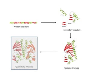
  • 2. Quaternary structure • Refers to the organization of subunits in a protein with multiple subunits • Subunits may be identical or different • Associate to form dimers (TIM, HIV protease, DNA binding proteins), trimers (MS2 viral capsid protein), tetramers (Haemoglobin, Proteasome, Bacterial photosynthetic reaction center), etc. • Subunits have a defined stoichiometry and arrangement • Subunits are held together by weak, noncovalent interactions (hydrophobic, electrostatic) • Typical Kd for two subunits: 10-8 to 10-16 M (tight association) – Entropy loss due to association - unfavorable – Entropy gain due to burying of hydrophobic groups - very favourable
  • 3. Why do proteins attain quaternary structures? • Morphological function:  Many proteins have functions that require creation of large, stable structures. These include long, thin structural elements and large, hollow capsids and rings. • Bringing catalytic sites together  More complex scaffolds may better support function e.g. by the introduction of a new active site at the interface between subunits. It has been estimated that roughly one sixth of oligomeric enzymes has an active site located at the inter-subunit interface. • Stability  Larger proteins are more resistant to degradation and denaturation. Indeed, an increase in oligomerization state is one of the protein stabilization strategies observed in thermophilic organisms. Protein stability involves a fine balance between the enthalpic stabilization by many weak nonbonded interactions and the competing effect of various entropic factors of conformational mobility and solvation.
  • 4. • Cooperativity (allostery):  Allostery and multivalent associations are other functions that create an evolutionary force selecting large proteins with several identical active sites rather than monomeric proteins with a single active site. • Reduction of surface area:  In general, it is preferable to reduce the protein surface area that is exposed to solvent, by creating a large protein with several identical active sites. Reduction of surface area reduces the amount of solvent needed to hydrate proteins. The reduced surface area provided by an oligomeric protein provides protection from degradation. Reduced surface area also improves the diffusion of substrates to enzyme active sites.
  • 5. Assume that the aqueous cytoplasm is composed entirely of 20 kDa subunits and that all oligomers are spherical in shape. Thus, a monomer would be a sphere of radius 1.8 nm, a dimer would have a radius of 2.2 nm, and so on. Assume that the cytoplasm is 20% protein and that the bound water of hydration is ~1.4 g/g of protein or a hydration shell ~0.6 nm thick. If the aqueous cytoplasm is composed entirely of monomers, the hydrated proteins occupy 47% of the total volume, over twice the 20% volume occupied by the protein alone. Assuming this same 0.6 nm layer of hydration, the volume of the hydrated protein drops to 40% for dimers, 35% for tetramers and 30% for dodecamers. Thus, oligomerization can significantly reduce the amount of water bound to protein surfaces. An analysis of surface area to volume ratio of oligomeric proteins
  • 6. How is large proteins built? The large proteins may be constructed in one of following ways: 1. As long single chains 2. As heterooligomers of several smaller chains 3. As homooligomers of identical chains
  • 7. 1. Error control: By building a large complex from many small subunits, translation errors may be reduced by discarding subunits with defects, providing an extra step for proofreading. 2. Coding efficiency: Homooligomers provide a genetically compact way to encode the information to build a large protein. It reduces the genetic space such as in viruses. 3. Genetic efficiency: Oligomeric proteins may be subjected to amplified evolutionary pressures, as deleterious mutations may be more pronounced and thus removed sooner from the gene pool. Conversely, the advantages of beneficial mutations may also be made evident sooner. 4. Regulation of assembly: Large assemblies built of many identical subunits have attractive regulatory properties, because they are subject to sensitive phase transitions. For instance, actin is involved in many dynamic processes at the cell surface. A collection of actin-binding proteins control the nucleation, growth, termination and disassembly of actin filaments allowing fine spatial and temporal control. What are the advantages of having multimers than large monomers?
  • 8. Protein surface is irregular. What does enable proteins to bind specific molecules? How does oligomerization of proteins occur in the cell? Shape complementarity is necessary for large number of weak interactions and to maximize the strength of interactions ((H-bonds and van der Waals).
  • 9.
  • 10. Finally, oligomerization can arise via fusion of a gene encoding a dimerization or oligomerization domain, such as a coiled-coil domain, onto a previously monomeric protein.
  • 13. Protein assemblies built of identical subunits are usually symmetric The homooligomeric proteins found in modern cells are also highly symmetrical with soluble oligomers forming closed complexes related by simple point groups and extended polymers showing helical symmetry.
  • 14. The human growth hormone-receptor complex Asymmetric complexSymmetric complex
  • 15. Why build symmetrical oligomeric proteins? 1. Stability of association: The stability of closed, symmetrical oligomers is a consequence of two factors: (a) the specificity of protein-protein interfaces favors symmetrical complexes, and (b) the maximum numbers of inter subunit interactions are formed in closed complexes. 2. Finite assembly: Proteins must avoid unwanted aggregation. Point group symmetry provides a method to create oligomers of defined copy number. Several disease states seem to be the result of pathological aggregation of mutant proteins such as sickle-cell anemia, Alzheimer’s disease and prion-related diseases. 3. Folding efficiency: Symmetric protein structures provide fewer kinetic barriers to folding than do asymmetric structures.
  • 16. Where does oligomerization of proteins occur inside the cell? The primary sites for protein synthesis and folding are the cytosol and the endoplasmic reticulum. Cytosolic proteins are synthesized, folded and oligomerized in the cytosol. Membrane and secretory proteins are synthesized in the endoplasmic reticulum (ER) and oligomerization typically occurs within the ER, although, in some cases, oligomerization takes place in the intermediate compartment and Golgi apparatus.
  • 17. What are the factors which can affect the oligomer formation? 1. Ligand binding: Many receptors undergo dimerization upon ligand binding. 2. Polymerization: Proteins such as actin can polymerize. Other proteins can polymerize after undergoing a conformational change giving rise to amyloid fibrils. 3. Concentration: The oligomerization state of a protein depends on the concentration of protein. At nM concentration the tendency to be in the monomeric state will be much higher than in the μM or mM range. 4. Environmental condition: Weak associations may happen due to conditions such as concentration, temperature, pH, solvent conditions (the ionic strength, metal cofactors and effectors concentrations) and have higher Kd values in the μM or mM range. 5. Domain linker: it is known that variation of inter-domain linker lengths can result in variations in oligomeric state. Some examples are the legume lectins, which can dimerize by various modes as well as tetramerize, the cystine-knot growth factors and lumazine synthase.
  • 18. How to determine the oligomeric states of proteins experimentally? In general, the following in vitro biophysical techniques can be used: 1. Size exclusion chromatography 2. Cross-linking 3. Analytical ultracentrifugation 4. Isothermal titration calorimetry 5. Mass spectrometry 6. Förster resonance energy transfer (FRET) 7. Scattering techniques 8. Yeast two hybrid assays 9. Fluorescence anisotropy 10. NMR spectroscopy
  • 19. Characteristics of oligomeric interfaces Interfacial residues tend to protrude from the surface of the protein and the interaction surface tends to be circular in shape. Protein–protein interaction interfaces are relatively planar as are many hetero-oligomer interfaces. The buried surface area in obligate homodimeric proteins is usually greater than 1400 Å2. In nonobligate complexes, the interface buried surface area is usually less than 2500 Å2, whereas for weak and transient associations the buried surface area of the interface is less than 1000 Å2 . It has been found that certain conserved residues or hot spots generally at the center of an interface are responsible for most of the binding energy of an oligomeric interaction.
  • 20. Inter-subunit interfaces are less non-polar, and have a greater proportion of hydrophilic and polar residues, than a typical protein hydrophobic core. Approximately one-fifth of the residues at oligomeric interfaces are polar, a greater proportion than is found in buried hydrophobic cores. Hydrogen bonds and salt bridges are important for the stabilization of oligomeric interfaces, as suggested by the prevalence of polar hot spot residues. Early studies suggested that there is about one hydrogen bond per 200 Å2 of subunit interface. Oligomeric interfaces often have significant electrostatic and geometrical shape complementarity that gives rise to the specificity of the interaction.
  • 21. It has been calculated that the average oligomeric state of cellular proteins is tetrameric and a survey suggests that 35% or more of the proteins in a cell are oligomeric. Most oligomeric proteins are homo-oligomers. Higher-order oligomers are less prevalent and a relatively small fraction of oligomeric structures have odd numbered stoichiometries. Most oligomeric proteins and essentially all homo-oligomeric proteins, are symmetrical. This symmetry is most frequently cyclic, dihedral, or cubic. Observations from the databases analysis (A) Isologous dimer (B) heterologous tetrame (C) Heterologous polymer.
  • 22. Oligomeric state No. of homo oligomers No. of hetero oligomers Percent Monomer 72 - 19.4 Dimer 115 27 38.2 Trimer 15 5 5.4 Tetramer 62 16 21.0 Pentamer 1 1 0.1 Hexamer 20 1 5.6 Heptamer 1 1 0.1 Goodsell and Olson, 2000, Annu. Rev. Biophys. Biomol. Struct. 29, 105-153. Natural occurrence of oligomeric proteins in E. coli Oligomeric state No. of homo oligomers No. of hetero oligomers Percent Octamer 3 6 2.4 Nonamer 0 0 0.0 Decamer 1 0 0.0 Undecamer 0 1 0.0 Dodecamer 4 2 1.6 Higher oligomers 8 - 2.2 Polymers 10 - 2.7
  • 23. So, do you think that all proteins should be multimer? In most cases, evolution appears to drive proteins to larger size and thus to symmetric, oligomeric complexes. In some specialized classes of proteins, however, functional considerations have the opposite effect, favoring small, monomeric proteins: 1. Rapid diffusion: Cytochrome c, ferredoxin, plastocyanin, and other soluble electron transport proteins must be small and streamlined to diffuse rapidly to their sites of action in the crowded environment inside cells. Extracellular hydrolases, hormones, and many toxins are small for the same reason. 2. Stability at low concentrations: Oligomeric proteins are unstable at very low concentrations, so secreted proteins are commonly monomeric. Apparently, the disulfide bridge serves primarily to hold the subunits together at the low concentrations found as the toxin diffuses to its target.
  • 24. Inappropriate quaternary interactions induce disease Sickel-cell hemoglobin: Hydrophobic patch from the mutation in b2 subunit (Gln  Val) Thick fiber
  • 25. 153 aa Alpha – 144 and Beta – 146 aa Hemoglobin Vs Myoglobin Hemoglobin is a dimer or tetramer?
  • 26. RMSD (α,β) of hemoglobin = 1.244 Å (43% sequence identity) RMSD (Hem_β, myoglobin) = 1.301 Å (no significant similarity) RMSD (Hem_α, myoglobin) = 1.397 Å (27% sequence identity)
  • 27. Myoglobin (dimer) interface area = 399 Å2. Why myoglobin itself is not multimeric? Why hemoglobin is not homotetramer? Hemoglobin (alpha, alpha) interface area = 276 Å2.
  • 28. Hemoglobin (α,β) interface area = 814 Å2.Hemoglobin (β,β) interface area = 240 Å2. Now, can you tell me whether hemoglobin is a dimer or tetramer?