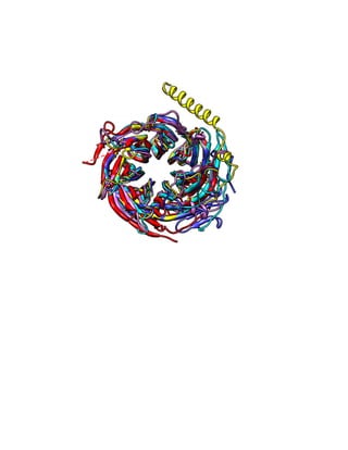
PAGE 1
REPORT: PROTIEN FOLDING AND MECHANISMS OF
PROTEOSTASIS
Presented by
MUFASSIRA RAHMAN
USN: 1PI14BT014
Semester: IV
Under the guidance of
Mr. SUNIL KUMAR
Asst. Professor
Department of biotechnology
Bangalore, May 2016
PAGE 2
CERTIFICATE
This is to certify that the report entitled “Protein folding and mechanism of proteostasis”
submitted by MUFASSIRA RAHMAN bearing USN 1PI14BT013, is in partial fulfillment of
curriculum of B.E (Biotechnology) program of PES Institute of Technology (Autonomous
under VTU) is an authentic record of my own work carried out during the period of January
2016 to April 2016 (IV semester) under the supervision of Prof.Sunil Kumar, Biotech
Department.
The matter presented in this Project Report is not submitted for any other course/degree in
any other department.
Signature of Student
(Mufassira)
Signature of Guide Signature of HOD
Date:
Place:
Signature of Internal Examiner:
Signature of External Examiner:
PAGE 3
ACKNOWLEDEGEMENT
I express my satisfaction on the completion of this paper presentation project report
submission as a part of the curriculum for the degree of Bachelor of Engineering,
Biotechnology. I express my deepest gratitude to my guide and mentor Mr. Sunil Kumar
for his kind help, support and consistent guidance in this project for entire period of this
course, which has helped me to complete this course successfully. I also thank Dr. Roshan
Makam, Head of the Department-Biotechnology, PESIT for giving me an opportunity to
present this work as a part of my IV semester course. I also appreciate the help of
Librarians for their kind support in providing the necessary papers required for References.
PAGE 4
CONTENTS
Sl.no Content Page no
1. Protein folding 6
2. Partially folded proteins 7
3. Mechanism of proteostasis 8
4. Chaperone and its classification 9
5. Chaperone assisted folding 9
6. GroES/GroEL complex 10
7. Clinical focus 11
8. Conclusion 12
9. Bibliography 13
PAGE 5
OBJECTIVE OF LEARNING:
⦁ Basic understanding of protein folding
⦁ Mechanism of Proteostasis (Chaperone protein function)
Almost each chemical process on which our lives depend is enthused or controlled by
protein molecules. Diverse proteins are distinguished by different order of amino acids in
the polymeric sequence. Protein folding is an intrinsic feature of normal folding within the
complex cellular environment, and its effects are minimized in living systems by the action
of a range of protective mechanisms, including molecular chaperons and quality control
system of cell which has evolved to maintain homeostasis and for preservation of the native
structure.
For example to increase the accessible conformational space, cells develop molecular
chaperons. These are the set of proteins that linked with unfolded polypeptides thereby
preventing aggregation and protein folding in an ATP-dependent manner.
Unfolded and misfolded polypeptides have a tendency to aggregate to form a variety of
species, including the highly ordered and kinetically stable amyloid fibrils. These species
signify a generic form of structure resulting from the innate polymer properties of
polypeptide chain, and their formation is associated with a wide range of debilitating human
diseases.
PAGE 6
PROTEIN FOLDING
A physical process by which a polypeptide chain (sequence of amino acids) folds into its
characteristic & functional native structure from a random coil or a linear sequence.
Proteins have four levels of structure: Primary, Secondary, Tertiary and Quaternary. But
with the increasing information available about proteins two additional levels of structures
have been distinguished by molecular biologists: motifs and domains.
Primary Structure: It is the specific linear sequence of amino acid residues making up) a
polypeptide chain. The sequence of amino acids is determined by the nucleotide sequence
of the gene that encodes the protein. Only peptide bonds are associated with the primary
structure.
Secondary structure results from the hydrogen bonding between the amino acids of
polypeptides. Two patterns of hydrogen bonding occur in proteins. In one pattern, hydrogen
bonds are formed along a single polypeptide chain, linking one amino acid to another
distant one down the chain. This tends to pull the chain into a coil, called alpha helix. In
another pattern, hydrogen bonds are formed between amino acids across two chains. Often
many chains are linked in this pattern, forming a pleated sheet like structure called beta (p)
sheet. The two chains may be oriented in a parallel or anti parallel manner forming parallel
P~ pleated sheets and anti parallel p-pleated sheets respectively.
Tertiary structure results from extensive folding of polypeptide into a complex, rigid right
handed a helix and globular structure. Most secondary structures fold compactly due to
interactions between amino acid residues present far apart. Ionic bonds between oppositely
charged side groups bring region into close proximity. Hydrogen bonds, hydrophobic
interactions and disulphide linkage also lock particular regions of proteins together.
In case of Quaternary structure, when two or more polypeptide chains associate to form a
functional protein, a quaternary structure results. Here, each polypeptide in known as a
subunit (protomer).
Motifs: In a protein the elements of secondary structure sometimes combine in
characteristic ways called motif or "Super secondary structure". The motifs are specific
regions on proteins to bind to other structures such as nucleic acids. Domains: Many
proteins are encoded in sections by different exons. These sections typically of 100 to 200
amino acids long encoded by different exons fold into structurally independent functional
units called domain.
It is thermodynamically feasible as free energy of this process is negative, usually driven by
enthalpy (the energy of bond formation and bond breaking) while entropy of the polypeptide
chain decreases dramatically as it goes from the millions of shapes that the unfolded
protein can adopt to the one properly folded shape, Therefore, the entropy change from the
standpoint of the protein alone is negative (ΔSsystem < 0). Accordingly we can understand
from folding funnel hypothesis that the Intermediates in the folding process are molecules in
lower free energy pockets along the sides of the funnel. If the pockets(free energy "wells")
PAGE 7
are deep, the folding process slows down; and if a particular well is too deep, the molecule
may get stuck in that intermediate state (misfolded state), reducing the ultimate yield of
correctly folded molecules at the end. For example, sometimes folding intermediates might
have hydrophobic groups on their surfaces, and several partly folded molecules might end
up sticking to each other by hydrophobic interactions to bury those hydrophobic(formation
of insoluble misfolded aggregates of multiple protein molecules), and be unable to
dissociate unless they're unfolded again.
PARTIALLY FOLDED PROTEINS
Partially folded, misfolded proteins are the deviation from the series of number of possible
conformation available and according to levinthal’s paradox we know that the no.of possible
conformation to a protein is astronomically very large. Therefore Protein misfolding is a
common and intrinsic property of proteins that occurs continuously.
Misfolding is influenced by the amino acid composition, and certain mutations are known to
accelerate the process. Moreover, it also depends on environmental conditions, because
once proteins are exposed to specific environmental changes such as increased
temperature, high or low pH, agitation, elevated glucose, or oxidative agents they can lose
their native conformation more rapidly. The process wherein the native state is disrupted is
called denaturation, and it generally results in the unfolding of the proteins. Because of the
lack of arrangement, unfolded proteins are nonfunctional. Importantly, the unfolded state is
thermodynamically unfavorable and unstable.
While the partially folded states in proteins are difficult to conceptualize and their
experimental study is challenging, a wide variety of roles for protein structure disorder has
been proposed. Disordered proteins are visualized as dynamic assemblies, wherein the
atom positions and Ramachandran angles axis vary significantly over time.
Thus larger degree of conformational sampling for partially folded or intrinsically disordered
protein (IDP) are given significant conformational entropy, usually restricted by inter or intra
molecular interactions. Moreover, it has been suggested that loss of conformational entropy
upon ligand binding originates a weaker binding for IDP, undergoing disorder-to-order
transitions in secondary structure upon ligand interactions. Structural disorder may span
from short segments in specific domains to entire proteins.
The importance of disorder in proteins is self-evident, as a large portion of molecular
interactions depend on the complementary interaction between structurally organized
proteins and IDPs. This structural condition could confer diverse advantages, such as rapid
and specific binding and it is also associated with many cell activities such as transcription,
cell signaling, RNA processing, ubiquitination, ion transport, cytoskeletal organization, cell
cycle control, and other highly regulated biological mechanisms.
PAGE 8
MECHANISM TO CONSERVE PROTEIN FOLDING
In order to maintain the native protein folding, cells have developed various strategies to
accomplish this task, through sophisticated chaperone and quality control networks that can
resolve damage at the level of protein, organelle, or cell For instance, on the smallest
scale, the integrity of individual proteins is monitored during their synthesis in ribosomes,
and coupled with co-translational chaperone function. On a larger scale, cells use
compartmentalized defenses and networks of communication capable of signaling between
cells, and so respond to changes in the proteome homeostasis. Together, these layered
defenses help protect cells from alterations in protein folding and degradation, avoiding the
appearance of misfolded proteins and deleterious events.
Autophagy mechanism: defined as the natural, destructive mechanism that disassembles,
through a regulated process, unnecessary or dysfunctional cellular components. Autophagy
allows the orderly degradation and recycling of cellular components
Endoplasmic reticulum stress: ER Stress is the imbalance of homeostasis, which can be
sensed even at the subcellular level. ER stress impairs many process and results in the
accumulation of unfolded or misfolded proteins, which leads to the activation of a specific
cellular process called the unfolded protein response (UPR).
Proteasomes: These are protein complexes inside all eukaryotes and archaea, and in some
bacteria. The main function of the proteasome is to degrade unneeded or damaged
proteins by proteolysis, a chemical reaction that breaks peptide bonds.
CHAPERONES
In extremely crowded cellular environment, the folding of polypeptide chains into precise
functional structures is a daunting task, so the cell typically requires the assistance of a set
of proteins termed molecular chaperones. Chaperones are an essential group of proteins
necessary under both normal and stress conditions. They assist in the efficient folding of
newly-translated proteins as these proteins are being synthesized on the ribosome and can
maintain pre-existing proteins in a stable conformation.
Chaperones can also promote the disaggregation of preformed protein aggregates and
capture unfolded polypeptides, stabilize intermediates, and prevent misfolded species from
accumulating in stressed cells. Many of the identified chaperones are called heat shock
proteins. Molecular chaperones have an essential role in the regulation of protein
conformation states - the process during which transient or stable interactions with client
proteins affects their conformation and activity. The general mechanism by which
chaperones carry out their function usually involves multiple rounds of regulated binding
and release of an unstable conformer of target polypeptides. The capacity of chaperones to
regulate these processes involves various co-chaperones combinations to interact with
chaperones to release folded proteins, to facilitate the assembly or disassembly or to affect
subcellular trafficking.
PAGE 9
CLASSIFICATION OF CHAPERONES
The principal heat-shock proteins that have chaperone activity belong to five conserved
classes: HSP33, HSP60, HSP70, HSP90, HSP100, and the small heat-shock proteins.
1. Hsp10 (GroES in E.coli)
2. HspB group (GrpE)
3. Hsp40 (DnaJ): Cofactor of Hsp70
4. Hsp60 (GroEL): Involved in protein folding after its post-translational import to the
mitochondrion/chloroplast
5. Hsp70 (Dank): It provides thermotolerance to cell on exposure to heat stress. Also
prevents protein folding during post-translational import into the mitochondria/chloroplast.
6. Hsp90 (HtpG): Maintenance of steroid receptors and transcription factors.
7. Hsp100 (Clp group): Tolerance of extreme temperature
Based on interactions they are also classified as:
1. Foldases: The ATP dependent foldases are directly involved in protein folding.
2. Holdases: Holdases are ATP-independent proteins assisting client proteins to
foldases by preventing aggregation and misfolding.
3. Disaggregases: Disaggregases are also ATP-dependent, which disaggregate the
client protein aggregates and transfer the partially folded proteins to holdases or
foldases.
CHAPERONE ASSISSTED FOLDING
Chaperones have multiple apical domains that are hydrophobic in nature which binds to
exposed hydrophobic side chains of unfolded proteins. Binding of chaperones to this
regions temporarily blocks protein aggregation. Process of binding to the apical domains is
achieved by ATP- independent chaperones such as holdases and co-chaperones followed
by ATP hydrolysis brings about the conformational change in the inner surface of
chaperones, allowing the folding of client proteins. Folding competes with chaperone
binding and the process offers the thermodynamic stability to proteins to acquire native
structure.
HSF1(heat shock transcription factor 1) is a key molecule induced under cellular stress
activating HSR(Heat shock response) leads to transcription of HSP genes(chaperones) by
binding to DNA containing heat shock elements. During the stress the level of unfolded or
misfolded protein concentration increases resulting in HSF1 to disassociate from HSP
family to bring the post -transcriptional modification by phosphorylation followed by
transcription.
PAGE 10
The two main HspB groups involved are Hsp60 and Hsp70 along with their co-chaperones
interaction. There is slight difference in the folding mechanism, Hsp70 is associated with
Hsp40 as co-chaperone. Folding takes place in the aqueous solution effectively reducing
the aggregation and accumulation of misfolds. Whereas Hsp60 forms a multimeric complex
within which protein folds and the co-chaperone Hsp10 act as lid and brings about folding
and refolding.
GroEL/GroES CHAPERONE COMPLEX
GroEL and GroES chaperones is the best studied system, whose function is relatively well
understood. GroEL chaperone consists of two rings – cis upper ring and Tran’s lower ring.
GroES co-chaperone binds to both GroEL rings. Each of the rings consists of seven
identical units. These units are arranged in a circular manner and form a cavity. GroES
upon binding to GroEL serves as “lid”, which encapsulates the volume inside the cavity for
protein folding. Each of the GroEL ring units is composed of three domains – apical (the
upper one), intermediate, and lower equatorial. The role of equatorial domain is to bind and
hydrolyze ATP and to transmit signals for concerted function of cis and trans rings. Apical
domains contain binding sites for GroES docking and capturing substrate protein.
Capture (T state): The cavity of GroEL is open and GroES is not yet bound. Exposed
hydrophobic sites in apical domains of GroEL non-specifically bind a non-native protein.
Also no sequence specificity is evident in binding substrate proteins; and only non-native
proteins can be bound, while native proteins are not recognized.
Encapsulation (R’ states): Binding of ATP by equatorial domain causes several structural
changes. T→R transition is due to clockwise rotation of all apical domains.. Because
substrate is bound to these sites, Binding of GroES encapsulates substrate protein and also
causes further structural rearrangement of apical domains. Likely this effectively displace
client protein deeper into the cavity, which are now engaged in the interactions with the
GroES molecule. All the conformational movements are highly cooperative and concerted.
Full encapsulation of substrate protein and binding of GroES create R’ state. GroES is
attached to GroEL for a total of about 7 sec.
ATP hydrolysis (R’’ state) and substrate release: ATP hydrolysis in the equatorial domain
which sends a signal to release substrate (R’→R’’ transition). Initial release of GroES is
released of substrate itself and as GroEL returns to the T state, the cavity shrinks back
causing walls to become again hydrophobic.
CLINICAL FOCUS
Misfolding of normally soluble peptides and proteins has been associated with about 50
disorders with a multitude of different symptoms in which mechanisms of non-native
interaction could form aggregates. Fortunately, all cells possess protein quality control
machinery that sequesters misfolded proteins, either by refolding or degrading. This activity
is largely performed by the stress response chaperones (i.e. Hsp70). However, the
expression level of molecular chaperones varies widely among cell types. Cell death or
PAGE 11
damage occurs likely only when misfolded and aggregates are prone superior overwhelm
the defensive mechanism , resulting in breakdown of proteostasis process which initiates
trigging series of reaction ad the pathological process to occur. Often the condition involves
formation of inappropriate aggregates, frequently as a result of misfolded conformations
involving b sheet structures that tend to stack to form cross beta structures; these cross
beta structures are so insoluble that it has been very hard to study the details of their
molecular structures.
In prion diseases, there's a normal cellular protein (function often unknown, involving
different proteins in different prion diseases) that also occurs in an abnormal conformation.
If concentration of the abnormally folded conformational state is high enough to have an
effect, interaction of the abnormal protein molecules with molecules in the normal
conformation converts the normal molecules to the abnormal structure (a shift in the protein
conformational equilibrium caused by the protein-protein interactions).
Neurodegenerative diseases: Neurodegenerative diseases were the first described protein
misfolding diseases. Here, Amyloid fibril formation is considered to be one of the important
cause for neurological disease wherein disturbances in proteostatsis leads to metastasis
and amyloid fibril formation associated with chronic degenerative disorders.
Alzheimer’s disease (AD) was first described by Alois Alzheimer in 1907, which is one of
the neurodegenerative disorders, succeeding from memory loss to thoughtful dementia and
leads to death for the average of eight years. Neuropathological features of these terrible
diseases are extracellular deposition of Aβ protein represent a significant step in AD
pathogenesis, which is over expressed in AD.
Metabolic disorder: In islet of Langerhans, accumulation of amylin increases ER stress
leading to dysfunctioning of pancreatic beta cells, apoptosis eventually loss of cell mass in
pancreases.
Currently, there is no cure for any of these diseases. however, it believes that the
concerned research effort in the areas of protein folding, combined with system biology
analysis of networks of protein quality control will provide the knowledge base for the
development of new therapeutic strategies
PAGE 12
CONCLUSION
Interpretation of studies of protein folding and proteostasis helps in critical assessment. A
rigorous analysis prevents misleading conclusions. Cellular machinery plays an important
role in regulating synthesis, translocation. The activity of highly organized molecules such
as chaperones, and the participation of pathways associated with complete degradation of
organelles, helps to understand and to maintain the homeostasis of proteins as a whole.
The insights into the features of the functional conformations of proteins, the environments
in which they work, and the ways that cellular defense mechanisms normally function so
effectively together to maintain protein homeostasis, can expand the possibilities for better
understanding the processes.. Folding and degradation of proteins seems to operate in the
very stringent manner to ensure that protein aggregation is minimized by various processes
to prevent neurodegenerative diseases.
PAGE 13
Bibliography
A mystery unfolds: Franz-Ulrich Hartl and Arthur L. Horwich 2011
J. Clin Invest.; 121(10): 3774–3777. doi: 10.1172/JCI60889
Arthur L. Horwich. 2013. “Chaperonin-mediated Protein Folding”,
J. Biol Chem.; 288(33): 23622–23632. doi: 10.1074/jbc.X113.497321
Brandvold KR, Morimoto RI.2015.”The Chemical Biology of Molecular Chaperones--
Implications for Modulation of Proteostasis.” J Mol Biol. 427(18): 2931–47vc
Dobson CM.2002; “Protein misfolding diseases: Getting out of shape.” Nature. 418
Enrique Reynaud, Ph.D. .; 2010; “Protein Misfolding and Degenerative Diseases” . Nature
Education 3(9):28
Jeng W, Lee S, Sung N, Lee J, Tsai FT .2015“Molecular chaperones: guardians of the
proteome in normal and disease states.” Doi: 10.12688/f1000research.7214.
Parsell DA, Kowal AS, and Singer MA, et al: 1994” Protein disaggregation mediated by
heat-shock protein Hsp104.” Nature. 372(6505); 475(8)
Rampelt H, Kirstein-Miles J, Nillegoda NB, et al.:2012; “Metazoan Hsp70 machines use
Hsp110 to power protein disaggregation.” J. EMBO; 31(21): 4221–35.
Chaudhuri TK1, Paul S. 2006; “Protein-misfolding diseases and chaperone-based
therapeutic approaches.” J. FEBS; 273(7):1331-49.
Ryabova NA, Marchenkov V V, Marchenkova SY, Kotova NV, Semisotnov GV. 2013;
“Molecular chaperone GroEL/ES: unfolding and refolding processes.” Biochemistry (Mosc);
78(13):1405-14. doi: 10.1134/S0006297913130038
PAGE 14

report

  • 2.
    PAGE 1 REPORT: PROTIENFOLDING AND MECHANISMS OF PROTEOSTASIS Presented by MUFASSIRA RAHMAN USN: 1PI14BT014 Semester: IV Under the guidance of Mr. SUNIL KUMAR Asst. Professor Department of biotechnology Bangalore, May 2016
  • 3.
    PAGE 2 CERTIFICATE This isto certify that the report entitled “Protein folding and mechanism of proteostasis” submitted by MUFASSIRA RAHMAN bearing USN 1PI14BT013, is in partial fulfillment of curriculum of B.E (Biotechnology) program of PES Institute of Technology (Autonomous under VTU) is an authentic record of my own work carried out during the period of January 2016 to April 2016 (IV semester) under the supervision of Prof.Sunil Kumar, Biotech Department. The matter presented in this Project Report is not submitted for any other course/degree in any other department. Signature of Student (Mufassira) Signature of Guide Signature of HOD Date: Place: Signature of Internal Examiner: Signature of External Examiner:
  • 4.
    PAGE 3 ACKNOWLEDEGEMENT I expressmy satisfaction on the completion of this paper presentation project report submission as a part of the curriculum for the degree of Bachelor of Engineering, Biotechnology. I express my deepest gratitude to my guide and mentor Mr. Sunil Kumar for his kind help, support and consistent guidance in this project for entire period of this course, which has helped me to complete this course successfully. I also thank Dr. Roshan Makam, Head of the Department-Biotechnology, PESIT for giving me an opportunity to present this work as a part of my IV semester course. I also appreciate the help of Librarians for their kind support in providing the necessary papers required for References.
  • 5.
    PAGE 4 CONTENTS Sl.no ContentPage no 1. Protein folding 6 2. Partially folded proteins 7 3. Mechanism of proteostasis 8 4. Chaperone and its classification 9 5. Chaperone assisted folding 9 6. GroES/GroEL complex 10 7. Clinical focus 11 8. Conclusion 12 9. Bibliography 13
  • 6.
    PAGE 5 OBJECTIVE OFLEARNING: ⦁ Basic understanding of protein folding ⦁ Mechanism of Proteostasis (Chaperone protein function) Almost each chemical process on which our lives depend is enthused or controlled by protein molecules. Diverse proteins are distinguished by different order of amino acids in the polymeric sequence. Protein folding is an intrinsic feature of normal folding within the complex cellular environment, and its effects are minimized in living systems by the action of a range of protective mechanisms, including molecular chaperons and quality control system of cell which has evolved to maintain homeostasis and for preservation of the native structure. For example to increase the accessible conformational space, cells develop molecular chaperons. These are the set of proteins that linked with unfolded polypeptides thereby preventing aggregation and protein folding in an ATP-dependent manner. Unfolded and misfolded polypeptides have a tendency to aggregate to form a variety of species, including the highly ordered and kinetically stable amyloid fibrils. These species signify a generic form of structure resulting from the innate polymer properties of polypeptide chain, and their formation is associated with a wide range of debilitating human diseases.
  • 7.
    PAGE 6 PROTEIN FOLDING Aphysical process by which a polypeptide chain (sequence of amino acids) folds into its characteristic & functional native structure from a random coil or a linear sequence. Proteins have four levels of structure: Primary, Secondary, Tertiary and Quaternary. But with the increasing information available about proteins two additional levels of structures have been distinguished by molecular biologists: motifs and domains. Primary Structure: It is the specific linear sequence of amino acid residues making up) a polypeptide chain. The sequence of amino acids is determined by the nucleotide sequence of the gene that encodes the protein. Only peptide bonds are associated with the primary structure. Secondary structure results from the hydrogen bonding between the amino acids of polypeptides. Two patterns of hydrogen bonding occur in proteins. In one pattern, hydrogen bonds are formed along a single polypeptide chain, linking one amino acid to another distant one down the chain. This tends to pull the chain into a coil, called alpha helix. In another pattern, hydrogen bonds are formed between amino acids across two chains. Often many chains are linked in this pattern, forming a pleated sheet like structure called beta (p) sheet. The two chains may be oriented in a parallel or anti parallel manner forming parallel P~ pleated sheets and anti parallel p-pleated sheets respectively. Tertiary structure results from extensive folding of polypeptide into a complex, rigid right handed a helix and globular structure. Most secondary structures fold compactly due to interactions between amino acid residues present far apart. Ionic bonds between oppositely charged side groups bring region into close proximity. Hydrogen bonds, hydrophobic interactions and disulphide linkage also lock particular regions of proteins together. In case of Quaternary structure, when two or more polypeptide chains associate to form a functional protein, a quaternary structure results. Here, each polypeptide in known as a subunit (protomer). Motifs: In a protein the elements of secondary structure sometimes combine in characteristic ways called motif or "Super secondary structure". The motifs are specific regions on proteins to bind to other structures such as nucleic acids. Domains: Many proteins are encoded in sections by different exons. These sections typically of 100 to 200 amino acids long encoded by different exons fold into structurally independent functional units called domain. It is thermodynamically feasible as free energy of this process is negative, usually driven by enthalpy (the energy of bond formation and bond breaking) while entropy of the polypeptide chain decreases dramatically as it goes from the millions of shapes that the unfolded protein can adopt to the one properly folded shape, Therefore, the entropy change from the standpoint of the protein alone is negative (ΔSsystem < 0). Accordingly we can understand from folding funnel hypothesis that the Intermediates in the folding process are molecules in lower free energy pockets along the sides of the funnel. If the pockets(free energy "wells")
  • 8.
    PAGE 7 are deep,the folding process slows down; and if a particular well is too deep, the molecule may get stuck in that intermediate state (misfolded state), reducing the ultimate yield of correctly folded molecules at the end. For example, sometimes folding intermediates might have hydrophobic groups on their surfaces, and several partly folded molecules might end up sticking to each other by hydrophobic interactions to bury those hydrophobic(formation of insoluble misfolded aggregates of multiple protein molecules), and be unable to dissociate unless they're unfolded again. PARTIALLY FOLDED PROTEINS Partially folded, misfolded proteins are the deviation from the series of number of possible conformation available and according to levinthal’s paradox we know that the no.of possible conformation to a protein is astronomically very large. Therefore Protein misfolding is a common and intrinsic property of proteins that occurs continuously. Misfolding is influenced by the amino acid composition, and certain mutations are known to accelerate the process. Moreover, it also depends on environmental conditions, because once proteins are exposed to specific environmental changes such as increased temperature, high or low pH, agitation, elevated glucose, or oxidative agents they can lose their native conformation more rapidly. The process wherein the native state is disrupted is called denaturation, and it generally results in the unfolding of the proteins. Because of the lack of arrangement, unfolded proteins are nonfunctional. Importantly, the unfolded state is thermodynamically unfavorable and unstable. While the partially folded states in proteins are difficult to conceptualize and their experimental study is challenging, a wide variety of roles for protein structure disorder has been proposed. Disordered proteins are visualized as dynamic assemblies, wherein the atom positions and Ramachandran angles axis vary significantly over time. Thus larger degree of conformational sampling for partially folded or intrinsically disordered protein (IDP) are given significant conformational entropy, usually restricted by inter or intra molecular interactions. Moreover, it has been suggested that loss of conformational entropy upon ligand binding originates a weaker binding for IDP, undergoing disorder-to-order transitions in secondary structure upon ligand interactions. Structural disorder may span from short segments in specific domains to entire proteins. The importance of disorder in proteins is self-evident, as a large portion of molecular interactions depend on the complementary interaction between structurally organized proteins and IDPs. This structural condition could confer diverse advantages, such as rapid and specific binding and it is also associated with many cell activities such as transcription, cell signaling, RNA processing, ubiquitination, ion transport, cytoskeletal organization, cell cycle control, and other highly regulated biological mechanisms.
  • 9.
    PAGE 8 MECHANISM TOCONSERVE PROTEIN FOLDING In order to maintain the native protein folding, cells have developed various strategies to accomplish this task, through sophisticated chaperone and quality control networks that can resolve damage at the level of protein, organelle, or cell For instance, on the smallest scale, the integrity of individual proteins is monitored during their synthesis in ribosomes, and coupled with co-translational chaperone function. On a larger scale, cells use compartmentalized defenses and networks of communication capable of signaling between cells, and so respond to changes in the proteome homeostasis. Together, these layered defenses help protect cells from alterations in protein folding and degradation, avoiding the appearance of misfolded proteins and deleterious events. Autophagy mechanism: defined as the natural, destructive mechanism that disassembles, through a regulated process, unnecessary or dysfunctional cellular components. Autophagy allows the orderly degradation and recycling of cellular components Endoplasmic reticulum stress: ER Stress is the imbalance of homeostasis, which can be sensed even at the subcellular level. ER stress impairs many process and results in the accumulation of unfolded or misfolded proteins, which leads to the activation of a specific cellular process called the unfolded protein response (UPR). Proteasomes: These are protein complexes inside all eukaryotes and archaea, and in some bacteria. The main function of the proteasome is to degrade unneeded or damaged proteins by proteolysis, a chemical reaction that breaks peptide bonds. CHAPERONES In extremely crowded cellular environment, the folding of polypeptide chains into precise functional structures is a daunting task, so the cell typically requires the assistance of a set of proteins termed molecular chaperones. Chaperones are an essential group of proteins necessary under both normal and stress conditions. They assist in the efficient folding of newly-translated proteins as these proteins are being synthesized on the ribosome and can maintain pre-existing proteins in a stable conformation. Chaperones can also promote the disaggregation of preformed protein aggregates and capture unfolded polypeptides, stabilize intermediates, and prevent misfolded species from accumulating in stressed cells. Many of the identified chaperones are called heat shock proteins. Molecular chaperones have an essential role in the regulation of protein conformation states - the process during which transient or stable interactions with client proteins affects their conformation and activity. The general mechanism by which chaperones carry out their function usually involves multiple rounds of regulated binding and release of an unstable conformer of target polypeptides. The capacity of chaperones to regulate these processes involves various co-chaperones combinations to interact with chaperones to release folded proteins, to facilitate the assembly or disassembly or to affect subcellular trafficking.
  • 10.
    PAGE 9 CLASSIFICATION OFCHAPERONES The principal heat-shock proteins that have chaperone activity belong to five conserved classes: HSP33, HSP60, HSP70, HSP90, HSP100, and the small heat-shock proteins. 1. Hsp10 (GroES in E.coli) 2. HspB group (GrpE) 3. Hsp40 (DnaJ): Cofactor of Hsp70 4. Hsp60 (GroEL): Involved in protein folding after its post-translational import to the mitochondrion/chloroplast 5. Hsp70 (Dank): It provides thermotolerance to cell on exposure to heat stress. Also prevents protein folding during post-translational import into the mitochondria/chloroplast. 6. Hsp90 (HtpG): Maintenance of steroid receptors and transcription factors. 7. Hsp100 (Clp group): Tolerance of extreme temperature Based on interactions they are also classified as: 1. Foldases: The ATP dependent foldases are directly involved in protein folding. 2. Holdases: Holdases are ATP-independent proteins assisting client proteins to foldases by preventing aggregation and misfolding. 3. Disaggregases: Disaggregases are also ATP-dependent, which disaggregate the client protein aggregates and transfer the partially folded proteins to holdases or foldases. CHAPERONE ASSISSTED FOLDING Chaperones have multiple apical domains that are hydrophobic in nature which binds to exposed hydrophobic side chains of unfolded proteins. Binding of chaperones to this regions temporarily blocks protein aggregation. Process of binding to the apical domains is achieved by ATP- independent chaperones such as holdases and co-chaperones followed by ATP hydrolysis brings about the conformational change in the inner surface of chaperones, allowing the folding of client proteins. Folding competes with chaperone binding and the process offers the thermodynamic stability to proteins to acquire native structure. HSF1(heat shock transcription factor 1) is a key molecule induced under cellular stress activating HSR(Heat shock response) leads to transcription of HSP genes(chaperones) by binding to DNA containing heat shock elements. During the stress the level of unfolded or misfolded protein concentration increases resulting in HSF1 to disassociate from HSP family to bring the post -transcriptional modification by phosphorylation followed by transcription.
  • 11.
    PAGE 10 The twomain HspB groups involved are Hsp60 and Hsp70 along with their co-chaperones interaction. There is slight difference in the folding mechanism, Hsp70 is associated with Hsp40 as co-chaperone. Folding takes place in the aqueous solution effectively reducing the aggregation and accumulation of misfolds. Whereas Hsp60 forms a multimeric complex within which protein folds and the co-chaperone Hsp10 act as lid and brings about folding and refolding. GroEL/GroES CHAPERONE COMPLEX GroEL and GroES chaperones is the best studied system, whose function is relatively well understood. GroEL chaperone consists of two rings – cis upper ring and Tran’s lower ring. GroES co-chaperone binds to both GroEL rings. Each of the rings consists of seven identical units. These units are arranged in a circular manner and form a cavity. GroES upon binding to GroEL serves as “lid”, which encapsulates the volume inside the cavity for protein folding. Each of the GroEL ring units is composed of three domains – apical (the upper one), intermediate, and lower equatorial. The role of equatorial domain is to bind and hydrolyze ATP and to transmit signals for concerted function of cis and trans rings. Apical domains contain binding sites for GroES docking and capturing substrate protein. Capture (T state): The cavity of GroEL is open and GroES is not yet bound. Exposed hydrophobic sites in apical domains of GroEL non-specifically bind a non-native protein. Also no sequence specificity is evident in binding substrate proteins; and only non-native proteins can be bound, while native proteins are not recognized. Encapsulation (R’ states): Binding of ATP by equatorial domain causes several structural changes. T→R transition is due to clockwise rotation of all apical domains.. Because substrate is bound to these sites, Binding of GroES encapsulates substrate protein and also causes further structural rearrangement of apical domains. Likely this effectively displace client protein deeper into the cavity, which are now engaged in the interactions with the GroES molecule. All the conformational movements are highly cooperative and concerted. Full encapsulation of substrate protein and binding of GroES create R’ state. GroES is attached to GroEL for a total of about 7 sec. ATP hydrolysis (R’’ state) and substrate release: ATP hydrolysis in the equatorial domain which sends a signal to release substrate (R’→R’’ transition). Initial release of GroES is released of substrate itself and as GroEL returns to the T state, the cavity shrinks back causing walls to become again hydrophobic. CLINICAL FOCUS Misfolding of normally soluble peptides and proteins has been associated with about 50 disorders with a multitude of different symptoms in which mechanisms of non-native interaction could form aggregates. Fortunately, all cells possess protein quality control machinery that sequesters misfolded proteins, either by refolding or degrading. This activity is largely performed by the stress response chaperones (i.e. Hsp70). However, the expression level of molecular chaperones varies widely among cell types. Cell death or
  • 12.
    PAGE 11 damage occurslikely only when misfolded and aggregates are prone superior overwhelm the defensive mechanism , resulting in breakdown of proteostasis process which initiates trigging series of reaction ad the pathological process to occur. Often the condition involves formation of inappropriate aggregates, frequently as a result of misfolded conformations involving b sheet structures that tend to stack to form cross beta structures; these cross beta structures are so insoluble that it has been very hard to study the details of their molecular structures. In prion diseases, there's a normal cellular protein (function often unknown, involving different proteins in different prion diseases) that also occurs in an abnormal conformation. If concentration of the abnormally folded conformational state is high enough to have an effect, interaction of the abnormal protein molecules with molecules in the normal conformation converts the normal molecules to the abnormal structure (a shift in the protein conformational equilibrium caused by the protein-protein interactions). Neurodegenerative diseases: Neurodegenerative diseases were the first described protein misfolding diseases. Here, Amyloid fibril formation is considered to be one of the important cause for neurological disease wherein disturbances in proteostatsis leads to metastasis and amyloid fibril formation associated with chronic degenerative disorders. Alzheimer’s disease (AD) was first described by Alois Alzheimer in 1907, which is one of the neurodegenerative disorders, succeeding from memory loss to thoughtful dementia and leads to death for the average of eight years. Neuropathological features of these terrible diseases are extracellular deposition of Aβ protein represent a significant step in AD pathogenesis, which is over expressed in AD. Metabolic disorder: In islet of Langerhans, accumulation of amylin increases ER stress leading to dysfunctioning of pancreatic beta cells, apoptosis eventually loss of cell mass in pancreases. Currently, there is no cure for any of these diseases. however, it believes that the concerned research effort in the areas of protein folding, combined with system biology analysis of networks of protein quality control will provide the knowledge base for the development of new therapeutic strategies
  • 13.
    PAGE 12 CONCLUSION Interpretation ofstudies of protein folding and proteostasis helps in critical assessment. A rigorous analysis prevents misleading conclusions. Cellular machinery plays an important role in regulating synthesis, translocation. The activity of highly organized molecules such as chaperones, and the participation of pathways associated with complete degradation of organelles, helps to understand and to maintain the homeostasis of proteins as a whole. The insights into the features of the functional conformations of proteins, the environments in which they work, and the ways that cellular defense mechanisms normally function so effectively together to maintain protein homeostasis, can expand the possibilities for better understanding the processes.. Folding and degradation of proteins seems to operate in the very stringent manner to ensure that protein aggregation is minimized by various processes to prevent neurodegenerative diseases.
  • 14.
    PAGE 13 Bibliography A mysteryunfolds: Franz-Ulrich Hartl and Arthur L. Horwich 2011 J. Clin Invest.; 121(10): 3774–3777. doi: 10.1172/JCI60889 Arthur L. Horwich. 2013. “Chaperonin-mediated Protein Folding”, J. Biol Chem.; 288(33): 23622–23632. doi: 10.1074/jbc.X113.497321 Brandvold KR, Morimoto RI.2015.”The Chemical Biology of Molecular Chaperones-- Implications for Modulation of Proteostasis.” J Mol Biol. 427(18): 2931–47vc Dobson CM.2002; “Protein misfolding diseases: Getting out of shape.” Nature. 418 Enrique Reynaud, Ph.D. .; 2010; “Protein Misfolding and Degenerative Diseases” . Nature Education 3(9):28 Jeng W, Lee S, Sung N, Lee J, Tsai FT .2015“Molecular chaperones: guardians of the proteome in normal and disease states.” Doi: 10.12688/f1000research.7214. Parsell DA, Kowal AS, and Singer MA, et al: 1994” Protein disaggregation mediated by heat-shock protein Hsp104.” Nature. 372(6505); 475(8) Rampelt H, Kirstein-Miles J, Nillegoda NB, et al.:2012; “Metazoan Hsp70 machines use Hsp110 to power protein disaggregation.” J. EMBO; 31(21): 4221–35. Chaudhuri TK1, Paul S. 2006; “Protein-misfolding diseases and chaperone-based therapeutic approaches.” J. FEBS; 273(7):1331-49. Ryabova NA, Marchenkov V V, Marchenkova SY, Kotova NV, Semisotnov GV. 2013; “Molecular chaperone GroEL/ES: unfolding and refolding processes.” Biochemistry (Mosc); 78(13):1405-14. doi: 10.1134/S0006297913130038
  • 15.