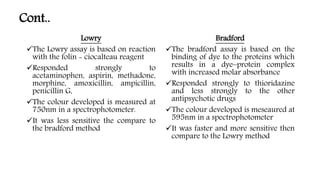
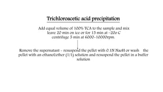
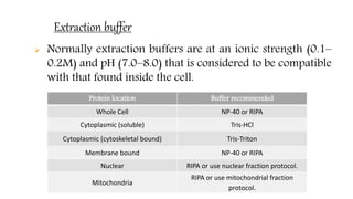
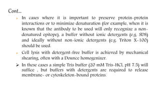
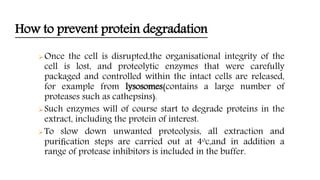
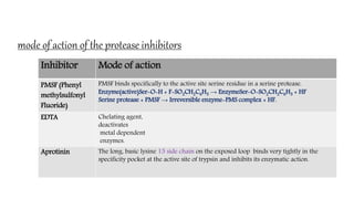
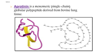
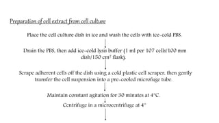
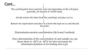
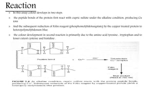
Preparation of tissue/cell(from cell culture)extracts
for protein separation. Comparision of protein
estimation by Lowry and Bradford methods,Principles
and advantages/disadvantages.
Presented by,
Yashwanth B S
FBT-MA7-05
introduction
 The initial step of any purification procedure must, of
course, be to disrupt the starting tissue to release proteins
from within the cell.
 The means of disrupting the tissue will depend on the cell
type.
 But thought must first be given to the composition of the
buffer used to extract the proteins.
Extraction buffer
 Normally extraction buffers are at an ionic strength (0.1–
0.2M) and pH (7.0–8.0) that is considered to be compatible
with that found inside the cell.
Protein location Buffer recommended
Whole Cell NP-40 or RIPA
Cytoplasmic (soluble) Tris-HCl
Cytoplasmic (cytoskeletal bound) Tris-Triton
Membrane bound NP-40 or RIPA
Nuclear RIPA or use nuclear fraction protocol.
Mitochondria
RIPA or use mitochondrial fraction
protocol.
RIPA buffer (RadioImmuno Precipitation Assay buffer)
 If there is concern that the protein of interest is not being
completely extracted from insoluble material or aggregates
,RIPA buffer may be more suitable, as it contains ionic
detergents that may more readily bring the proteins into
solution.
 Composition of RIPA buffer:
150 mM sodium chloride.
1.0% NP-40( Nonylphenoxypolyethoxylethanol ) or Triton X-100.
0.5% sodium deoxycholate.
0.1% SDS (sodium dodecyl sulphate).
50 mM Tris, pH 8.0
Cont…
 In cases where it is important to preserve protein-protein
interactions or to minimize denaturation (for example, when it is
known that the antibody to be used will only recognize a non-
denatured epitope), a buffer without ionic detergents (e.g. SDS)
and ideally without non-ionic detergents (e.g. Triton X-100)
should be used.
 Cell lysis with detergent-free buffer is achieved by mechanical
shearing, often with a Dounce homogenizer.
 In these cases a simple Tris buffer (20 mM Tris-HCl, pH 7.5) will
suffice , but buffers with detergents are required to release
membrane- or cytoskeleton-bound proteins.
How to prevent protein degradation
Once the cell is disrupted,the organisational integrity of the
cell is lost, and proteolytic enzymes that were carefully
packaged and controlled within the intact cells are released,
for example from lysosomes(contains a large number of
proteases such as cathepsins).
Such enzymes will of course start to degrade proteins in the
extract, including the protein of interest.
To slow down unwanted proteolysis, all extraction and
purification steps are carried out at 4oc,and in addition a
range of protease inhibitors is included in the buffer.
Protease inhibitors
Inhibitor Protease Inhibited
Final concentration in lysis
buffer
Stock (store at -20oC)
Aprotinin
Trypsin, Chymotrypsin,
Plasmin
2 μg/ml
Dilute in water, 10 mg/ml.
Do not re-use thawed
aliquots
Leupeptin
Lysosomal (inhibits serine
and cysteine proteases)
5-10 μg/ml
Dilute in water. Doo not
re-use once defrosted.
Pepstatin A
Aspartic proteases
(inhibits acid proteases)
1 μg/ml Dilute in methanol, 1mM.
PMSF (Phenyl
methylsulfonyl
Fluoride)
Chymotrypsin , thrombin
and papin
1 mM
Dilute in ethanol. You can
re-use the same aliquot.
EDTA.
Metalloproteases that
require Mg++ and Mn++
5 mM
Dilute in dH2O, 0.5M.
Adjust pH to 8.0.
mode of action of the protease inhibitors
Inhibitor Mode of action
PMSF (Phenyl
methylsulfonyl
Fluoride)
PMSF binds specifically to the active site serine residue in a serine protease.
Enzyme(active)Ser-O-H + F-SO2CH2C6H5 → EnzymeSer-O-SO2CH2C6H5 + HF
Serine protease + PMSF → Irreversible enzyme-PMS complex + HF.
EDTA Chelating agent,
deactivates
metal dependent
enzymes.
Aprotinin The long, basic lysine 15 side chain on the exposed loop binds very tightly in the
specificity pocket at the active site of trypsin and inhibits its enzymatic action.
…
 Aprotinin is a monomeric (single-chain)
globular polypeptide derived from bovine lung
tissue.
Anti oxidant
 Within the cell the protein is in a highly reducing environment,
but when released into the buffer it is exposed to a more
oxidising environment.
 Since most proteins contain a number of free sulphydryl
groups(from the amino acid cysteine) these can undergo
oxidation to give inter and intra molecular disulphide bridges.
 To prevent this,reducing agents such as dithiothreitol, b-
mercaptoethanol,are often included in the buffer.
Preparation of cell extract from cell culture
Place the cell culture dish in ice and wash the cells with ice-cold PBS.
Drain the PBS, then add ice-cold lysis buffer (1 ml per 107 cells/100 mm
dish/150 cm2 flask).
Scrape adherent cells off the dish using a cold plastic cell scraper, then gently
transfer the cell suspension into a pre-cooled microfuge tube.
Maintain constant agitation for 30 minutes at 4°C.
Centrifuge in a microcentrifuge at 4°
Cont…
The centrifugation force and time may vary depending on the cell type;(
generally, 20 minutes at 12,000 rpm).
Gently remove the tubes from the centrifuge and place on ice.
Remove the supernatant and place in a fresh tube kept on ice and discard
the pellet.
Determination protein concentration ( By Lowry’s method)
Once determination of the concentration of each sample, we can
freeze them at -20°C or -80°C for later use/repare for
immunoprecipitation or for loading onto a gel.
Lowry method of protein estimation
Principle
 The principle behind the Lowry method of determining protein concentrations lies in the
reactivity of the peptide nitrogen with the cupric sulfate under alkaline conditions and the
subsequent reduction of the Folin- Ciocalteau phosphomolybdic phosphotungstic acid to
heteropolymolybdenum blue by the copper-catalyzed oxidation of aromatic acids.
 The Lowry method is sensitive to pH changes and therefore the pH of assay solution should
be maintained at 9 - 10.5 .
 The intensity of the colour formed is directly proportional to the amount of these
compounds present in the protein.
 The colour developed is measured at 750nm in a spectrophotometer.
Reaction
 In this assay colour develops in two steps.
 the peptide bonds of the protein first react with cupric sulfate under the alkaline condition, producing Cu
ions
 And the subsequent reduction of Folin reagent (phosphomolybdotungstate) by the copper treated protein to
heteropolymolybdenum blue.
 the colour development in second reaction is primarily due to the amino acid tyrosine , tryptophan and to
lesser extent cysteine and histidine .
Advantages and disadvantages
Advantages
 Sensitive over a wide range
 The most commonly referenced procedure for protein determination
 Can be performed at room temperature
 10-20 times more sensitive than UV detection
 Can be performed in a microplate format
Disadvantages
 Many substances interfere with the assay
 Alkaline copper reagent is laborious to prepare and will develop carbonate scales over
storage which interfere with optical activity, thus it must be prepared fresh daily
 Takes a considerable amount of time to perform
 The assay is photosensitive, so illumination during the assay must be kept consistent for all
samples
 Amount of color varies with different proteins
 The major disadvantage of the Lowry method is the narrow Ph range within which it is
accurate.
Bradford method of protein estimation
:Principle
The assay is based on the observation that the absorbance maximum for an
acidic solution of Coomassie Brilliant Blue G-250 shifts from 465 nm to 595 nm
when binding to protein occurs. Both hydrophobic and ionic interactions
stabilize the anionic form of the dye, causing a visible color change. The assay is
useful since the extinction coefficient of a dye-albumin complex solution is
constant over a 10-fold concentration range. Within the linear range ofthe assay
(~5-25 µg/ ml), the more protein present, the more Coomassie binds. The
protein concentration of a test sample is determined by comparison to that of a
series of protein standards known to reproducibly exhibit a linear absorbance
profile in this assay.
Formation of protein dye complex in Bradford assay
• Samples treated with the Bradford
assay. The brown sample (lower
absorbance) contains no protein,
while the blue sample (higher
absorbance) contains protein.
• The amount of protein in the second
sample can be determined by
comparison to a standard curve
Advantages and disadvantages
Advantages
 Fast and inexpensive
 Highly specific for protein
 Very sensitive [1-20 µg (micro assay) 20-200 µg (macro assay)]
 Compatible with a wide range of substances
 Extinction co-efficient for the dye-protein complex is stable over 10 orders of
magnitude (assessed in albumin)
 Dye reagent complex is stable for approximately one hour
Disadvantages
 Absorbance spectra of the two coomassie brilliant blue G-250 species partially
overlap making the standard curve.
 Non-linear standard curve over wide ranges
 Response to different proteins can vary widely, choice of standard is very
important
 It is also inhibited by the presence of detergents.
 The Bradford assay is linear over a short range, typically from 0 µg/ml to
2000 µg/ml, often making dilutions of a sample necessary before analysis.
Comparision of lowry and Bradford method
 A comparison of the dye-binding method of Bradford (1976) with the
Folin phenol method of Lowry et al. (1951) for the assay of protein.
 The dye-binding method is fast, simple, and more sensitive, but the
Lowry procedure gives better linearity at high protein concentrations
and better stability once color has developed.
 Lowry is based on copper complex formation with the peptide bond
while Bradford is on dye binding onto charged residues. Lowry is then
influenced by the presence of some compounds with amine group like
ammonium sulphate (common used for fractionation), Bradford is
influenced by ph of your protein solution (not only due to the
protonation state of your charge residue side chain but also the dye has
different absorbance maxima at different ph). So, first look at the
content of your sample.
 Bradford and Lowry assays are really only quantitative if they are
calibrated not with BSA but with the protein with you are working with.
This is especially a problem for small proteins with a non-standard
distribution of amino acids.
Cont..
Lowry
The Lowry assay is based on reaction
with the folin - ciocalteau reagent
Responded strongly to
acetaminophen, aspirin, methadone,
morphine, amoxicillin, ampicillin,
penicillin G,
The colour developed is measured at
750nm in a spectrophotometer.
It was less sensitive the compare to
the bradford method
Bradford
The bradford assay is based on the
binding of dye to the proteins which
results in a dye–protein complex
with increased molar absorbance
Responded strongly to thioridazine
and less strongly to the other
antipsychotic drugs
The colour developed is meseaured at
595nm in a spectrophotometer
It was faster and more sensitive then
compare to the Lowry method
Importance of TCA precipitation of protein
TCA is a good choice for some precipitation as it can be used at reasonably
low concentrations(<20%). This means that the volume can be kept low, and
therefore the protein concentration high.
 A precipitation step that allows the separation of the protein sample from
interfering substances and also consequently concentrates the protein
sample, allowing the determination of proteins in dilute solution.
 Trichloroacetic acid (TCA) precipitation can be used to precipitate proteins
away from TCA-soluble, low-molecular-weight compounds that may
interfere with assays for total protein.
Cont…
Trichloroacetic acid (TCA) denatures protein by driving hydrophobic aggregation.
TCA acts primarily through two mechanisms
1) Denaturation of the hydration shells around the protein:-These are structured regions of
water that form around hydrophobic patches on the surface of the folded protein. The
waters in the shell are stolen into the hydration shell around the precipitant, effectively
increasing the hydrophobic effect.
2) In addition ,the anionic TCA may trigger partial protein unfolding through disruption of
the electrostatic interactions which determine the native tertiary structure of the protein.
As a result , the usually well – hidden hydrophobic interior of the protein becomes
exposed to the solvent.
Both of these results in the intermolecular coalescence of single protein molecules into
larger aggregates, to minimise the exposure of hydrophobic regions to the solvent. If the
aggregates grows sufficiently large in size, it will displace more water and increase in density-
forming an opaque precipitate.
Trichloroacetic acid precipitation
Add equal volume of 100% TCA to the sample and mix
leave 20 min on ice or for 15 min at –20o C
centrifuge 5 min at 6000-10000rpm.
Remove the supernatant - resuspend the pellet with 0.1N NaoH or wash the
pellet with an ethanol/ether (1/1) solution and resuspend the pellet in a buffer
solution
References;
• Principles and Techniques of Biochemistry and
Molecular Biology - Wilson and Walker.
• Molecular Cloning : A Laboratory Manual - Joseph
Sambrook and David Russell.
• www.wikipedia.com
PREPERATION F CELL EXTRACT

PREPERATION F CELL EXTRACT

  • 1.
    Preparation of tissue/cell(fromcell culture)extracts for protein separation. Comparision of protein estimation by Lowry and Bradford methods,Principles and advantages/disadvantages. Presented by, Yashwanth B S FBT-MA7-05
  • 2.
    introduction  The initialstep of any purification procedure must, of course, be to disrupt the starting tissue to release proteins from within the cell.  The means of disrupting the tissue will depend on the cell type.  But thought must first be given to the composition of the buffer used to extract the proteins.
  • 3.
    Extraction buffer  Normallyextraction buffers are at an ionic strength (0.1– 0.2M) and pH (7.0–8.0) that is considered to be compatible with that found inside the cell. Protein location Buffer recommended Whole Cell NP-40 or RIPA Cytoplasmic (soluble) Tris-HCl Cytoplasmic (cytoskeletal bound) Tris-Triton Membrane bound NP-40 or RIPA Nuclear RIPA or use nuclear fraction protocol. Mitochondria RIPA or use mitochondrial fraction protocol.
  • 4.
    RIPA buffer (RadioImmunoPrecipitation Assay buffer)  If there is concern that the protein of interest is not being completely extracted from insoluble material or aggregates ,RIPA buffer may be more suitable, as it contains ionic detergents that may more readily bring the proteins into solution.  Composition of RIPA buffer: 150 mM sodium chloride. 1.0% NP-40( Nonylphenoxypolyethoxylethanol ) or Triton X-100. 0.5% sodium deoxycholate. 0.1% SDS (sodium dodecyl sulphate). 50 mM Tris, pH 8.0
  • 5.
    Cont…  In caseswhere it is important to preserve protein-protein interactions or to minimize denaturation (for example, when it is known that the antibody to be used will only recognize a non- denatured epitope), a buffer without ionic detergents (e.g. SDS) and ideally without non-ionic detergents (e.g. Triton X-100) should be used.  Cell lysis with detergent-free buffer is achieved by mechanical shearing, often with a Dounce homogenizer.  In these cases a simple Tris buffer (20 mM Tris-HCl, pH 7.5) will suffice , but buffers with detergents are required to release membrane- or cytoskeleton-bound proteins.
  • 6.
    How to preventprotein degradation Once the cell is disrupted,the organisational integrity of the cell is lost, and proteolytic enzymes that were carefully packaged and controlled within the intact cells are released, for example from lysosomes(contains a large number of proteases such as cathepsins). Such enzymes will of course start to degrade proteins in the extract, including the protein of interest. To slow down unwanted proteolysis, all extraction and purification steps are carried out at 4oc,and in addition a range of protease inhibitors is included in the buffer.
  • 7.
    Protease inhibitors Inhibitor ProteaseInhibited Final concentration in lysis buffer Stock (store at -20oC) Aprotinin Trypsin, Chymotrypsin, Plasmin 2 μg/ml Dilute in water, 10 mg/ml. Do not re-use thawed aliquots Leupeptin Lysosomal (inhibits serine and cysteine proteases) 5-10 μg/ml Dilute in water. Doo not re-use once defrosted. Pepstatin A Aspartic proteases (inhibits acid proteases) 1 μg/ml Dilute in methanol, 1mM. PMSF (Phenyl methylsulfonyl Fluoride) Chymotrypsin , thrombin and papin 1 mM Dilute in ethanol. You can re-use the same aliquot. EDTA. Metalloproteases that require Mg++ and Mn++ 5 mM Dilute in dH2O, 0.5M. Adjust pH to 8.0.
  • 8.
    mode of actionof the protease inhibitors Inhibitor Mode of action PMSF (Phenyl methylsulfonyl Fluoride) PMSF binds specifically to the active site serine residue in a serine protease. Enzyme(active)Ser-O-H + F-SO2CH2C6H5 → EnzymeSer-O-SO2CH2C6H5 + HF Serine protease + PMSF → Irreversible enzyme-PMS complex + HF. EDTA Chelating agent, deactivates metal dependent enzymes. Aprotinin The long, basic lysine 15 side chain on the exposed loop binds very tightly in the specificity pocket at the active site of trypsin and inhibits its enzymatic action.
  • 9.
    …  Aprotinin isa monomeric (single-chain) globular polypeptide derived from bovine lung tissue.
  • 10.
    Anti oxidant  Withinthe cell the protein is in a highly reducing environment, but when released into the buffer it is exposed to a more oxidising environment.  Since most proteins contain a number of free sulphydryl groups(from the amino acid cysteine) these can undergo oxidation to give inter and intra molecular disulphide bridges.  To prevent this,reducing agents such as dithiothreitol, b- mercaptoethanol,are often included in the buffer.
  • 11.
    Preparation of cellextract from cell culture Place the cell culture dish in ice and wash the cells with ice-cold PBS. Drain the PBS, then add ice-cold lysis buffer (1 ml per 107 cells/100 mm dish/150 cm2 flask). Scrape adherent cells off the dish using a cold plastic cell scraper, then gently transfer the cell suspension into a pre-cooled microfuge tube. Maintain constant agitation for 30 minutes at 4°C. Centrifuge in a microcentrifuge at 4°
  • 12.
    Cont… The centrifugation forceand time may vary depending on the cell type;( generally, 20 minutes at 12,000 rpm). Gently remove the tubes from the centrifuge and place on ice. Remove the supernatant and place in a fresh tube kept on ice and discard the pellet. Determination protein concentration ( By Lowry’s method) Once determination of the concentration of each sample, we can freeze them at -20°C or -80°C for later use/repare for immunoprecipitation or for loading onto a gel.
  • 13.
    Lowry method ofprotein estimation Principle  The principle behind the Lowry method of determining protein concentrations lies in the reactivity of the peptide nitrogen with the cupric sulfate under alkaline conditions and the subsequent reduction of the Folin- Ciocalteau phosphomolybdic phosphotungstic acid to heteropolymolybdenum blue by the copper-catalyzed oxidation of aromatic acids.  The Lowry method is sensitive to pH changes and therefore the pH of assay solution should be maintained at 9 - 10.5 .  The intensity of the colour formed is directly proportional to the amount of these compounds present in the protein.  The colour developed is measured at 750nm in a spectrophotometer.
  • 14.
    Reaction  In thisassay colour develops in two steps.  the peptide bonds of the protein first react with cupric sulfate under the alkaline condition, producing Cu ions  And the subsequent reduction of Folin reagent (phosphomolybdotungstate) by the copper treated protein to heteropolymolybdenum blue.  the colour development in second reaction is primarily due to the amino acid tyrosine , tryptophan and to lesser extent cysteine and histidine .
  • 15.
    Advantages and disadvantages Advantages Sensitive over a wide range  The most commonly referenced procedure for protein determination  Can be performed at room temperature  10-20 times more sensitive than UV detection  Can be performed in a microplate format Disadvantages  Many substances interfere with the assay  Alkaline copper reagent is laborious to prepare and will develop carbonate scales over storage which interfere with optical activity, thus it must be prepared fresh daily  Takes a considerable amount of time to perform  The assay is photosensitive, so illumination during the assay must be kept consistent for all samples  Amount of color varies with different proteins  The major disadvantage of the Lowry method is the narrow Ph range within which it is accurate.
  • 16.
    Bradford method ofprotein estimation :Principle The assay is based on the observation that the absorbance maximum for an acidic solution of Coomassie Brilliant Blue G-250 shifts from 465 nm to 595 nm when binding to protein occurs. Both hydrophobic and ionic interactions stabilize the anionic form of the dye, causing a visible color change. The assay is useful since the extinction coefficient of a dye-albumin complex solution is constant over a 10-fold concentration range. Within the linear range ofthe assay (~5-25 µg/ ml), the more protein present, the more Coomassie binds. The protein concentration of a test sample is determined by comparison to that of a series of protein standards known to reproducibly exhibit a linear absorbance profile in this assay.
  • 17.
    Formation of proteindye complex in Bradford assay • Samples treated with the Bradford assay. The brown sample (lower absorbance) contains no protein, while the blue sample (higher absorbance) contains protein. • The amount of protein in the second sample can be determined by comparison to a standard curve
  • 18.
    Advantages and disadvantages Advantages Fast and inexpensive  Highly specific for protein  Very sensitive [1-20 µg (micro assay) 20-200 µg (macro assay)]  Compatible with a wide range of substances  Extinction co-efficient for the dye-protein complex is stable over 10 orders of magnitude (assessed in albumin)  Dye reagent complex is stable for approximately one hour Disadvantages  Absorbance spectra of the two coomassie brilliant blue G-250 species partially overlap making the standard curve.  Non-linear standard curve over wide ranges  Response to different proteins can vary widely, choice of standard is very important  It is also inhibited by the presence of detergents.  The Bradford assay is linear over a short range, typically from 0 µg/ml to 2000 µg/ml, often making dilutions of a sample necessary before analysis.
  • 19.
    Comparision of lowryand Bradford method  A comparison of the dye-binding method of Bradford (1976) with the Folin phenol method of Lowry et al. (1951) for the assay of protein.  The dye-binding method is fast, simple, and more sensitive, but the Lowry procedure gives better linearity at high protein concentrations and better stability once color has developed.  Lowry is based on copper complex formation with the peptide bond while Bradford is on dye binding onto charged residues. Lowry is then influenced by the presence of some compounds with amine group like ammonium sulphate (common used for fractionation), Bradford is influenced by ph of your protein solution (not only due to the protonation state of your charge residue side chain but also the dye has different absorbance maxima at different ph). So, first look at the content of your sample.  Bradford and Lowry assays are really only quantitative if they are calibrated not with BSA but with the protein with you are working with. This is especially a problem for small proteins with a non-standard distribution of amino acids.
  • 20.
    Cont.. Lowry The Lowry assayis based on reaction with the folin - ciocalteau reagent Responded strongly to acetaminophen, aspirin, methadone, morphine, amoxicillin, ampicillin, penicillin G, The colour developed is measured at 750nm in a spectrophotometer. It was less sensitive the compare to the bradford method Bradford The bradford assay is based on the binding of dye to the proteins which results in a dye–protein complex with increased molar absorbance Responded strongly to thioridazine and less strongly to the other antipsychotic drugs The colour developed is meseaured at 595nm in a spectrophotometer It was faster and more sensitive then compare to the Lowry method
  • 21.
    Importance of TCAprecipitation of protein TCA is a good choice for some precipitation as it can be used at reasonably low concentrations(<20%). This means that the volume can be kept low, and therefore the protein concentration high.  A precipitation step that allows the separation of the protein sample from interfering substances and also consequently concentrates the protein sample, allowing the determination of proteins in dilute solution.  Trichloroacetic acid (TCA) precipitation can be used to precipitate proteins away from TCA-soluble, low-molecular-weight compounds that may interfere with assays for total protein.
  • 22.
    Cont… Trichloroacetic acid (TCA)denatures protein by driving hydrophobic aggregation. TCA acts primarily through two mechanisms 1) Denaturation of the hydration shells around the protein:-These are structured regions of water that form around hydrophobic patches on the surface of the folded protein. The waters in the shell are stolen into the hydration shell around the precipitant, effectively increasing the hydrophobic effect. 2) In addition ,the anionic TCA may trigger partial protein unfolding through disruption of the electrostatic interactions which determine the native tertiary structure of the protein. As a result , the usually well – hidden hydrophobic interior of the protein becomes exposed to the solvent. Both of these results in the intermolecular coalescence of single protein molecules into larger aggregates, to minimise the exposure of hydrophobic regions to the solvent. If the aggregates grows sufficiently large in size, it will displace more water and increase in density- forming an opaque precipitate.
  • 23.
    Trichloroacetic acid precipitation Addequal volume of 100% TCA to the sample and mix leave 20 min on ice or for 15 min at –20o C centrifuge 5 min at 6000-10000rpm. Remove the supernatant - resuspend the pellet with 0.1N NaoH or wash the pellet with an ethanol/ether (1/1) solution and resuspend the pellet in a buffer solution
  • 24.
    References; • Principles andTechniques of Biochemistry and Molecular Biology - Wilson and Walker. • Molecular Cloning : A Laboratory Manual - Joseph Sambrook and David Russell. • www.wikipedia.com