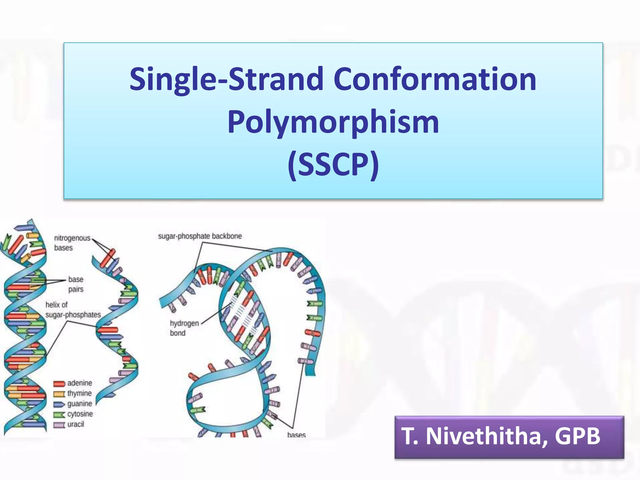
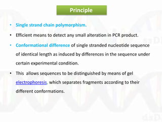
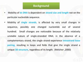
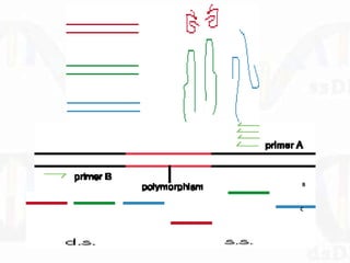
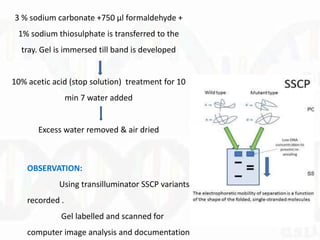
Single-Strand Conformation
Polymorphism
(SSCP)
T. Nivethitha, GPB
• Single strand chain polymorphism.
• Efficient means to detect any small alteration in PCR product.
• Conformational difference of single stranded nucleotide sequence
of identical length as induced by differences in the sequence under
certain experimental condition.
• This allows sequences to be distinguished by means of gel
electrophoresis, which separates fragments according to their
different conformations.
Principle
Background
• Mobility of ds DNA is dependent on strand size and length not on the
particular nucleotide sequence.
• Mobility of single strands, is affected by very small changes in
sequence, possibly one changed nucleotide out of several
hundred. Small changes are noticeable because of the relatively
unstable nature of single-stranded DNA; in the absence of a
complementary strand, the single strand experience intrastrand base
pairing, resulting in loops and folds that give the single strand a
unique 3D structure, regardless of its length. (Melcher ,2000)
A
B
C
Steps
DNA extraction
Amplification of DNA fragments – PCR
Denaturation followed by rapid Cooling
Separation on non denaturing PAGE gel
Visualization by Silver staining or autoradiography
Preparation of SSCP gel
Composition of 12% non dentauring PAGE
• Gel mixture is kept dissolved completely and stored @ -20 degree
Celsius till it is used.
Components Volume
50% Acrylamide - bisacrylamide 24ml
10X TBE 5 ml
Glycerol 10 ml
Water 61 ml
PCR - SSCP
SSCP – analysis of amplified gene fragment is carried out using BIO RAD
protein II xi Cell vertical gel electrophoresis unit.
Gel sandwich is prepared and the cleaned comb is inserted from
topside of sandwich and clamps applied over the plates comb to create sharp
wells.
Bottom side of gel sandwich is
Sealed using 10 ml gel mix. Kept in slanting
position 50 µl APS + 20 µl TEMED is injected
b/w glass plates and polymerised.
• After polymerisation the assembled gel sandwich is placed in casting slot.
And 12% PAGE gel prepared by adding gel mix.(45 min polymerisation)
• Comb removed, well flushed with 10x buffer.
• Gel placed in electrophoretic tank (filled with 0.5x TBE) & pre run @200
volts.
PCR tube (4µl PCR product + 12 µl formamide dye )- denature
@95◦C for 10 min.
Kept in ice chilled box (-20◦C deep freeze for 10 min)
After pre run samples loaded in gel & electrophoresis
performed in 0.5 X Tris Borate (pH 8.3)- EDTA bufer @ 10-
12.5 V/cm for 3-24 hr@ room T depending on optimized
condition for primer.
Gel – SILVER STAINING to visualize bands
Immersed in 10% acetic acid (30 min) to fix DNA bands
Acetic acid decanted & rinsed with water and decanted
Gel stained in 0.1% silver nitrate 500 ml+ 750 µl
37% formaldehyde(45 min)
Rinsed with water & water decanted
3 % sodium carbonate +750 µl formaldehyde +
1% sodium thiosulphate is transferred to the
tray. Gel is immersed till band is developed
10% acetic acid (stop solution) treatment for 10
min 7 water added
Excess water removed & air dried
OBSERVATION:
Using transilluminator SSCP variants
recorded .
Gel labelled and scanned for
computer image analysis and documentation
Features of SSCP
The sensitivity of PCR-SSCP depends on
 The mutation pattern in the target sequence.
 Size of DNA fragments and its GC content.
 Gel temperature.
 Gel matrix composition.
 Buffer composition (ionic strength and pH).
 DNA
Advantage
SSCP screening allows to determine:
 if the gene contains sufficient polymorphism.
 which portion of the gene is most polymorphic
 if there is polymorphism among multicopy genes with individuals.
 what level of intraspecific variation exists.
Many individual PCR products are screened simultaneously.
Disadvantage
• The amount of mobility difference have little if any correlation to
amount of sequence differences.
• Only information gained is whether the PCR amplicon is “identical”
or “different “.
• Absence of mutation cannot be proven, since some mutation may
remain undetected.
• Sequence data needed for primer construction.
• High standardized electrophoretic condition is needed.
SSCP LIMITATIONS AND CONSIDERATIONS
• Single-stranded DNA mobilities are dependent on
temperature. For best results, gel electrophoresis must be run in a
constant temperature.
• Sensitivity of SSCP is affected by pH. ds DNA fragments are
denatured by exposure to basic conditions: a high pH. (Kukita et al).
found that adding glycerol to the polyacrylamide gel lowers the pH
of the electrophoresis buffer--more specifically, the Tris-borate
buffer--and the result is increased SSCP sensitivity and clearer data.
• Fragment length - 150-300 bp.
Application
• Scientific method to detect mutation without sequencing.
• To discover new DNA Polymorphism apart from sequencing.
• Diagnostic tools in Molecular Biology.
• In virology to detect various strains in viruses.
• For detecting microlesions, such as single-base substitutions, small
deletions, small insertions, or microinversions
• To detect mutations in DNA or reverse-transcribed RNA
(complementary DNA) samples in a large number of human disease
genes as well as in tumor-derived material.
Reference
• Molecular Markers & Plant Biotechnology – Rukam S Tomar et.al,
• Single-Strand Conformation Polymorphism (SSCP) Analysis - Igor
Vorechovsky
• PCR–SSCP: A Method for the Molecular Analysis of Genetic Diseases
Kakavas V. Konstantinos, Plageras Panagiotis

Single strand conformation polymorphism

  • 1.
  • 2.
    • Single strandchain polymorphism. • Efficient means to detect any small alteration in PCR product. • Conformational difference of single stranded nucleotide sequence of identical length as induced by differences in the sequence under certain experimental condition. • This allows sequences to be distinguished by means of gel electrophoresis, which separates fragments according to their different conformations. Principle
  • 3.
    Background • Mobility ofds DNA is dependent on strand size and length not on the particular nucleotide sequence. • Mobility of single strands, is affected by very small changes in sequence, possibly one changed nucleotide out of several hundred. Small changes are noticeable because of the relatively unstable nature of single-stranded DNA; in the absence of a complementary strand, the single strand experience intrastrand base pairing, resulting in loops and folds that give the single strand a unique 3D structure, regardless of its length. (Melcher ,2000)
  • 4.
  • 5.
    Steps DNA extraction Amplification ofDNA fragments – PCR Denaturation followed by rapid Cooling Separation on non denaturing PAGE gel Visualization by Silver staining or autoradiography
  • 6.
    Preparation of SSCPgel Composition of 12% non dentauring PAGE • Gel mixture is kept dissolved completely and stored @ -20 degree Celsius till it is used. Components Volume 50% Acrylamide - bisacrylamide 24ml 10X TBE 5 ml Glycerol 10 ml Water 61 ml
  • 7.
    PCR - SSCP SSCP– analysis of amplified gene fragment is carried out using BIO RAD protein II xi Cell vertical gel electrophoresis unit. Gel sandwich is prepared and the cleaned comb is inserted from topside of sandwich and clamps applied over the plates comb to create sharp wells. Bottom side of gel sandwich is Sealed using 10 ml gel mix. Kept in slanting position 50 µl APS + 20 µl TEMED is injected b/w glass plates and polymerised.
  • 8.
    • After polymerisationthe assembled gel sandwich is placed in casting slot. And 12% PAGE gel prepared by adding gel mix.(45 min polymerisation) • Comb removed, well flushed with 10x buffer. • Gel placed in electrophoretic tank (filled with 0.5x TBE) & pre run @200 volts. PCR tube (4µl PCR product + 12 µl formamide dye )- denature @95◦C for 10 min. Kept in ice chilled box (-20◦C deep freeze for 10 min)
  • 10.
    After pre runsamples loaded in gel & electrophoresis performed in 0.5 X Tris Borate (pH 8.3)- EDTA bufer @ 10- 12.5 V/cm for 3-24 hr@ room T depending on optimized condition for primer. Gel – SILVER STAINING to visualize bands Immersed in 10% acetic acid (30 min) to fix DNA bands Acetic acid decanted & rinsed with water and decanted Gel stained in 0.1% silver nitrate 500 ml+ 750 µl 37% formaldehyde(45 min) Rinsed with water & water decanted
  • 11.
    3 % sodiumcarbonate +750 µl formaldehyde + 1% sodium thiosulphate is transferred to the tray. Gel is immersed till band is developed 10% acetic acid (stop solution) treatment for 10 min 7 water added Excess water removed & air dried OBSERVATION: Using transilluminator SSCP variants recorded . Gel labelled and scanned for computer image analysis and documentation
  • 12.
  • 13.
    The sensitivity ofPCR-SSCP depends on  The mutation pattern in the target sequence.  Size of DNA fragments and its GC content.  Gel temperature.  Gel matrix composition.  Buffer composition (ionic strength and pH).  DNA
  • 14.
    Advantage SSCP screening allowsto determine:  if the gene contains sufficient polymorphism.  which portion of the gene is most polymorphic  if there is polymorphism among multicopy genes with individuals.  what level of intraspecific variation exists. Many individual PCR products are screened simultaneously.
  • 15.
    Disadvantage • The amountof mobility difference have little if any correlation to amount of sequence differences. • Only information gained is whether the PCR amplicon is “identical” or “different “. • Absence of mutation cannot be proven, since some mutation may remain undetected. • Sequence data needed for primer construction. • High standardized electrophoretic condition is needed.
  • 16.
    SSCP LIMITATIONS ANDCONSIDERATIONS • Single-stranded DNA mobilities are dependent on temperature. For best results, gel electrophoresis must be run in a constant temperature. • Sensitivity of SSCP is affected by pH. ds DNA fragments are denatured by exposure to basic conditions: a high pH. (Kukita et al). found that adding glycerol to the polyacrylamide gel lowers the pH of the electrophoresis buffer--more specifically, the Tris-borate buffer--and the result is increased SSCP sensitivity and clearer data. • Fragment length - 150-300 bp.
  • 17.
    Application • Scientific methodto detect mutation without sequencing. • To discover new DNA Polymorphism apart from sequencing. • Diagnostic tools in Molecular Biology. • In virology to detect various strains in viruses. • For detecting microlesions, such as single-base substitutions, small deletions, small insertions, or microinversions • To detect mutations in DNA or reverse-transcribed RNA (complementary DNA) samples in a large number of human disease genes as well as in tumor-derived material.
  • 18.
    Reference • Molecular Markers& Plant Biotechnology – Rukam S Tomar et.al, • Single-Strand Conformation Polymorphism (SSCP) Analysis - Igor Vorechovsky • PCR–SSCP: A Method for the Molecular Analysis of Genetic Diseases Kakavas V. Konstantinos, Plageras Panagiotis